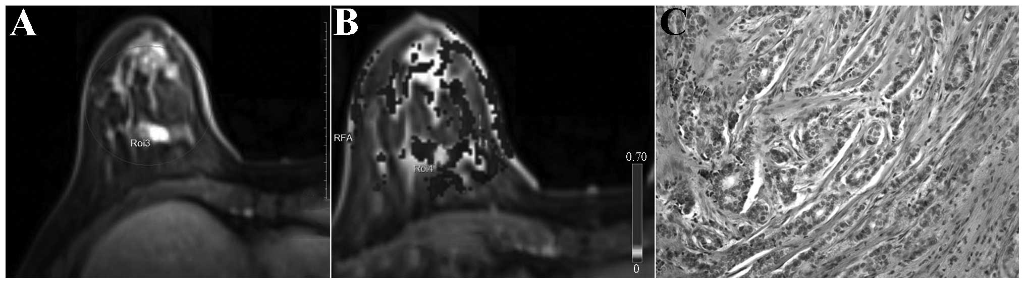
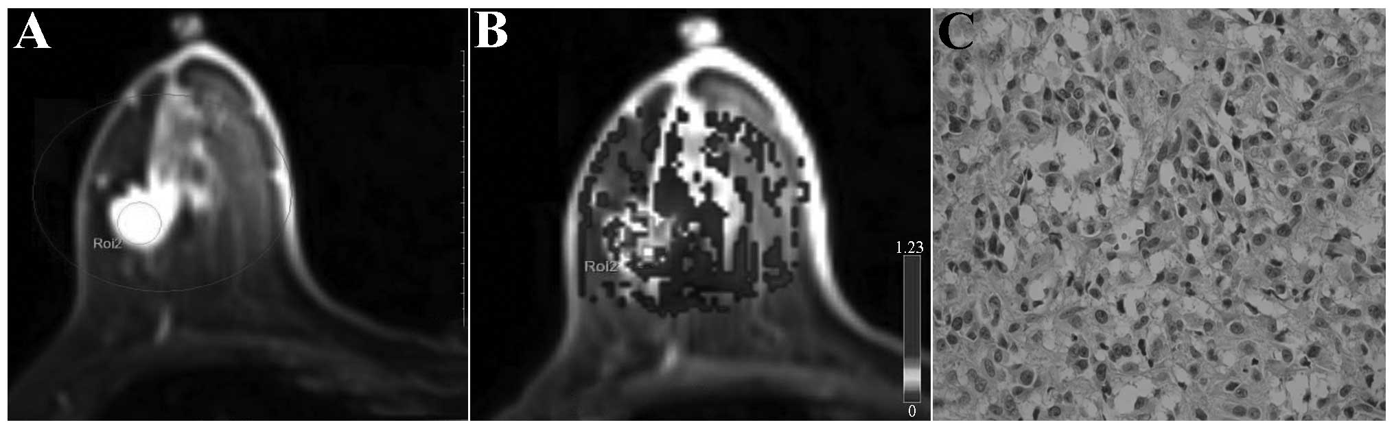
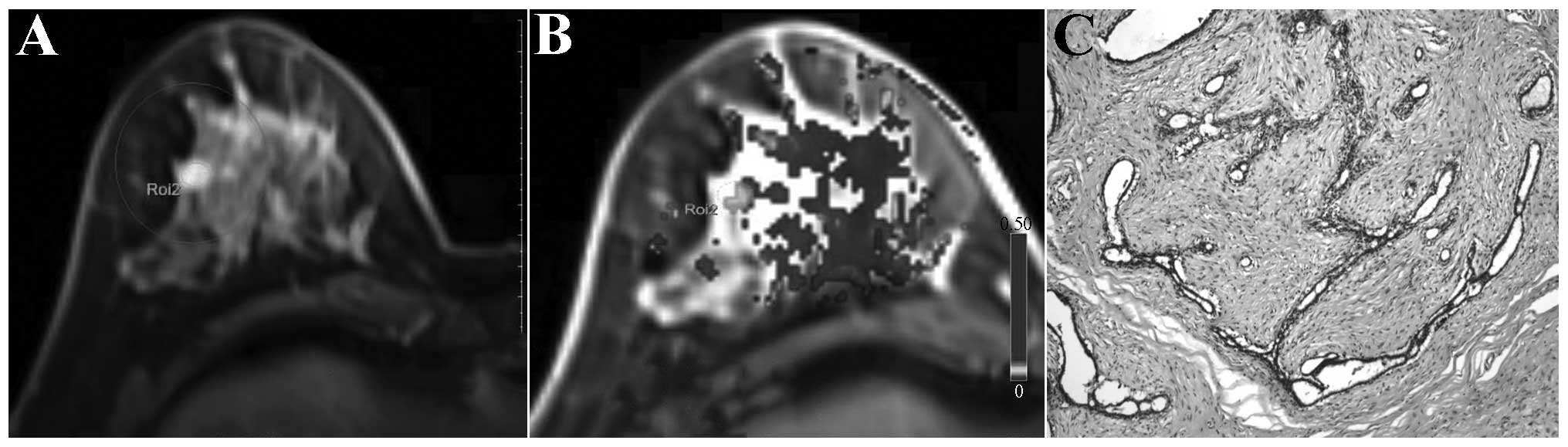
|
1
|
Berg WA, Gutierrez L, NessAiver MS, Carter
WB, Bhargavan M, Lewis RS and Ioffe OB: Diagnostic accuracy of
mammography, clinical examination, US, and MR imaging in
preoperative assessment of breast cancer. Radiology. 233:830–849.
2004. View Article : Google Scholar : PubMed/NCBI
|
|
2
|
Liberman L: Breast cancer screening with
MRI - what are the data for patients at high risk? N Engl J Med.
351:497–500. 2004. View Article : Google Scholar : PubMed/NCBI
|
|
3
|
Folkman J: Tumor angiogenesis: therapeutic
implications. N Engl J Med. 285:1182–1186. 1971. View Article : Google Scholar : PubMed/NCBI
|
|
4
|
Weidner N, Folkman J, Pozza F, et al:
Tumor angiogenesis: a new significant and independent prognostic
indicator in early-stage breast carcinoma. J Natl Cancer Inst.
84:1875–1887. 1992. View Article : Google Scholar : PubMed/NCBI
|
|
5
|
DeVries A, Griebel J, et al:
Perfusion-index values evaluated by dynamic magnetic resonance
imaging in advanced rectal carcinoma. A new predictor of response
to preoperative radiochemotherapy? Strahlenther Onkol. 176:567–572.
2000.(In German).
|
|
6
|
Kremer S, Grand S, Rémy C, et al:
Contribution of dynamic contrast MR imaging to the differentiation
between dural metastasis and meningioma. Neuroradiology.
46:642–648. 2004. View Article : Google Scholar : PubMed/NCBI
|
|
7
|
Haris M, Gupta RK, Singh A, et al:
Differentiation of infective from neoplastic brain lesions by
dynamic contrast-enhanced MRI. Neuroradiology. 50:531–540. 2008.
View Article : Google Scholar : PubMed/NCBI
|
|
8
|
Zhang H, Rödiger LA, et al: Perfusion MR
imaging for differentiation of benign and malignant meningiomas.
Neuroradiology. 50:525–530. 2008. View Article : Google Scholar : PubMed/NCBI
|
|
9
|
Pickles MD, Manton DJ, Lowry M and
Turnbull LW: Prognostic value of pre-treatment DCE-MRI parameters
in predicting disease free and overall survival for breast cancer
patients undergoing neoadjuvant chemotherapy. Eur J Radiol.
71:498–505. 2009. View Article : Google Scholar
|
|
10
|
Kang TW, Kim ST, Byun HS, et al:
Morphological and functional MRI, MRS, perfusion and diffusion
changes after radiosurgery of brain metastasis. Eur J Radiol.
72:370–380. 2009. View Article : Google Scholar
|
|
11
|
Hauser T, Essig M, Jensen A, et al:
Characterization and therapy monitoring of head and neck carcinomas
using diffusion-imaging-based intravoxel incoherent motion
parameters-preliminary results. Neuroradiology. 55:527–536. 2013.
View Article : Google Scholar : PubMed/NCBI
|
|
12
|
Larsen VA, Simonsen HJ, Law I, Larsson HB
and Hansen AE: Evaluation of dynamic contrast-enhanced T1-weighted
perfusion MRI in the differentiation of tumor recurrence from
radiation necrosis. Neuroradiology. 55:361–369. 2013. View Article : Google Scholar
|
|
13
|
Bäuerle T, Seyler L, Münter M, et al:
Diffusion-weighted imaging in rectal carcinoma patients without and
after chemoradiotherapy: a comparative study with histology. Eur J
Radiol. 82:444–452. 2013. View Article : Google Scholar
|
|
14
|
Jones EF, Sinha SP, Newitt DC, Klifa C,
Kornak J, Park CC and Hylton NM: Mri enhancement in stromal tissue
surrounding breast tumors: association with recurrence free
survival following neoadjuvant chemotherapy. PLoS One.
8:e619692013. View Article : Google Scholar : PubMed/NCBI
|
|
15
|
Li X, Arlinghaus LR, Ayers GD, et al:
Dce-mri analysis methods for predicting the response of breast
cancer to neoadjuvant chemotherapy: pilot study findings. Magn
Reson Med. 71:1592–1602. 2014. View Article : Google Scholar
|
|
16
|
Schnall MD, Blume J, Bluemke DA, DeAngelis
GA, et al: Diagnostic architectural and dynamic features at breast
MR imaging: multicenter study. Radiology. 238:42–53. 2006.
View Article : Google Scholar
|
|
17
|
Rykala J, Przybylowska K, Majsterek I, et
al: Angiogenesis markers quantification in breast cancer and their
correlation with clinicopathological prognostic variables. Pathol
Oncol Res. 17:809–817. 2011. View Article : Google Scholar : PubMed/NCBI
|
|
18
|
Medeiros LR, Duarte CS, Rosa DD, Edelweiss
MI, et al: Accuracy of magnetic resonance in suspicious breast
lesions: a systematic quantitative review and meta-analysis. Breast
Cancer Res Treat. 126:273–285. 2011. View Article : Google Scholar : PubMed/NCBI
|
|
19
|
Tofts PS, Brix G, Buckley DL, et al:
Estimating kinetic parameters from dynamic contrast-enhanced
t1-weighted mri of a diffusable tracer: standardized quantities and
symbols. J Magn Reson Imaging. 10:223–232. 1999. View Article : Google Scholar : PubMed/NCBI
|
|
20
|
Padhani AR and Husband JE: Dynamic
contrast-enhanced mri studies in oncology with an emphasis on
quantification, validation and human studies. Clin Radiol.
56:607–620. 2001. View Article : Google Scholar : PubMed/NCBI
|
|
21
|
Preda A, van Vliet M, Krestin GP, Brasch
RC and van Dijke CF: Magnetic resonance macromolecular agents for
monitoring tumor microvessels and angiogenesis inhibition. Invest
Radiol. 41:325–331. 2006. View Article : Google Scholar : PubMed/NCBI
|
|
22
|
Ocak I, Bernardo M, Metzger G, Barrett T,
Pinto P, Albert PS and Choyke PL: Dynamic contrast-enhanced mri of
prostate cancer at 3 t: a study of pharmacokinetic parameters. AJR
Am J Roentgenol. 189:849–858. 2007. View Article : Google Scholar : PubMed/NCBI
|
|
23
|
Yao WW, Zhang H, Ding B, et al: Rectal
cancer: 3d dynamic contrast-enhanced mri; correlation with
microvascular density and clinicopathological features. Radiol med.
116:366–374. 2011. View Article : Google Scholar : PubMed/NCBI
|
|
24
|
Li X, Arlinghaus LR, Ayers GD, et al:
dce-mri analysis methods for predicting the response of breast
cancer to neoadjuvant chemotherapy: pilot study findings. Magn
Reson Med. 71:1592–1602. 2014. View Article : Google Scholar
|
|
25
|
Nilsen LB, Fangberget A, Geier OM,
Engebraaten O, Borgen E, Olsen DR and Seierstad T: Associations
between tumor vascularization assessed by in vivo dce-mri and the
presence of disseminated tumor cells in bone marrow in breast
cancer patients at the time of diagnosis. J Magn Reson Imaging. Jan
27–2014.(Epub ahead of print). View Article : Google Scholar : PubMed/NCBI
|
|
26
|
Jain RK: Normalization of tumor
vasculature: an emerging concept in antiangiogenic therapy.
Science. 307:58–62. 2005. View Article : Google Scholar : PubMed/NCBI
|
|
27
|
Tofts PS: Modeling tracer kinetics in
dynamic gd-dtpa mr imaging. J Magn Reson Imaging. 7:91–101. 1997.
View Article : Google Scholar : PubMed/NCBI
|
|
28
|
Wang Y1, Huang W, Panicek DM, et al:
Feasibility of using limited-population-based arterial input
function for pharmacokinetic modeling of osteosarcoma dynamic
contrast-enhanced MRI data. Magn Reson Med. 59:1183–1189. 2008.
View Article : Google Scholar : PubMed/NCBI
|
|
29
|
Koo HR, Cho N, Song IC, et al: Correlation
of perfusion parameters on dynamic contrast-enhanced MRI with
prognostic factors and subtypes of breast cancers. J Magn Reson
Imaging. 36:145–151. 2012. View Article : Google Scholar : PubMed/NCBI
|
|
30
|
Siegmann KC, Müller-Schimpfle M, Schick F,
et al: Mr imaging-detected breast lesions: histopathologic
correlation of lesion characteristics and signal intensity data. Am
J Roentgenol. 178:1403–1409. 2002. View Article : Google Scholar
|
|
31
|
Baek HM, Chen JH, Nie K, et al: Predicting
pathologic response to neoadjuvant chemotherapy in breast cancer by
using mr imaging and quantitative 1h mr spectroscopy. Radiology.
251:653–662. 2009. View Article : Google Scholar : PubMed/NCBI
|