Introduction
Sepsis is a combination of clinical manifestations
of systemic inflammation specifically associated with an infectious
insult (1). The condition is the
most frequent cause of mortality in the majority of intensive care
units and is responsible for >250,000 mortalities in the United
States annually (2). Despite
advances in basic and clinical research, there is no effective
therapeutic intervention against the disease. Lipopolysaccharide
(LPS), a component of the outer cell wall in Gram-negative bacteria
(3), activates intracellular
signaling pathways via Toll-like receptor (TLR) 4. CpG DNA,
contained in microbial DNA sequences, is recognized by TLR9. LPS
and CpG DNA act as pathogen-associated molecular patterns (PAMPs)
to develop effects independently or synergistically and are potent
triggers of inflammation, eventually causing systemic inflammatory
response syndrome (SIRS) and sepsis (4–7). LPS
and CpG DNA therefore play a key role in the initial cause of
sepsis and may be targets for the treatment of sepsis. Treatment of
sepsis has previously been attempted by targeting one of either of
the two PAMPs, for example through the use of lipid A analogues or
antibodies against endotoxin (8).
The benefits of these drugs are still debatable as they may not be
effective for all types of sepsis (9). It is therefore necessary to develop a
more efficacious treatment for sepsis, and targeting LPS and CpG
DNA simultaneously may be a suitable strategy.
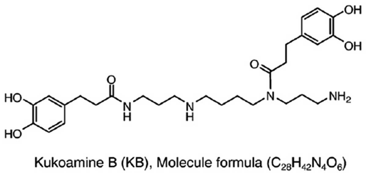
Kukoamine B (KB,
C28H42N4O6), a pure
spermine alkaloid with polyamine backbone and dihydrocaffeic acid
appendage, is extracted from a traditional Chinese herb, cortex
Lycii, via a rapid screening technique based on a herb affinity
assay. KB can immobilize LPS or CpG DNA separately on the reacting
surfaces of a biosensor (10,11)
and has been found to exhibit a high affinity with both LPS and CpG
DNA (12,13); however, little is known regarding
whether the in vivo administration of KB can effectively
inhibit inflammation in septic mice. The aim of the present study
was therefore to investigate the inhibitory effects of KB on the
inflammatory response in the livers of LPS-induced septic mice.
Materials and methods
Ethics statement
The present study was approved by the Council on
Animal Care and the Protection and Welfare of Animals at Jiangsu
University (Zhenjiang, China). All experimental protocols followed
the National Institutes of Health of China guidelines for the care
and use of experimental animals.
Reagents and materials
KB was kindly provided by Dr Zhen Jiang (Third
Military Medical University, Chongqing, China). The chemical
structure of KB is shown in Fig.
1. LPS, from Escherichia coli 055:B5, was purchased from
Sigma Chemicals (St. Louis, MO, USA). LPS was diluted in normal
saline and was administered to the animals intraperitoneally.
Interleukin-1β (IL-1β) and tumor necrosis factor α (TNF-α) ELISA
kits were purchased from Joyee Biotechnics Co., Ltd. (Shanghai,
China). The myeloperoxidase (MPO) assay kit was obtained from
Nanjing Jiancheng Bioengineering Institute (Nanjing, China).
Limulus amebocyte lysat (LAL) reagents were obtained from Xiamen
Houshiji Co., Ltd. (Xiamen, China). All other chemicals were of
reagent grade and obtained from Sigma Chemicals, unless otherwise
stated.
Animals and experimental sepsis
Institute of Cancer Research mice (body weight, 20±2
g; Experimental Animal Center of the Jiangsu University) were kept
in the animal house in a temperature-controlled room and were
allowed free access to normal animal diets and tap water. The mice
were left for a four-day acclimation period prior to the beginning
of the experiments. Thirty-two mice were randomly divided into
three groups: Control (n=8), LPS (n=12) and LPS + KB (n=12). Mice
in the LPS group were injected with LPS (10 mg/kg,
intraperitoneally) (14). In the
LPS + KB group, KB (20 μg/kg, intravenously) was administered 4 h
after the LPS challenge for a further 4 h. Control animals received
only a vehicle.
Measurement of plasma LPS
concentration
At 0, 2 and 4 h after KB treatment, the
concentration of LPS in the plasma was determined using the LAL
test. Briefly, each sample (100 μl) was added into 100 μl
quantitative LAL reagents dissolved in LPS-free water and reacted
at 37°C for 2 h. The gel clotting formation of the LAL products
induced by the existence of non-neutralized LPS was measured
through the kinetic turbidimetric assay, in which endodoxin
triggers a cascade of enzymatic reactions to activate the clotting
enzyme. The formation of the gel clot is proportional to the
concentration of endotoxin in the sample. Aliquots (100 μl) of all
samples, standards and negative controls were seeded into a 96-well
plate (non-pyrogens) and incubated at 37°C in a BioTek ELx808
reader (BioTek Instruments, Inc., Winooski, VT, USA). for 10 min.
Following incubation, ~100 μl quantitative LAL reagents, dissolved
in LPS-free water, rotate up and down until the solution turned
clear prior to use, was added to each well (Chinese Horsehoe Crab
Reagent Manufacturery Co., Ltd., Xiamen, China). Following gentle
vibration for 10 sec, the absorbance at 630 nm was measured and
readings were repeated every 30 sec for 2 h. The results were
calculated using Gen5™ data analysis software (BioTek Instruments,
Inc.).
Measurement of serum levels of
transaminases
At 4 h after KB treatment, the mice were
anesthetized with spontaneous inhalation of
isoflurane-N2O (Abbott Laboratories, Missisauga, ON,
Canada) in a 60% O2/40% N2 mixture. Blood
samples were obtained by cardiac puncture of the left ventricle.
The samples were stored in serum tubes (Capiject®;
Terumo Medical Corporation, Somerset, NJ, USA) and immediately
centrifuged at 6,500 × g for 5 min. The serum levels of alanine
aminotransferase (ALT) and aspartate aminotransferase (AST) were
measured using commercially available clinical assay kits according
to the manufacturers’ instructions and determined by a serum
autoanalyzer (AU2700; Olympus Corp., Tokyo, Japan). ALT catalyzes
the transfer of the amino group from L-alanine to α-ketoglutarate
resulting in the formation of pyruvate and L-glutamate. Lactate
dehydrogenase catalyzes the reduction of pyruvate and the
simultaneous oxidation of NADH to NAD decreasing the absorbance,
directly proportional to ALT activity. AST catalyzes the transfer
of an amino group between L-aspartate and 2-oxoglutarate. The
oxalacetate formed in the first reaction then reacts with NADH in
the presence of malate dehydrogenase (MDH) to form NAD. The
resulting decrease in absorbance is directly proportional to AST
activity.
For ALT, reagent 1 (R1) contained 100 mM Tris
buffer, 0.18 mM NADH 15 mM α-ketoglutaric acid, 1,200 U/l LDH. R2
contained 240 mM L-alanine. For AST, R1 contained 100 mM Tris
buffer, 12 mM α-ketoglutaric acid, 0.18 mM NADH, >2,000 U/l LDH
and 1,2000 U/l MDH; R2 contained 240 mM L-aspartate. A 4:1 mixture
of R1 reagent (100 mM Tris buffer, 0.18 mM NADH 15 mM
α-ketoglutaric acid, 1,200 U/l LDH) and R2 reagent (240 mM
L-alanine) was pipietted (1.0 ml) into appropriate tubes.
Subsequently, 40 μl of the samples, calibrator and negative control
to the reagent was mixed and incubated at 37°C for 1 min. The
absorbance was measured using a spectrophotometer at 340 nm and the
mixture was returned to 37°C. The readings were repeated every 1
min for a total of 9 min. The mean absorbance difference/minute
(ΔA/min.) was calculated and expressed as activity U/l. This was
calculated using the formula: Activity = ΔAu/min / ΔAc/min * Cc.
ΔAu/min deontes the mean absorbance difference/minute of the
sample, ΔAc/min denotes the mean absorbance difference/minute of
the calibrator, Cc denotes the concentration of the calibrator. The
commercially available clinical assay kits were from Sichuan Maker
Biotechnology Co., Ltd (Sichuan, China).
Histopathological examination
To characterize any histological alterations, the
livers were harvested from the animals of the different groups 4 h
after KB treatment and fixed in 4% formaldehyde solution. The
tissue was dehydrated with graded alcohol and embedded in paraffin,
and the sections were then stained with hematoxylin and eosin. The
tissue morphological characteristics were examined using light
microscopy.
Preparation of tissue homogenates
Mice were sacrificed 4 h after KB treatment. The
livers were immediately collected and stored at −80°C. Equal
weights of liver tissue from the three groups were homogenized in
ice-cold 0.9% NaCl to yield a 10% (w/v) homogenate. The homogenates
were then cleared by centrifuging at 9,000 × g at 4°C. The
supernatants were obtained and stored at −70°C.
ELISA
The levels of TNF-α and IL-1β in the plasma and
tissue homogenates were measured using ELISA kits in accordance
with the manufacturer’s instructions (Joyee Biotecnics Co., Ltd.,
Shanghai, China.).
MPO activity
To assess the neutrophil infiltration, the MPO
activity in the tissue homogenates was determined by utilizing a
commercially available kit in accordance with the manufacturer’s
instruction (Nanjing Jiancheng Bioengineering Institute, Nanjing,
China) (15). Aliquots (0.3 ml)
were added to a 2.3-ml reaction mixture containing 50 mM potassium
phosphate buffer, o-dianisidine and 20 mM
H2O2 solution. One unit of enzyme activity
(expressed as U/g tissue) was defined as the amount of MPO required
to cause a change in absorbance measured at 460 nm for 3 min.
Immunohistochemical examination
The paraffin-embedded liver tissue sections were
subjected to immunohistochemical staining. Briefly, paraffin
sections of 5 μm were prepared and mounted on SuperFrost™ Plus
glass slides (Kaihong Healthcare Co., Ltd., Nanjing, China), and
were then deparaffinized in xylene and rehydrated in a graded
series of ethanol baths. Endogenous peroxidase activity in the
deparaffinized sections was blocked through treatment with 3%
H2O2. Following the termination of endogenous
peroxidase activity, the nonspecific proteins were blocked with 3%
solcoseryl for 30 min at room temperature. The sections were
subsequently incubated with goat polyclonal immunoglobulin (Ig)G
primary antibodies against intercellular adhesion molecule-1
(ICAM-1; sc-1511) and vascular cell adhesion molecule-1 (VCAM-1;
sc-1504) (Santa Cruz Biotechnology, Inc., Santa Cruz, CA, USA)
diluted 1:200 in Tris buffered saline containing 0.1% Tween-20
(TBST) at 4°C overnight. The following morning the secondary
antibodies [rabbit anti-goat immunoglobulin (Ig) G conjugated with
horseradish peroxidase], obtained from Maixin Biotech Co., Ltd.
(Fuzhou, China) were used to bind with the primary antibodies. The
location of the stained proteins was subsequently determined by
reaction with 3′-diaminobenzidine tetrahydrochloride solution
according to the manufacturer’s instructions (Sigma Chemicals) and
examined by light microscopy.
Western blot analysis
Total nucleic protein was extracted with a nuclear
protein extraction buffer kit (Vazyme Biotech, Nanjing, China).
Protein concentration was assayed using a bicinchoninic acid
protein assay kit (Beyotime Institute of Biotechnology, Haimen,
China). SDS-PAGE was performed on equivalent amounts of protein
samples using precast 7% resolving/3% stacking Tris-HCl gels
(Bio-Rad, Hercules, CA, USA). Following electrophoresis, the
separated proteins were transferred to polyvinylidene fluoride
membranes (Amersham Pharmacia Biotech, Inc., Piscataway, NJ, USA),
prior to the membranes being blocked in 5% non-fat milk in TBST for
1 h at room temperature. The blocked membranes were incubated in
rabbit polyclonal IgG primary antibodies specific for mouse nuclear
factor-κ-gene binding (NF-κB)-p56 (sc-372 at a dilution of
1:1,000), in TBST overnight at 4°C. The membranes were then washed
and probed with horseradish peroxidase-conjugated secondary
antibody (Amersham Pharmacia Biotech, Inc.) for 1 h at room
temperature. Chemiluminescence detection was performed with the
Amersham enhanced chemiluminescence detection kit according to the
manufacturer’s instructions (Amersham Pharmacia Biotech, Inc.). To
ensure a similar quantity of protein in each sample, the membranes
were ‘stripped off’, reprobed with β-actin, developed with
horseradish peroxidase-conjugated secondary antibody and visualized
using enhanced chemiluminescence. The specific bands were
quantified by densitometry (Bio-Rad GS-710 densitometer; Bio-Rad
Laboratories, Des Moines, IA, USA).
Electrophoretic mobility shift assay
(EMSA)
Whole-tissue (medial lobe of the liver) nuclear
protein was extracted as described previously (16,17).
For EMSA, 10 μg total nuclear protein was incubated with 1.0 pmol
double-stranded γ-[32P]-adenosine triphosphate
end-labeled oligonucleotides containing consensus-binding sequences
for NF-κB (sense strand 5′-AGGGACTTCCGCTGGGGACTTTCC-3′) in a
binding buffer (10 mM HEPES, pH 7.9, 80 mM NaCl, 3 mM
MgCl2, 0.1 mM EDTA, 1 mM dithiothreitol, 1 mM
phenylmethylsulfonyl fluoride and 10% glycerol). The samples were
incubated for 30 min at room temperature and then run through a 4%
non-denaturing polyacrylamide gel (0.5X Tris-borate-EDTA buffer) at
280 V for 1 h. Once dry, the gel was exposed to X-ray film (Kodak,
Rochester, NY, USA) for 4–6 h in cassettes at −80°C. Signal
detection and quantification was performed by computer-assisted
densitometry.
Statistical analysis
All values are expressed as the mean ± standard
deviation and were analyzed using one-way analysis of variance,
with a post hoc Dunnett’s t-test, and a two-tailed Student’s
t-test. Differences between groups were considered to be
statistical significant at P<0.05.
Results
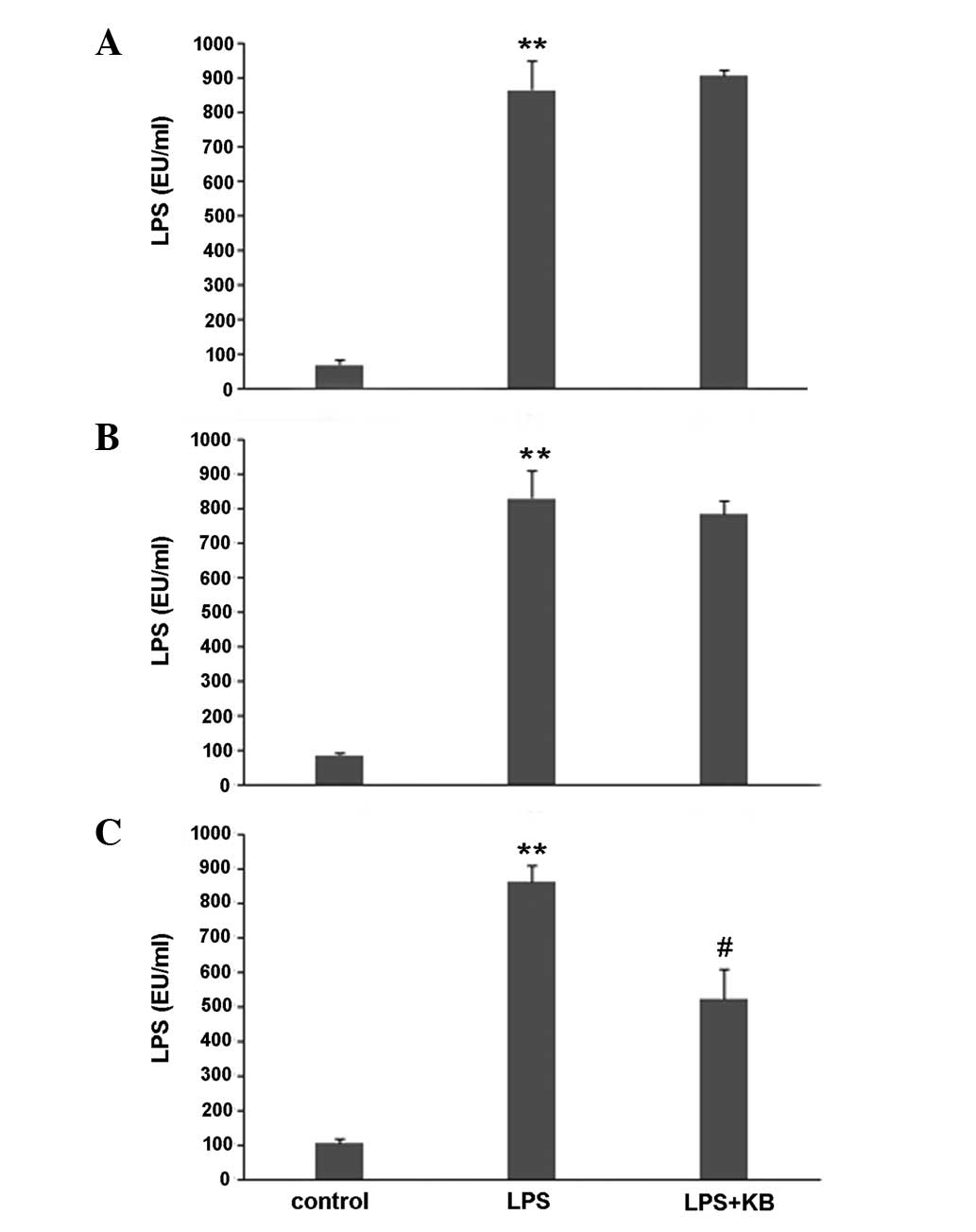
Effect of KB on LPS concentration in the
plasma of LPS-induced septic mice
After a 4-h challenge with LPS, mice were treated
with KB for 0, 2 and 4 h. The LPS concentrations in the plasma were
determined. As shown in Fig. 2,
mice challenged with LPS had a significantly higher LPS
concentration compared with the control group. The LPS
concentration declined following KB treatment for 2 h (Fig. 2B). The decrease in LPS
concentration was significant following KB treatment for 4 h
(Fig. 2C).
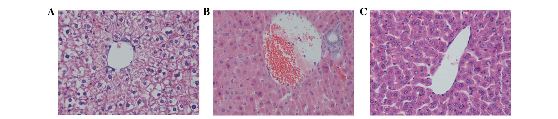
Histology
Histological analysis showed that the livers from
the control mice exhibited the normal architecture, while LPS
challenge induced irregularity in hepatocyte arrangement and
central vein congestion, as well as the infiltration of
inflammatory cells into the tissue. Administration of KB
significantly decreased the granulocyte infiltration and
inflammatory response (Fig.
3).
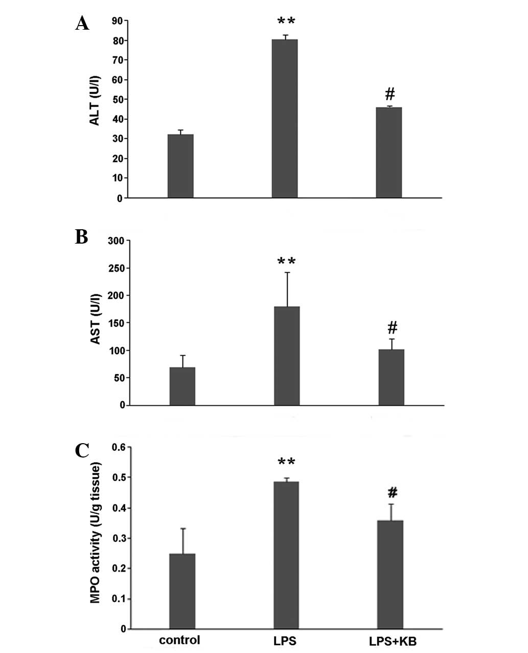
Effect of KB on serum aminotransferase
levels and liver MPO activity in LPS-induced septic mice
Hepatocyte injury was evaluated by determining the
serum concentrations of ALT and AST. As shown in Fig. 4A and B, the levels of ALT and AST
in LPS-induced septic mice were found to be markedly increased.
Following the administration of KB, this elevation was
significantly attenuated (compared with the LPS group, P<0.05).
To determine whether the LPS-induced increase in polymorphonuclear
leukocyte (PMN) accumulation in the liver was effectively prevented
by KB, the activity of MPO, an enzyme in the azurophilic granules
of neutrophils, was assessed. The mean MPO levels are shown in
Fig. 4C. The MPO activity in the
livers obtained from LPS-induced septic mice was markedly increased
compared with that in the control animals (P<0.01), whereas the
activity was significantly decreased by treatment with KB
(P<0.05).
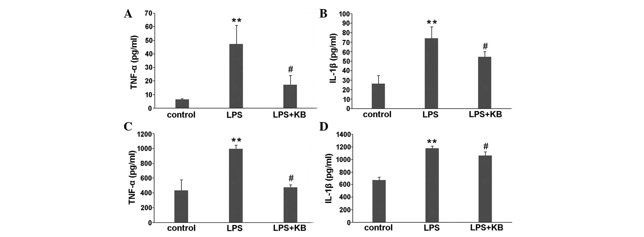
Effect of KB on cytokine expression in
LPS-induced septic mice
To evaluate the inflammatory response, levels of
TNF-α and IL-1β in the plasma and liver homogenates were detected.
The expression of TNF-α and IL-1β in the plasma of LPS-induced
septic mice was markedly elevated compared with that in the plasma
of the control mice. Following the administration of KB for 4 h,
the increases in TNF-α and IL-1β expression were significantly
reduced (Fig. 5A and B). Similar
results for TNF-α and IL-1β were found in the liver homogenates
(Fig. 5C and D).
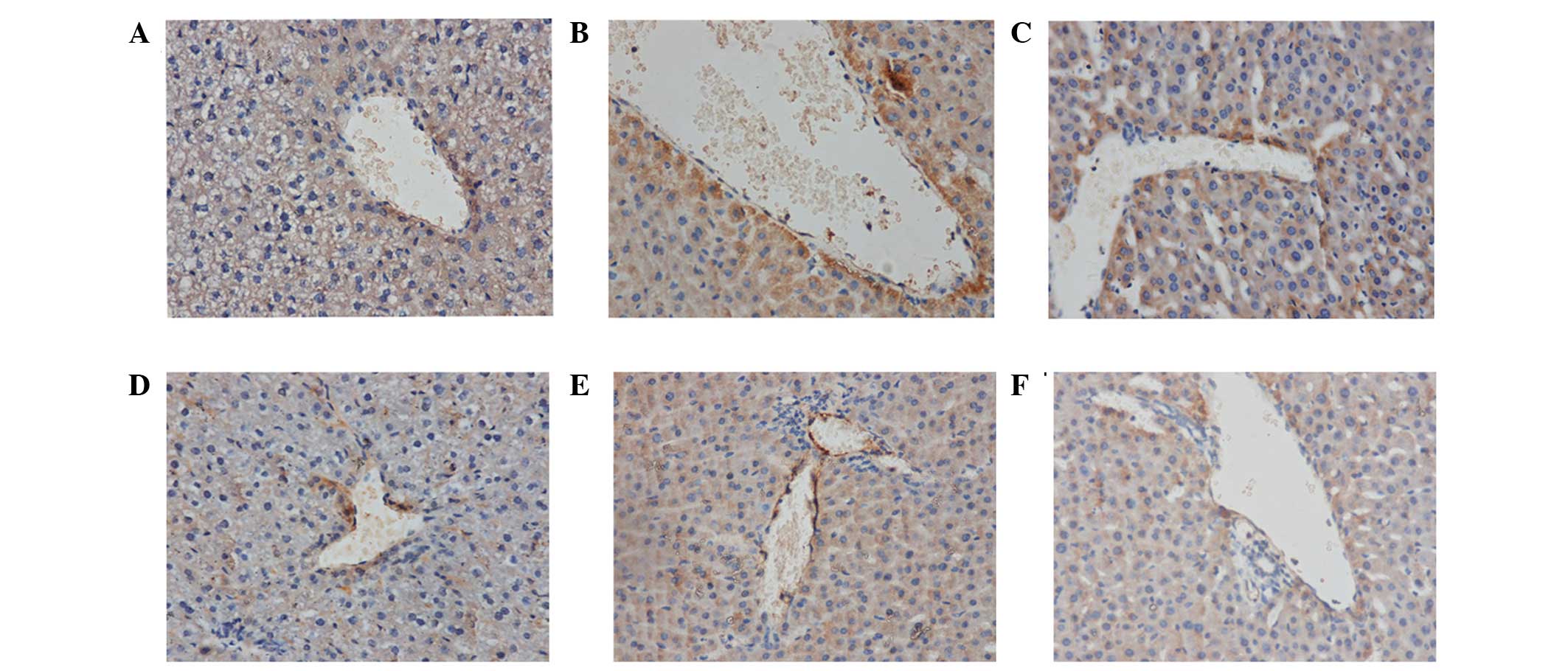
Effect of KB on ICAM-1 and VCAM-1
expression in the livers of LPS-induced septic mice
Following LPS stimulation, the expression of ICAM-1
and VCAM-1 in the liver tissue was significantly increased compared
with that in the control. With the in vivo administration of
KB, the expression of ICAM-1 and VCAM-1 significantly decreased
(Fig. 6).
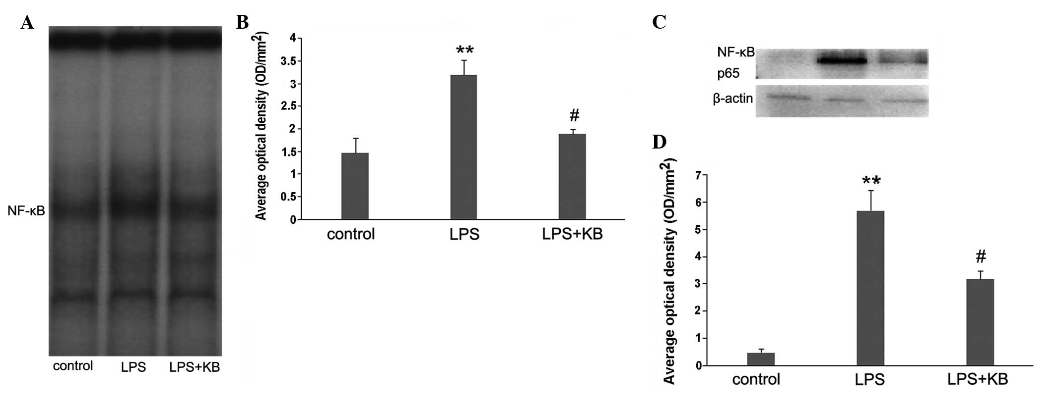
Effect of KB on NF-κB activity in
LPS-induced septic mice
The binding activity of nuclear protein to the
radio-labeled consensus binding sequences of NF-κB was assessed
using EMSA. Following LPS challenge, the NF-κB activation in the
liver was markedly increased, and this activity was inhibited by
the in vivo administration of KB (Fig. 7A and B). The nuclear translocation
of the p65 subunit of NF-κB was subsequently investigated using
western blot analysis to conform the effect of KB on NF-κB-p65
translocation from the cytosol to the nucleus. The data indicated
that levels of NF-κB-p65 were markedly elevated in the nuclear
protein of LPS-challenged mice; KB treatment attenuated this
elevation (Fig. 7C and D).
Discussion
KB, an active alkaloid compound isolated from the
traditional Chinese herb cortex Lycii, is considered to be a novel
and promising candidate for the treatment of sepsis (12). KB is firstly characterized as a
selective dual inhibitor of LPS and CpG DNA (13). KB possesses anti-inflammatory
activity, as demonstrated by its ability to inhibit the
inflammatory response in mouse macrophages, rescue mice from
heat-killed E. coli-induced sepsis and prevent the
upregulation of TLR4 and TLR9 expression (12,13).
These data provide evidence that, as a potential LPS-neutralizer,
KB affects the signal transduction pathway activation stimulated by
LPS; however, to the best of our knowledge, no studies have
assessed the anti-LPS ability of KB following its administration in
LPS-challenged mice. The aim of the present study was therefore to
focus on the anti-inflammatory effects of KB in the livers of
LPS-challenged mice and to explore a potential mechanism. In the
study it was found that the plasma LPS concentration reached a high
level in LPS-challenged mice. As expected, the LPS level was
significantly decreased in the LPS-challenged mice treated with KB
for 4 h. These data are consistent with those in previously
published studies (12).
Exerting important roles in metabolism, homeostasis
and host defense mechanisms, the liver has been investigated
extensively and is believed to be a major organ responsible for
SIRS or sepsis (18). Liver injury
in LPS-challenged mice is characterized by neutrophil infiltration
in the liver parenchyma, irregularity in the arrangement of
numerous hepatocytes and central and portal vein congestion. In the
present study, it was observed that these histological changes were
slight in the LPS + KB group. In parallel, the elevated levels of
the hepatic enzymes AST and ALT were effectively reduced in the
LPS-challenged mice treated with KB, indicating that KB
administration plays a key role in the anti-inflammatory response
and the protection of organ functions.
The excessive production of certain cytokines, such
as TNF-α and IL-1β, released by macrophages and other mononuclear
cells in response to LPS is understood to be one of the earliest
events in hepatic inflammation. This cytokine production triggers a
cascade of other cytokines that act in coordination to cause the
death of hepatocytes and the recruitment of inflammatory cells
(19–21). To investigate whether suppression
of the LPS-induced liver inflammatory response by KB was due to a
downregulation in the expression of systemic and local
proinflammatory cytokines, the expression of TNF-α and IL-1β was
measured in plasma and liver tissue in the experimental mice of the
present study. In vivo application of KB in the
LPS-challenged mice markedly decreased the production of TNF-α and
IL-1β in the plasma and liver tissue. These data indicated that the
activation and release of proinflammatory mediators in systemic and
local tissue could be, at least partly, inhibited by KB, and that
this process could be associated with the decreased LPS level
following neutralization by KB.
In addition to the expression of proinflammatory
cytokines, neutrophil sequestration was investigated in the present
study. Neutrophil-mediated parenchymal cell damage in the liver is
initiated by the accumulation of neutrophils in the hepatic tissue
(22). MPO is an enzyme that can
predominantly be found in the azurophilic granules of PMNs. The
measurement of tissue MPO activity is frequently utilized to
reflect the PMN accumulation in damaged tissues, since MPO activity
correlates significantly with the number of PMNs determined
histochemically in tissues (23,24).
The present results showed that MPO activity in the liver was
markedly enhanced following LPS stimulation. In vivo
administration of KB led to a significant decrease in MPO activity,
subsequently preventing PMN chemotaxis and infiltration in the
liver, and decreasing the production of oxidants and tissue
oxidative injury.
ICAM-1 and VCAM-1 are two members of the Ig-like
supergene family of adhesion molecules, which are responsible for
mediating the tight adhesion of PMNs to endothelial cells and
assisting leukocyte transmigration (25–27).
In the present study, the expression of ICAM-1 and VCAM-1 was
detected in the livers of experimental mice. The data showed that
KB inhibited the increases in ICAM-1 and VCAM-1 expression in the
livers of LPS-induced septic mice. These results were in accord
with the changes in MPO activity, and strongly indicated that KB is
associated with the inhibition of leukocyte sequestration and
adhesion, and may consequently effectively decrease the
inflammatory response in livers stimulated by LPS.
The binding of LPS to host cells induces a
receptor-mediated (TLR4) signaling cascade that leads to the
activation of NF-κB. Following activation, NF-κB translocates to
the nucleus and causes rapid gene induction, resulting in the
expression of inflammatory mediators, including cytokines,
chemokines and adhesion molecules (28–30).
To further explore the underlying mechanism by which KB achieves
its beneficial effects, NF-κB translocation and activity, which
play an important role in the pathogenesis of liver injury, were
investigated. The present study demonstrated that NF-κB
translocation was strongly increased, and accompanied by enhanced
NF-κB activity, in the liver tissue of LPS-induced septic mice.
Notably, KB effectively attenuated the nuclear translocation and
activation of NF-κB, indicating that KB has a pivotal role in the
inhibition of NF-κB via its ability to bind to LPS, subsequently
leading to the alleviation of leukocyte infiltration and the
LPS-induced proinflammatory response.
In combination, the results of the present study
have demonstrated that KB inhibits inflammation in septic mice by
its unique property of combining with LPS, leading to a reduction
in the concentration of plasma LPS. Decreases in the plasma LPS
levels were accompanied by reductions in the expression of ICAM-1
and VCAM-1 and leukocyte sequestration in the livers of LPS-induced
septic mice. In parallel, KB was shown to exert its protective
effects against the inflammatory response by interfering with NF-κB
activation, and, therefore, suppressing the pro-adhesive phenotype
of the endothelial cells. Further studies are required in extension
of the present observations in order to investigate the detailed
mechanism(s) regarding KB in the treatment of sepsis.
Acknowledgements
This study was supported by the National Natural
Science Foundation of China (no. 30772256, 81071546 and 81272148)
and the Jiangsu Provincial Natural Science Foundation (no.
BK2012703).
References
|
1
|
Annane D, Bellissant E and Cavaillon JM:
Septic shock. Lancet. 365:63–78. 2005. View Article : Google Scholar : PubMed/NCBI
|
|
2
|
Martin GS, Mannino DM and Moss M: The
effect of age on the development and outcome of adult sepsis. Crit
Care Med. 34:15–21. 2006. View Article : Google Scholar
|
|
3
|
Roberts LA, Glenn HL, Whitfield RA and
Jacobson BS: Regulation of cell-substrate adhesion by the
lipoxygenase and cyclooxygenase branches of arachidonic acid
metabolism. Adv Exp Med Biol. 507:525–529. 2002. View Article : Google Scholar
|
|
4
|
Hume DA, Underhill DM, Sweet MJ, Ozinsky
AO, Liew FY and Aderem A: Macrophages exposed continuously to
lipopolysaccharide and other agonists that act via toll-like
receptors exhibit a sustained and additive activation state. BMC
Immunol. 2:112001. View Article : Google Scholar : PubMed/NCBI
|
|
5
|
De Nardo D, De Nardo CM, Nguyen T,
Hamilton JA and Scholz GM: Signaling crosstalk during sequential
TLR4 and TLR9 activation amplifies the inflammatory response of
mouse macrophages. J Immunol. 183:8110–8118. 2009. View Article : Google Scholar : PubMed/NCBI
|
|
6
|
Alexander C and Rietschel ET: Bacterial
lipopolysaccharides and innate immunity. J Endotoxin Res.
7:167–202. 2001.PubMed/NCBI
|
|
7
|
Sparwasser T, Miethke T, Lipford G, et al:
Bacterial DNA causes septic shock. Nature. 386:336–337. 1997.
View Article : Google Scholar : PubMed/NCBI
|
|
8
|
Angus DC, Birmingham MC, Balk RA, et al:
E5 murine monoclonal antiendotoxin antibody in gram-negative
sepsis: a randomized controlled trial. E5 Study Investigators.
JAMA. 283:1723–1730. 2000. View Article : Google Scholar
|
|
9
|
Nahra R and Dellinger RP: Targeting the
lipopolysaccharides: still a matter of debate? Curr Opin
Anaesthesiol. 21:98–104. 2008. View Article : Google Scholar : PubMed/NCBI
|
|
10
|
Liu X, Cheng J, Zheng X, et al: Targeting
CpG DNA to screen and isolate anti-sepsis fraction and monomers
from traditional Chinese herbs using affinity biosensor technology.
Int Immunopharmacol. 9:1021–1031. 2009. View Article : Google Scholar : PubMed/NCBI
|
|
11
|
Jiang Z, Hong Z, Guo W, et al: A synthetic
peptide derived from bactericidal/permeability-increasing protein
neutralizes endotoxin in vitro and in vivo. Int Immunopharmacol.
4:527–537. 2004. View Article : Google Scholar : PubMed/NCBI
|
|
12
|
Liu X, Zheng X, Wang N, et al: Kukoamine
B, a novel dual inhibitor of LPS and CpG DNA, is a potential
candidate for sepsis treatment. Br J Pharmacol. 162:1274–1290.
2011. View Article : Google Scholar :
|
|
13
|
Liu X, Zheng X, Long Y, et al: Dual
targets guided screening and isolation of Kukoamine B as a novel
natural anti-sepsis agent from traditional Chinese herb Cortex
lycii. Int Immunopharmacol. 11:110–120. 2011. View Article : Google Scholar
|
|
14
|
Nishioku T, Dohgu S, Takata F, et al:
Detachment of brain pericytes from the basal lamina is involved in
disruption of the blood-brain barrier caused by
lipopolysaccharide-induced sepsis in mice. Cell Mol Neurobiol.
29:309–316. 2009. View Article : Google Scholar
|
|
15
|
Hillegass LM, Griswold DE, Brickson B and
Albrightson-Winslow C: Assessment of myeloperoxidase activity in
whole rat kidney. J Pharmacol Methods. 24:285–295. 1990. View Article : Google Scholar : PubMed/NCBI
|
|
16
|
Sun BW, Chen ZY, Chen X and Liu C:
Attenuation of leukocytes sequestration by carbon
monoxide-releasing molecules: liberated carbon monoxide in the
liver of thermally injured mice. J Burn Care Res. 28:173–181. 2007.
View Article : Google Scholar : PubMed/NCBI
|
|
17
|
Sun B, Sun H, Liu C, Shen J, Chen Z and
Chen X: Role of CO-releasing molecules liberated CO in attenuating
leukocytes sequestration and inflammatory responses in the lung of
thermally injured mice. J Surg Res. 139:128–135. 2007. View Article : Google Scholar : PubMed/NCBI
|
|
18
|
El-Agamy DS, Makled MN and Gamil NM:
Protective effects of BML-111 against acetaminophen-induced acute
liver injury in mice. J Physiol Biochem. 70:141–149. 2014.
View Article : Google Scholar
|
|
19
|
Kim SH, Kim YS, Kang SS, Bae K, Hung TM
and Lee SM: Anti-apoptotic and hepatoprotective effects of gomisin
A on fulminant hepatic failure induced by D-galactosamine and
lipopolysaccharide in mice. J Pharmacol Sci. 106:225–233. 2008.
View Article : Google Scholar : PubMed/NCBI
|
|
20
|
Fukuda T, Mogami A, Tanaka H, Yoshikawa T,
Hisadome M and Komatsu H: Y-40138, a multiple cytokine production
modulator, protects against D-galactosamine and
lipopolysaccharide-induced hepatitis. Life Sci. 79:822–827. 2006.
View Article : Google Scholar : PubMed/NCBI
|
|
21
|
Zhu J, Wang J, Sheng Y, et al: Baicalin
improves survival in a murine model of polymicrobial sepsis via
suppressing inflammatory response and lymphocyte apoptosis. PLoS
One. 7:e355232012. View Article : Google Scholar : PubMed/NCBI
|
|
22
|
Jaeschke H and Hasegawa T: Role of
neutrophils in acute inflammatory liver injury. Liver Int.
26:912–919. 2006. View Article : Google Scholar : PubMed/NCBI
|
|
23
|
Odobasic D, Kitching AR, Yang Y, et al:
Neutrophil myeloperoxidase regulates T-cell-driven tissue
inflammation in mice by inhibiting dendritic cell function. Blood.
121:4195–4204. 2013. View Article : Google Scholar : PubMed/NCBI
|
|
24
|
Zhang N, Francis KP, Prakash A and Ansaldi
D: Enhanced detection of myeloperoxidase activity in deep tissues
through luminescent excitation of near-infrared nanoparticles. Nat
Med. 19:500–505. 2013. View
Article : Google Scholar : PubMed/NCBI
|
|
25
|
Barreiro O, Zamai M, Yáñez-Mó M, et al:
Endothelial adhesion receptors are recruited to adherent leukocytes
by inclusion in preformed tetraspanin nanoplatforms. J Cell Biol.
183:527–542. 2008. View Article : Google Scholar : PubMed/NCBI
|
|
26
|
Vestweber D: Adhesion and signaling
molecules controlling the transmigration of leukocytes through
endothelium. Immunol Rev. 218:178–196. 2007. View Article : Google Scholar : PubMed/NCBI
|
|
27
|
Ley K, Laudanna C, Cybulsky MI and
Nourshargh S: Getting to the site of inflammation: the leukocyte
adhesion cascade updated. Nat Rev Immunol. 7:678–689. 2007.
View Article : Google Scholar : PubMed/NCBI
|
|
28
|
Schmidt A, Oberle N, Weiss EM, et al:
Human regulatory T cells rapidly suppress T cell receptor-induced
Ca(2+), NF-κB, and NFAT signaling in conventional T cells. Sci
Signal. 4:ra902011. View Article : Google Scholar
|
|
29
|
Madonna R and De Caterina R: Relevance of
new drug discovery to reduce NF-κB activation in cardiovascular
disease. Vascul Pharmacol. 57:41–47. 2012. View Article : Google Scholar : PubMed/NCBI
|
|
30
|
Han EH, Yang JH, Kim HK, et al:
1-Bromopropane up-regulates cyclooxygenase-2 expression via NF-κB
and C/EBP activation in murine macrophages. Food Chem Toxicol.
50:1616–1622. 2012. View Article : Google Scholar : PubMed/NCBI
|