A novel method of combining Periodic Acid Schiff staining with Wright-Giemsa staining to identify the pathogens Penicillium marneffei, Histoplasma capsulatum, Mucor and Leishmania donovani in bone marrow smears

  • Authors:
    • Lingyan Qin
    • Ligang Zhao
    • Chunyan Tan
    • Xu Chen
    • Zheng Yang
    • Wuning Mo
  • View Affiliations

  • Published online on: March 16, 2015     https://doi.org/10.3892/etm.2015.2357
  • Pages: 1950-1954
Metrics: Total Views: 0 (Spandidos Publications: | PMC Statistics: )
Total PDF Downloads: 0 (Spandidos Publications: | PMC Statistics: )


Abstract

Penicillium marneffei, Histoplasma capsulatum, Mucor and Leishmania donovani can lead to penicilliosis marneffei, histoplasmosis, mucormycosis and leishmaniasis, respectively, which, to a certain extent, share similar clinical manifestations. These pathogens are approximately the same size, therefore it is relatively difficult to rapidly diagnose the diseases. The aim of the present study was to explore a novel method that attempts to rapidly identify the pathogens of these diseases. In the Wright‑Giemsa staining, the four pathogens were approximately the same size and mainly existed in macrophages. The multiplying P. marneffei had two nuclei, which were on both sides of the fungus, and had light cross‑walls in the middle. H. capsulatum had a purplish nucleus, which occupied between one‑third and one‑half of the spore. The cytoplasm was light blue. Peripheral spores were observed in the form of an empty, bright ring without color, like a capsule. Generally, Mucor were observed to have a long and lightly stained area, which could be easily confused with the Wright staining of dinuclear P. marneffei. L. donovani exhibited a deep‑staining kinetoplast near the nucleus. In the Periodic Acid Schiff (PAS) staining, the pathogens of P. marneffei and H. capsulatum were distinct and stained red. Differentiation between P. marneffei and H. capsulatum relied on their modes of reproduction: P. marneffei depends on fission, when the pathogens stretch into sausage‑shapes and are split by a cross‑wall, while H. capsulatum depends on budding so that narrow‑necked, single spores can be formed. With PAS staining, the cell walls and intracellular contents of Mucor and L. donovani were not stained, lightly stained or granulated and discontinuous. In conclusion, this method, combining PAS and Wright‑Giemsa staining, is simple and rapid, and may contribute to the effective identification of the four pathogens.
View Figures
View References

Related Articles

Journal Cover

May-2015
Volume 9 Issue 5

Print ISSN: 1792-0981
Online ISSN:1792-1015

Sign up for eToc alerts

Recommend to Library

Copy and paste a formatted citation
x
Spandidos Publications style
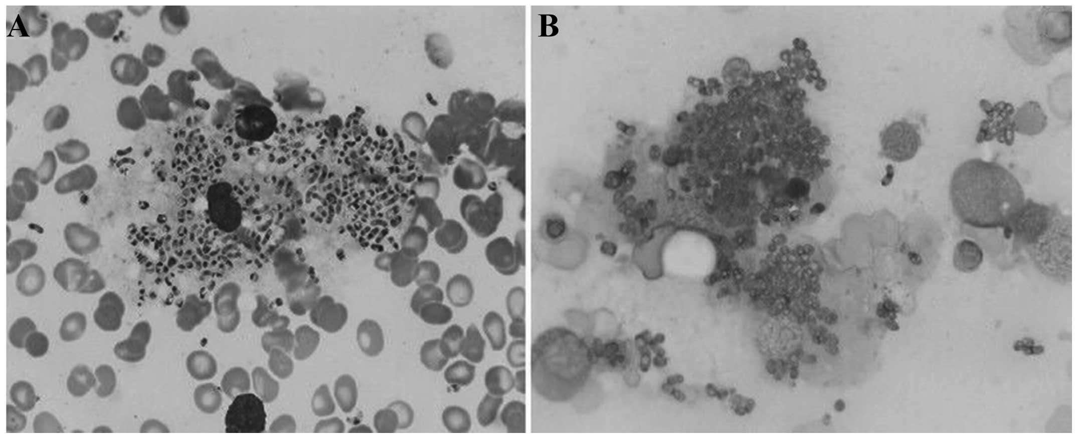
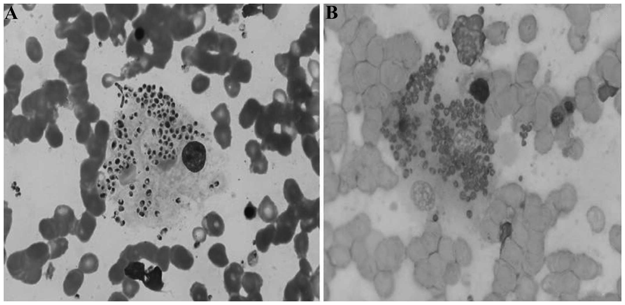
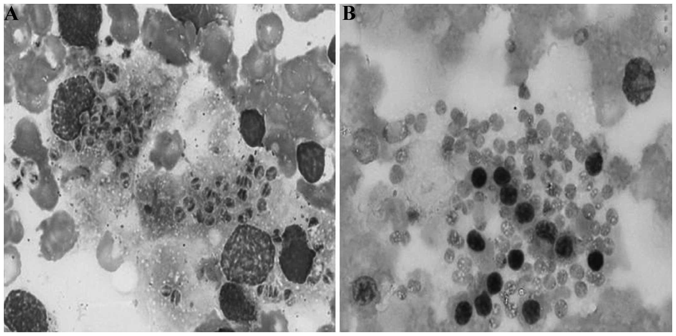
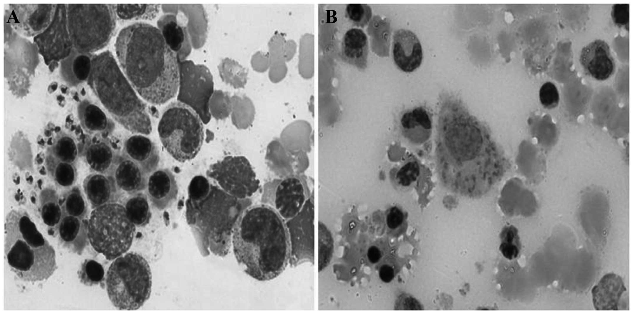
Qin L, Zhao L, Tan C, Chen X, Yang Z and Mo W: A novel method of combining Periodic Acid Schiff staining with Wright-Giemsa staining to identify the pathogens Penicillium marneffei, Histoplasma capsulatum, Mucor and Leishmania donovani in bone marrow smears. Exp Ther Med 9: 1950-1954, 2015
APA
Qin, L., Zhao, L., Tan, C., Chen, X., Yang, Z., & Mo, W. (2015). A novel method of combining Periodic Acid Schiff staining with Wright-Giemsa staining to identify the pathogens Penicillium marneffei, Histoplasma capsulatum, Mucor and Leishmania donovani in bone marrow smears. Experimental and Therapeutic Medicine, 9, 1950-1954. https://doi.org/10.3892/etm.2015.2357
MLA
Qin, L., Zhao, L., Tan, C., Chen, X., Yang, Z., Mo, W."A novel method of combining Periodic Acid Schiff staining with Wright-Giemsa staining to identify the pathogens Penicillium marneffei, Histoplasma capsulatum, Mucor and Leishmania donovani in bone marrow smears". Experimental and Therapeutic Medicine 9.5 (2015): 1950-1954.
Chicago
Qin, L., Zhao, L., Tan, C., Chen, X., Yang, Z., Mo, W."A novel method of combining Periodic Acid Schiff staining with Wright-Giemsa staining to identify the pathogens Penicillium marneffei, Histoplasma capsulatum, Mucor and Leishmania donovani in bone marrow smears". Experimental and Therapeutic Medicine 9, no. 5 (2015): 1950-1954. https://doi.org/10.3892/etm.2015.2357