Introduction
Stroke, also known as cerebrovascular accident, is a
type of acute cerebral blood circulation disorder with a sudden
onset that is caused by cerebral artery stenosis, occlusion or
rupture, which may result from a variety of predisposing factors.
The clinical manifestations of stroke are indicative of temporary
or permanent brain dysfunction (1).
Stroke is a common disease among the elderly. According to
epidemiological investigation in China, the incidence rate of
stroke in middle-aged adults is 200/1,000,000 per year, and the
annual mortality rate is 800,000–1,000,000. There are 1,500,000 new
cases of stroke per year, and ~75% of the survivors are left
disabled. In addition, the recurrence rate in 5 years can reach 40%
(2). In most cases, patients who
have been left with a disability following a stroke will also
experience further stroke-induced complications; therefore,
considerable focus has been placed on researching the recurrence of
post-stroke complications (3).
A number of studies have shown that depression is
one of the most common post-stroke complications (4,5). The
occurrence of depression significantly hinders the rehabilitation
of limb function in stroke patients; therefore, over the past two
decades, post-stroke depression (PSD) has become an area of
significant interest (3,6). Mimicking the pathological and
physiological processes of PSD in vivo, by simulating animal
models of PSD, is important to enable the thorough elucidation of
the occurrence, development and characteristics of PSD and to
explore new therapeutic methods for the disease. Several studies
have established an animal model of PSD (7,8), but
only a few studies have focused on the behavioral changes in a PSD
rat model (9). The aim of the
present study, therefore, was to examine and evaluate these changes
in a PSD rat model.
Materials and methods
Experimental animals and grouping
Forty-eight adult Wistar rats (male and female),
weighing 200±20 g, were purchased from the Experimental Animal
Center Of Peking University Medical School (Beijing, China). The
rats were randomly divided into three groups of 16: Group A (normal
group), group B (stroke group) and group C (PSD group). The rats in
group A were not treated, the rats in group B were subjected to
middle cerebral artery occlusion (MCAO) to generate a stroke model
and the rats in group C were used to establish a PSD model
following right MCAO. This study was carried out in strict
accordance with the guidelines stated in the Guide for the Care and
Use of Laboratory Animals of the National Institutes of Health. The
animal use protocol was reviewed and approved by the Institutional
Animal Care and Use Committee of Henan University Huaihe Hospital
(Kaifeng, China).
Establishment of the rat models in
groups B and C
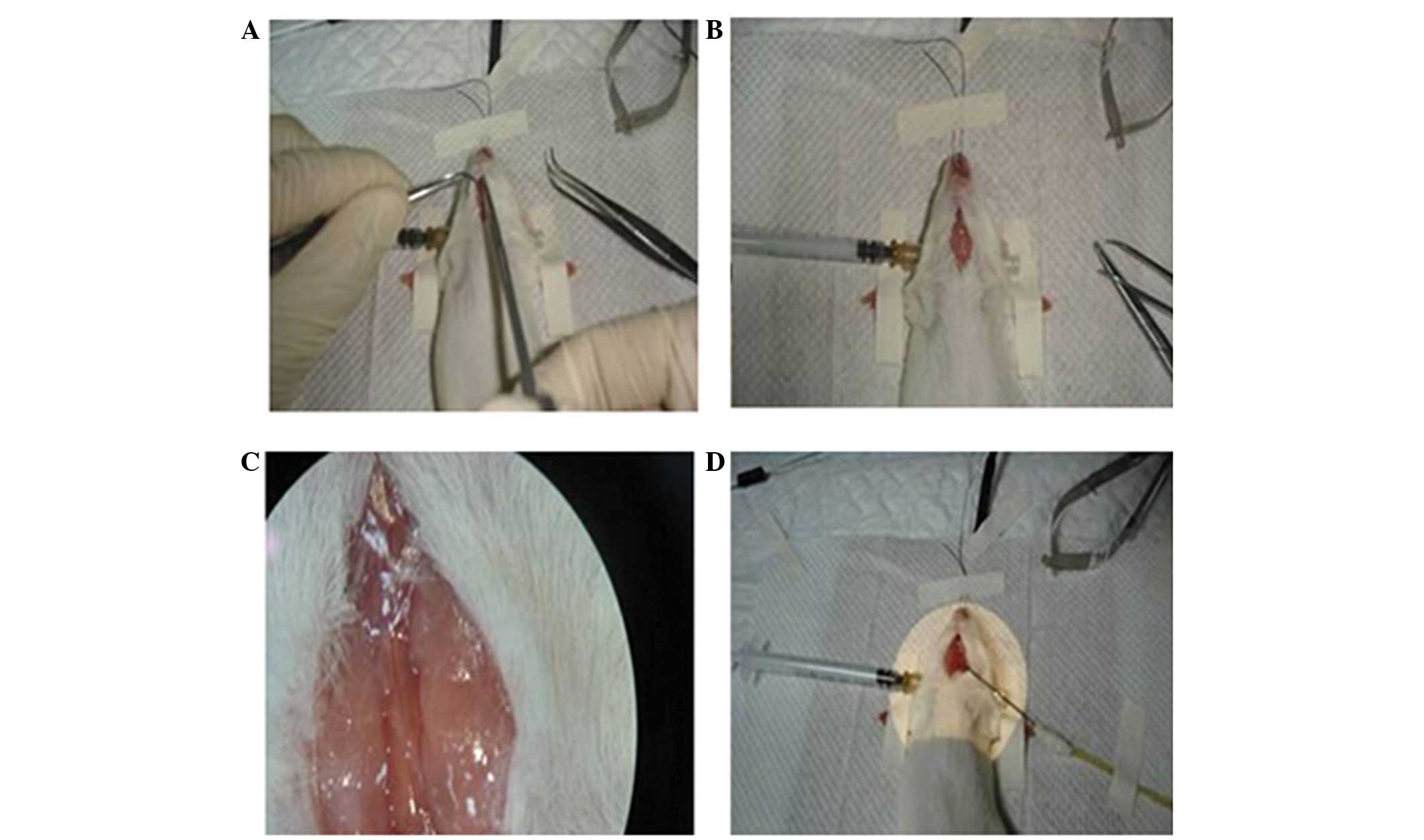
Right MCAO was performed in the 32 rats from groups
B and C. All surgeries were performed by the same individual.
Wistar rats underwent preoperative fasting for 12 h. Following
intraperitoneal anesthesia with amobarbital sodium (Sinopharm
Chemical Reagent Co., Shanghai, China), the rats were fixed in a
supine position on the operating board and the shaved skin was
disinfected. A 2-cm oblique skin incision was made on the midpoint
of the ligature between the right lateral canthus and posterior
auricular sulcus to expose the temporal arch, and a bone window
with a diameter of 0.5 cm was then opened in front of the temporal
arch root to expose the right MCA. Following the exposure of the
MCA, the dura mater was opened and the end of the main trunk of the
MCA was subjected to bipolar electrocoagulation (Hoboat, Nanjing,
China). Soon afterwards the bone window was filled with sterile
gelfoam (Hoboat) and the incision was sutured layer by layer. The
intraoperative body temperature was maintained at ~37°C. A
postoperative intraperitoneal injection of 40,000 units of
penicillin sodium (Shijiazhuang Pharma Group ZhongNuo
Pharmaceutical Co. Ltd., Shijiazhuang, China) was performed to
prevent infection. The postoperative general state, limb motor
function and blood pressure of the rats were closely observed
(10) (Fig. 1).
The criteria for a successful MCAO were as follows:
i) The reaction to pain was dulled or had disappeared in the
affected limbs; ii) the rat was unable to extend its upper limbs
when hanging upside down by the tail; iii) the rat slanted toward
the affected side when crawling. Motor function was examined weekly
following the MCAO surgery, and neurological function was measured
using the Bederson score, as follows: i) Score 3, the rat could not
crawl forward and exhibited a unidirectional circling motion; ii)
score 2, the rat could move forward but was easily overturned when
subjected to a lateral external force on the affected side, and the
affected limbs were flexed when the tail was lifted; iii) score 1,
the affected forelimbs were flexed when the tail was lifted; iv)
score 0, normal activity (11).
To prepare the rat model of PSD the 16 rats in group
C were subjected to MCAO surgery and then fed in isolation and
exposed to chronic, unpredictable, mild stress for 2 weeks, as
described previously (11).
Behavioral assessment of the rats
Three and 7 days after successful modeling, the
forced swimming, open-field and sucrose solution consumption tests
were performed on the three groups of rats, and the results were
recorded.
For the forced swimming test, water (depth, 18 cm;
temperature, 25°C) was injected into a cylindrical container
(height, 140 cm; diameter, 17.4 cm). Each rat was placed by itself
into the container for 5 min. The total length of time that each
rat maintained an immobile state in the water was recorded. For the
open-field test, an open cylindrical box (height, 40 cm; diameter,
80 cm) with a black peripheral wall was used. The bottom surface
was composed of 25 blocks of equal area. The rats were placed into
the box and their horizontal and vertical activity was assessed.
The number of blocks crossed by the rat was counted to give a score
of horizontal activity, while the score of vertical activity was
determined by counting the number of times the rat reared up on its
hind legs. Each rat was assessed once, in isolation, for 5 min. In
the sucrose solution consumption experiment the volume of 1%
sucrose solution drunk by each rat in 24 h was calculated (12,13).
The sucrose solution consumption test was used to
evaluate the reward behavior, the open-field test was used to
evaluate the vertical and horizontal activity scores and the state
of immobility, measured by the forced swimming test, reflected the
degree of despair in the rats. All 48 rats of the three groups were
tested and no rat died during the tests.
Statistical analysis
Quantitative analysis of all data was performed
using SPSS 14.0 software (SPSS Inc., Chicago, IL, USA). The
χ2 test was performed to test the significance of
heterogeneity in the present study. Correlation analysis for the
results of the three tests was carried out. P<0.05 was
considered to indicate a statistically significant difference.
Results
Comparison of the test results of the
three groups of rats on day 3
The sucrose solution consumption of the rats in
group B was lower than that in group A, but the difference was not
statistically significant (P>0.05), whereas the sucrose solution
consumption of the rats in group C was significantly lower than
that in groups A and B (P<0.05). In the open-field test, the
scores of the rats' horizontal and vertical activity were
significantly lower in group B than those in group A (P<0.05);
the same scores of the rats in group C were significantly lower
than those of the rats in groups A and B (P<0.05). The results
of the forced swimming test showed that the state of immobility of
the rats in group B lasted longer than that of the rats in group A,
but the difference was not statistically significant (P>0.05);
the state of immobility of the rats in group C also lasted longer
than that of the rats in groups A and B, but with a statistically
significant difference (P<0.01) (Table I).
 | Table I.Test results of the three groups of
rats after 3 days. |
Table I.
Test results of the three groups of
rats after 3 days.
| Index | Group A | Group B | Group C |
|---|
| Sucrose solution
consumption (g) |
28.48±5.51 |
25.59±5.44 |
19.49±4.37a,c |
| Horizontal motion
(blocks) |
30.74±6.59 |
17.31±4.32a |
11.98±3.77b,d |
| Upright motion
(times) |
23.38±6.11 |
14.26±4.81a |
9.27±2.70b,d |
| Forced swimming test
(sec) |
19.74±7.15 |
25.28±10.47 |
48.53±18.17b,d |
Comparison of the test results of the
three groups of rats on day 7
The sucrose solution consumption of the rats in
group B was significantly lower than that of the rats in group A
(P<0.05), and the sucrose solution consumption of the rats in
group C was significantly lower than that of the rats in groups A
and B (P<0.05). In the open-field test, the scores of the
horizontal and vertical activity of the rats in group B were
significantly lower than those of the rats in group A (P<0.05),
and the same scores of the rats in group C were significantly lower
than those of the rats in groups A and B, (P<0.05). The results
of the forced swimming test showed that the state of immobility of
the rats in group B lasted longer than that of the rats in group A,
but the difference was not statistically significant (P>0.05).
By contrast, the state of immobility of the rats in group C lasted
longer than that of the rats in groups A and B but with a
statistically significant difference (P<0.01) (Table II).
| Table II.Test results of the three groups of
rats after 7 days. |
Table II.
Test results of the three groups of
rats after 7 days.
| Index | Group A | Group B | Group C |
|---|
| Sucrose solution
consumption (g) |
29.58±5.76 |
20.64±5.12a |
15.79±3.78b,c |
| Horizontal motion
(blocks) |
29.53±6.37 |
16.57±4.10a |
9.13±3.25b,c |
| Upright motion
(times) |
22.87±6.02 |
14.58±4.69a |
7.59±2.36b,c |
| Forced swimming test
(sec) |
20.31±7.95 |
28.77±13.18a |
57.41±22.08b,c |
Correlation of scores
The immobility time of the rats in groups B and C in
the forced swimming test increased between the 3rd and 7th days,
while the scores in the other tests decreased; therefore,
correlation analysis was performed between the forced swimming test
and the other test results of cognitive and motor functions. The
results showed that there was a strongly negative correlation
between the results of the forced swimming test and the results for
the sucrose solution consumption and horizontal and vertical motion
tests (R=-0.695, −0.758 and −0.713, respectively; P<0.05), which
showed that these tests results were highly correlated. The
selected tests therefore proved to be suitable indicators for the
determination of the behavioral changes in rat models (Table III).
| Table III.Analysis of the correlation between
the forced swimming test and other indexes. |
Table III.
Analysis of the correlation between
the forced swimming test and other indexes.
| Statistic | Sucrose solution
consumption | Horizontal
motion | Vertical motion |
|---|
| R | −0.695 | −0.758 | −0.713 |
| P-value | <0.05 | <0.05 | <0.05 |
Discussion
Stroke is a frequently occurring disease among the
elderly, although there has been a clear increase in the incidence
rate of stroke in young and middle-aged patients (14). The rate of stroke-induced
disabilities can reach as high as 80% (15). Stroke-induced disabilities can have a
serious effect on the daily life of the patient, bring great
suffering and place a heavy burden on the family and society
(16). A number of previous clinical
studies have reported that at least 40–50% of stroke patients are
likely to suffer from PSD, lasting between 2 months and 1 year
after the stroke. In total, 45.4% of patients have been found to
suffer from PSD in the first month after the stroke, with mild to
moderate depression accounting for 91.8% of all cases of PSD
(17,18). The elderly are more susceptible to
suffering PSD than young people. An understanding of the prevention
and treatment of PSD can improve the self-care ability of stroke
patients, reduce the sequelae and complications and help patients
integrate back into society; therefore, stroke and its associated
complications warrant considerable research.
The causes of post-stroke depression can be
biological, psychological or social and are closely associated with
genetic factors (19). The
pathogenesis may be related to non-specific systemic reactions
which are stimulated by various factors, in particular, severe,
persistent and unmanageable stimuli can lead to obvious symptoms of
disease (20).
There have been several studies on the establishment
of stroke and PSD animal models (5,8);
however, only a few studies have focused on the behavioral changes
in PSD rat models (9). The present
study was therefore performed to investigate the behavioral changes
in a PSD rat model. We hypothesized that rats with PSD would
exhibit reduced activity and interest, despair and other
manifestations of depression. Out of the three evaluation indexes
in this study, the sucrose solution intake reflected the euphoria
associated with food, a natural reward. The reduction of the
sucrose solution consumption could not merely be interpreted as a
reduction in the animal's sensation of thirst, but was suggestive
of anhedonia, one of the core symptoms of human depression. The
open-field test was used to detect the directed and exploratory
behavior, as well as the state of fear and alertness, of the rats
in the new environment. The score of horizontal motion reflected
the activity of the animals, while the score of vertical motion
reflected the degree of curiosity of the animals toward novel
environmental conditions. The open-field test and sucrose solution
consumption indexes are the most common behavioral indexes with
which depression is studied, and in this case they were able to
prove whether or not our hypothesis was correct. The results
indicated that on the 3rd and 7th days, the sucrose solution
consumption of the PSD rat model was significantly reduced,
suggesting low sensitivity to food reward stimulation, which
resembles the anhedonia that occurs in patients with depression.
The results of the open-field and forced swimming tests showed that
prolonged stimulation caused the activities of the rats in the PSD
and stroke groups to decrease more notably. The reduction in the
horizontal motion suggested a decrease in the animal activity,
while the decrease in the upright motion revealed a reduced
curiosity toward the novel environment in the rats of the two
groups. These results are evidence that the animals were suffering
from depression. The immobility time in the forced swimming test
reflected the degree of despair in the rats. The immobility time in
the PSD group was far greater than that in the other two groups,
suggesting the desperate state exhibited by the rats with PSD. The
study results confirmed our hypothesis.
The reduced activity, decreased interest and
manifestations of depression in rats with PSD are based on solid
physiological biochemical and anatomical evidence. It has
previously been indicated that, when a patient suffers from
depression, the activity of biogenic amines (serotonin,
norepinephrine, dopamine) decreases; by contrast, when a patient
suffers from mania, the activity of biogenic amines increases
(21). The metabolites of biogenic
amines, including 5-hydroxyindoleacetic acid from serotonin,
homovanillic acid from dopamine and 3-methoxy-4-hydroxyphenylglycol
from norepinephrine, have been shown to vary in the blood, urine
and cerebrospinal fluid (22).
Recent anatomical studies have shown that damage to the hippocampus
is a common pathological change in depression (23,24).
Furthermore, animal experiments have shown that, in the rat model
of stroke, the survival of new cells in situ in the dentate
gyrus and the proportion of cells differentiating into neurons are
reduced and the proportion of cells differentiating into glial
cells is increased, as compared with the normal rats. It has also
been shown that stress can influence the neurogenesis rate in the
subgranular zone of the dentate gyrus and that antidepressants can
promote neuronal remodeling of the hippocampus to resist depression
(25). Furthermore, it has been
suggested that anhedonia, decreased interest, psychomotor
retardation and other symptoms of depression are associated with
the damage to the reward system function in the central nervous
system. It has additionally been indicated that the PSD rat model
induced by isolation in combination with chronic, unpredictable,
mild stress exhibits a series of changes in emotional behavior
that, to a certain extent, resembles depression in human.
The most significant finding of the present study is
that, as indicated by the results of the behavioral tests, model
rats exhibited certain behavioral characteristics similar to those
of anhedonia in human patients, such as reduced interest and
psychomotor inhibition. These behavioral changes were sustained,
suggesting that the depression model was successful and hence could
be used as a model of depression for further research. This
successful animal model could be used for drug intervention
experiments, to determine the variation in behavior at the level of
gene regulation or to observe changes in brain tissue structure or
pathophysiological changes in the PSD rat model, so as to lay the
foundations for the improvement of the diagnosis and treatment of
clinical depression. Such findings would play an important role in
the recovery of the cerebral function of stoke patients and enable
the evaluation of the curative effect of intervention methods.
The present study did, however, have certain
limitations, such as the small number of rat models included into
the study and the relatively short observation time (7 days).
Studying PSD requires the examination of a larger number of cases
and a longer follow-up period, and therefore a precise conclusion
cannot be drawn from the present results. In addition, this study
was not a completely randomized, prospective, double-blind study;
these issues will be addressed in our next study. Despite these
limitations, the findings of the present study have served as a
reminder that PSD has a serious negative impact on the health of
patients and that the treatment of stroke warrants further
investigation.
References
|
1
|
Jauch EC, Saver JL, Adams HP Jr, et al:
American Heart Association Stroke Council; Council on
Cardiovascular Nursing; Council on Peripheral Vascular Disease;
Council on Clinical Cardiology: Guidelines for the early management
of patients with acute ischemic stroke: A guideline for healthcare
professionals from the American Heart Association/American Stroke
Association. Stroke. 44:870–947. 2013. View Article : Google Scholar : PubMed/NCBI
|
|
2
|
Wei YL, Liu ZL and Duan XQ: Analysis of
prognostic factors of feeding-swallowing disorder patients in
stroke unit. Zhong Guo Kang Fu Yi Xue Za Zhi. 25:322–325. 2010.(In
Chinese).
|
|
3
|
Bradberry JC, Fagan SC, Gray DR and Moon
YS: New perspectives on the pharmacotherapy of ischemic stroke. J
Am Pharm Assoc (2003). 44:(2 Suppl 1). S46–56. 2004. View Article : Google Scholar : PubMed/NCBI
|
|
4
|
Kim JM, Stewart R, Kang HJ, et al: A
prospective study of statin use and poststroke depression. J Clin
Psychopharmacol. 34:72–79. 2014. View Article : Google Scholar : PubMed/NCBI
|
|
5
|
Schneider MA and Schneider MD: Recognizing
poststroke depression. Nursing. 42:60–63. 2012. View Article : Google Scholar : PubMed/NCBI
|
|
6
|
Del Zoppo GJ, Saver JL, Jauch EC and Adams
HP Jr: American Heart Association Stroke Council: Expansion of the
time window for treatment of acute ischemic stroke with intravenous
tissue plasminogen activator: A science advisory from the American
Heart Association/American Stroke Association. Stroke.
40:2945–2948. 2009. View Article : Google Scholar : PubMed/NCBI
|
|
7
|
Videbech P and Ravnkilde B: Hippocampal
volume and depression: A meta-analysis of MRI studies. Am J
Psychiatry. 161:1957–1966. 2004. View Article : Google Scholar : PubMed/NCBI
|
|
8
|
Kunze A, Zierath D, Drogomiretskiy O and
Becker K: Strain differences in fatigue and depression after
experimental stroke. Transl Stroke Res. 5:604–611. 2014. View Article : Google Scholar : PubMed/NCBI
|
|
9
|
Wang SH, Zhang ZJ, Guo YJ, Sui YX and Sun
Y: Involvement of serotonin neurotransmission in hippocampal
neurogenesis and behavioral responses in a rat model of post-stroke
depression. Pharmacol Biochem Behav. 95:129–137. 2010. View Article : Google Scholar : PubMed/NCBI
|
|
10
|
Tuglu C, Kara SH, Caliyurt O, Vardar E and
Abay E: Increased serum tumor necrosis factor-alpha levels and
treatment response in major depressive disorder. Psychopharmacology
(Berl). 170:429–433. 2003. View Article : Google Scholar : PubMed/NCBI
|
|
11
|
Basso AM, Bratcher NA, Harris RR, Jarvis
MF, Decker MW and Rueter LE: Behavioral profile of P2X7 receptor
knockout mice in animal models of depression and anxiety: Relevance
for neuropsychiatric disorders. Behav Brain Res. 198:83–90. 2008.
View Article : Google Scholar : PubMed/NCBI
|
|
12
|
Spalletta G, Bossù P, Ciaramella A, Bria
P, Caltagirone C and Robinson RG: The etiology of poststroke
depression: A review of the literature and a new hypothesis
involving inflammatory cytokines. Mol Psychiatry. 11:984–991. 2006.
View Article : Google Scholar : PubMed/NCBI
|
|
13
|
Craft TK and DeVries AC: Role of IL-1 in
poststroke depressive-like behavior in mice. Biol Psychiatry.
60:812–818. 2006. View Article : Google Scholar : PubMed/NCBI
|
|
14
|
Aarnio K, Haapaniemi E, Melkas S, Kaste M,
Tatlisumak T and Putaala J: Long-term mortality after first-ever
and recurrent stroke in young adults. Stroke. 45:2670–2676. 2014.
View Article : Google Scholar : PubMed/NCBI
|
|
15
|
Zweckberger K, Juettler E, Bösel J and
Unterberg WA: Surgical aspects of decompression craniectomy in
malignant stroke: Review. Cerebrovasc Dis. 38:313–323. 2014.
View Article : Google Scholar : PubMed/NCBI
|
|
16
|
Diener HC, Bogousslavsky J, Brass LM, et
al: MATCH investigators: Aspirin and clopidogrel compared with
clopidogrel alone after recent ischaemic stroke or transient
ischaemic attack in high-risk patients (MATCH): Randomised,
double-blind, placebo-controlled trial. Lancet. 364:331–337. 2004.
View Article : Google Scholar : PubMed/NCBI
|
|
17
|
Park GY, Im S, Oh CH, Lee SJ and Pae CU:
The association between the severity of poststroke depression and
clinical outcomes after first-onset stroke in Korean patients. Gen
Hosp Psychiatry. Mar 11–2015.(Epub ahead of print). View Article : Google Scholar : PubMed/NCBI
|
|
18
|
Henn FA and Vollmayr B: Neurogenesis and
depression: Etiology or epiphenomenon? Biol Psychiatry. 56:146–150.
2004. View Article : Google Scholar : PubMed/NCBI
|
|
19
|
Tharwani HM, Yerramsetty P, Mannlli P,
Patkar A and Masand P: Recent advances in poststroke depression.
Curr Psychiatry Rep. 9:225–231. 2007. View Article : Google Scholar : PubMed/NCBI
|
|
20
|
Boggio PS, Amancio EJ, Correa CF, et al:
Transcranial DC stimulation coupled with TENS for the treatment of
chronic pain: A preliminary study. Clin J Pain. 25:691–695. 2009.
View Article : Google Scholar : PubMed/NCBI
|
|
21
|
Morrissette DA and Stahl SM: Modulating
the serotonin system in the treatment of major depressive disorder.
CNS Spectr. 19:(Suppl 1). 57–67. 2014. View Article : Google Scholar : PubMed/NCBI
|
|
22
|
Hoffman RE, Gueorguieva R, Hawkins KA, et
al: Temporoparietal transcranial magnetic stimulation for auditory
hallucinations: Safety, efficacy and moderators in a fifty patient
sample. Biol Psychiatry. 58:97–104. 2005. View Article : Google Scholar : PubMed/NCBI
|
|
23
|
Holmes J: An attachment model of
depression: Integrating findings from the mood disorder laboratory.
Psychiatry. 76:68–86. 2013.PubMed/NCBI
|
|
24
|
Yang A, Hua P, Shang X, Cui Z, Zhong S,
Gong G and William Humphreys G: Deficiency of brain structural
sub-network underlying post-ischemic stroke apathy. Eur J Neurol.
22:341–347. 2015. View Article : Google Scholar : PubMed/NCBI
|
|
25
|
Lefaucheur JP: Stroke recovery can be
enhanced by using repetitive transcranial magnetic stimulation
(rTMS). Neurophysiol Clin. 36:105–115. 2006. View Article : Google Scholar : PubMed/NCBI
|