Introduction
Atherosclerosis is a complex process with multiple
risk factors. Accumulating evidence suggests that endothelial cell
dysfunction initiates and accelerates the development and
progression of atherosclerosis, and plays a pivotal role in this
process. In the past decade, endothelial dysfunction has been used
as a target for the prevention of cardiovascular disease (1). The association between inflammation and
endothelial dysfunction has been shown in metabolic syndrome,
diabetes, inflammatory bowel disease and experimental hypertension
(2,3). Furthermore, inflammation has been
identified as an independent risk factor for cardiovascular
diseases, and has a major role in all phases of atherosclerosis
(4,5). Vascular inflammation is thus a key
factor in the initiation and progression of atherosclerotic plaque.
Atherosclerosis is an inflammatory process that may result in the
formation of unstable coronary lesions vulnerable to disruption and
subsequent thrombosis. Anti-inflammatory treatment strategies may
improve endothelial function and play an important role in the
prevention and treatment of atherosclerosis.
The overexpression of pro-inflammatory cellular
adhesion molecules (CAMs) has been reported in the cardiovascular
system of animal models of atherosclerosis (6). CAMs are involved in the recruitment of
inflammatory cells, cell-cell and cell-matrix interactions and
signal transduction within the developing atherosclerotic lesion.
Furthermore, patients with carotid artery atherosclerosis have been
found to exhibit enhanced circulating levels of soluble E-selectin
and intracellular adhesion molecule 1 (ICAM-1) (7), which may serve as molecular markers for
atherosclerosis. Systemic levels of ICAM-1 and VCAM-1 are
associated with intimal-medial thickness and the degree of
atherosclerosis in hypertensive type-2 diabetic patients (8). Several anti-inflammatory interleukins
(ILs), however, have been suggested to possess anti-atherosclerotic
effects, including IL-4, IL-10, IL-19 and IL-33 (9–11).
Statins have demonstrated not only an inhibition of
plasma lipid levels, but also anti-inflammatory and antioxidant
activities on endothelial cells, resulting in the beneficial
reduction of atherosclerotic processes and cardiovascular risk in
animal models and humans (12–15).
These effects are believed to occur through the suppression of
monocytes and the expression of ICAM-1, monocyte chemoattractant
protein-1, IL-8, IL-6 and cyclooxygenase-2 (16). Of note, the protective effect of
high-density lipoprotein against the development of atherosclerosis
may be partly due to its anti-inflammatory and antioxidant
properties. Since inflammation contributes to the formation and
progression of atherosclerosis, certain anti-inflammatory drugs
have been evaluated for possible anti-atherosclerotic effects
(17). Numerous herbs and
supplements possess anti-inflammatory activities, such as curcumin,
Boswellia, white willow bark and ginger root extracts,
bromelain, quercetin and taraxasterol.
Quercetin, a bioflavonoid commonly found in a
variety of plants, is known to exert several biological effects,
including anti-inflammatory, antiviral and antitumor activities,
inhibition of platelet aggregation and adhesion, and vascular
endothelial cell-dependent vasodilation. These effects are
beneficial in numerous cardiovascular diseases, such as coronary
artery disease, hypertension, atherosclerosis and stroke. The
highest levels of quercetin are found in plant-based foods, such as
apples, onions, berries and red wine. Chronic intake of quercetin
can decrease blood pressure (18–20),
reduce plasma oxidized low-density lipoprotein (LDL) concentrations
in overweight subjects with a high cardiovascular disease risk
phenotype (19) and inhibit LDL
oxidation (21). Quercetin exerts
cardiovascular protective effects (22) and inhibits platelet aggregation and
essential components of the collagen-stimulated platelet activation
pathway in humans (23). Although a
previous study has shown that the flavonoid quercetin protects
H2O2-injured vascular endothelial cells by an
antioxidant mechanism (18), much
remains unclear about the mechanisms involved in the protection.
Taraxasterol, a pentacyclic-triterpene isolated from the Chinese
medicinal herb Taraxacum officinale, also exhibits an
anti-inflammatory effect (24).
Taraxasterol has been suggested to perform its cardiovascular
protection through the restoration of endothelial cell function
(25). To clarify this point, it is
necessary to carefully explore and compare the direct contributions
of taraxasterol and quercetin to the protection against endothelial
dysfunction and the inhibition of the proinflammatory vascular
events that initiate the atherosclerotic process. Of note, however,
is that the potential interaction between these two agents and
inflammatory cytokines, which can promote vascular inflammation,
has yet to be fully elucidated and should be taken into
consideration. For this purpose, the aim of the present study was
to explore the respective effects of taraxasterol and quercetin in
H2O2-induced endothelial injury.
Materials and methods
Chemicals and reagents
H2O2 was purchased from
Sinopharm Chemical Reagent Co., Ltd. (Shanghai, China).
4′,6′-Diamidino-2-phenylindole (DAPI) and paraformaldehyde were
obtained from Sigma-Aldrich (St. Louis, MO, USA), and quercetin and
taraxasterol were purchased from Sigma (St. Louis, MO, USA).
Phycoerythrin (PE)-conjugated anti-human ICAM-1 and anti-human
cluster of differentiation (CD)106 (VCAM-1) were obtained from
eBioscience (San Diego, CA, USA). Quercetin and taraxasterol were
dissolved in dimethyl sulfoxide (DMSO) and diluted to different
concentrations by cell culture medium according to the requirement
of the experiment (the final concentration of DMSO did not exceed
0.5%). H2O2 (30%) was diluted into different
concentrations by cell culture medium if necessary.
Cell viability
Cell viability was assessed using a cell counting
kit-8 (CCK-8) assay (Dojindo Laboratories, Kumamoto, Japan). Human
umbilical vein endothelial cells (HUVECs; American Type Culture
Collection, Rockville, MD, USA) were seeded in 96-well plates at an
initial density of 3×10⁴ cells/well. After 12 h of
incubation, the cells were treated in the presence or absence of
quercetin or taraxasterol for 12 h at concentrations of 0–210 µM
(30, 60, 90, 120, 150, 180 or 210 µM), or treated with different
concentration of H2O2 (0, 200, 400, 800 or
1,600 µM) for 4 h. The medium was removed and the cells were washed
twice with fresh media. Following washing, 100 µl fresh serum-free
Dulbecco's modified Eagle's medium (Gibco Life Technologies,
Carlsbad, CA, USA) containing 1/10 (v/v) CCK-8 reagent was added to
each well and incubated for an additional 4 h. Following
incubation, the viability of the HUVECs was assessed using a
96-well plate reader (DG5032; Huadong, Nanjing, China) at 450 nm.
The survival rate of the cells was calculated using the following
formula: Survival rate (%) = optical density (OD) of the treated
cells - OD of blank control/OD of negative control - OD of blank
control x100.
Determination of the ability of
quercetin and taraxasterol to attenuate the cytotoxic effect of
H2O2
HUVECs were seeded in 96-well plates at an initial
density of 3×10⁴ cells/well. After 12 h of incubation,
the cells were treated in the presence or absence of quercetin or
taraxasterol for 12 h at concentrations of 0–210 µM (30, 60, 90,
120, 150, 180 or 210 µM), followed by the addition of 800 µM
H2O2 for 4 h. The CCK-8 assay was performed
as described earlier.
Flow cytometric detection of apoptosis
and necrosis
Cell death was analyzed by flow cytometry, for which
the cells were stained with annexin V labeled with the fluorescent
probe fluorescein isothiocyanate (FITC) and propidium iodide (PI).
Cells cultured in the absence or in the presence of
H2O2 were incubated for 10 min at 4°C in 440
µl annexin buffer (Immunotech kit; Beckman Coulter, Marseilles,
France) containing 5 µl FITC-labeled annexin V and 5 µl PI. The
cells were then washed with phosphate-buffered saline and
resuspended in the same buffer. The HUVECs were also examined with
a fluorescence-activated cell sorting (FACS)-based terminal
deoxynucleotidyl transferase dUTP nick end labeling (TUNEL) assay
(APO-Direct™ kit; BD Pharmingen, San Diego, CA, USA). Samples were
analyzed using a flow cytometer (FACSCalibur; BD Biosciences,
Franklin Lakes, NJ, USA) and data were processed using CellQuest
software (BD Biosciences).
Flow cytometry
Anti-ICAM-1 (CD54; clone, HA58), -VCAM-1 (CD106;
clone, STA) and -CD80 (clone, 2D10.4) (1:50 dilution; all
eBioscience) mouse monoclonal antibodies (mAbs) conjugated with PE
were used for the flow cytometry. Mouse immunoglobulin G isotype
control mAbs were additionally purchased from eBioscience. The flow
cytometer (FACSCalibur; BD Biosciences) utilized was equipped with
an argon laser tuned at 488 nm, and mean fluorescence intensity
(MFI) was measured; all FACS data are expressed as the MFI.
Statistical analysis
All data are presented as the mean ± standard
deviation. Data were analyzed using a one-way analysis of variance
followed by Fisher's least significant difference post hoc test.
Calculations were performed using PASW® Statistics version 18 (IBM
SPSS, Armonk, NY, USA). P<0.05 was considered to indicate a
statistically significant difference.
Results
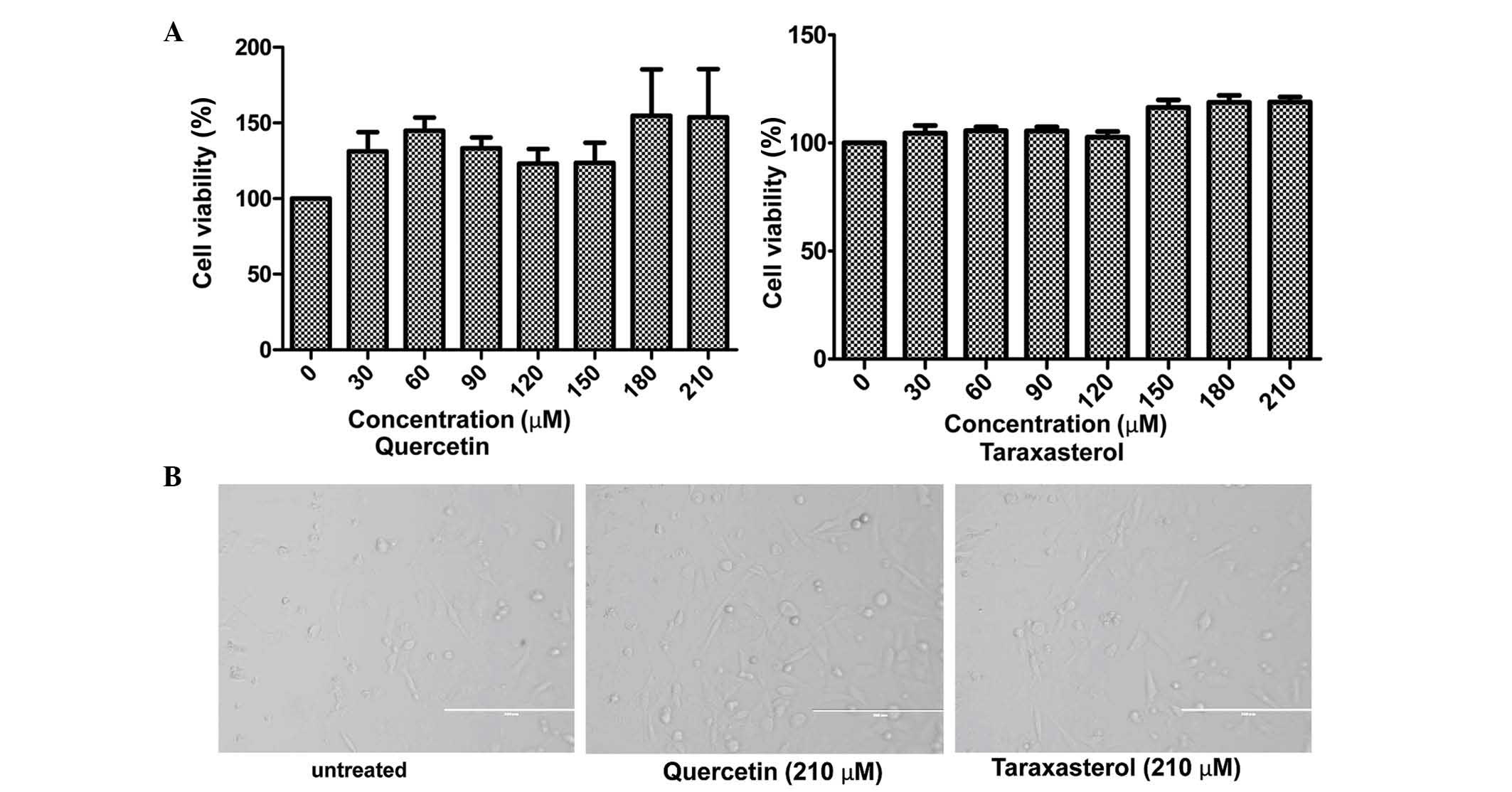
Effects of quercetin and taraxasterol
on cell viability
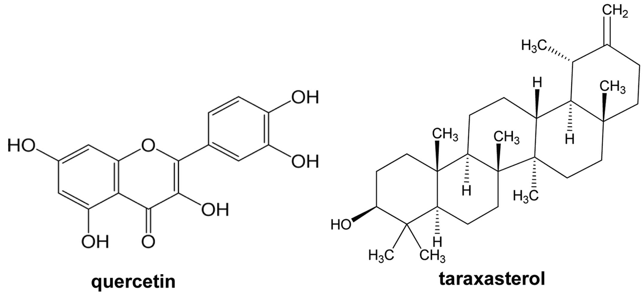
Quercetin is a well-known flavonoid, while
taraxasterol, a pentacyclic-triterpene, is isolated from
Taraxacum officinale (Fig.
1). The potential cytotoxicity of quercetin and taraxasterol
was evaluated by CCK-8 assay. HUVECs were incubated with quercetin
and taraxasterol, respectively, at concentrations ranging between 0
and 210 µM (0, 30, 60, 90, 120, 150, 180 and 210 µM) for 12 h.
Compared with untreated cells, the HUVEC morphology was not
affected and the viability was not reduced by treatment with either
agent (Fig. 2A); however, the agents
both stimulated the growth and proliferation of the HUVECs. Of the
two compounds, the pro-proliferative effect of quercetin was
stronger than that of taraxasterol. The representative
microphotographs are shown in Fig.
2B.
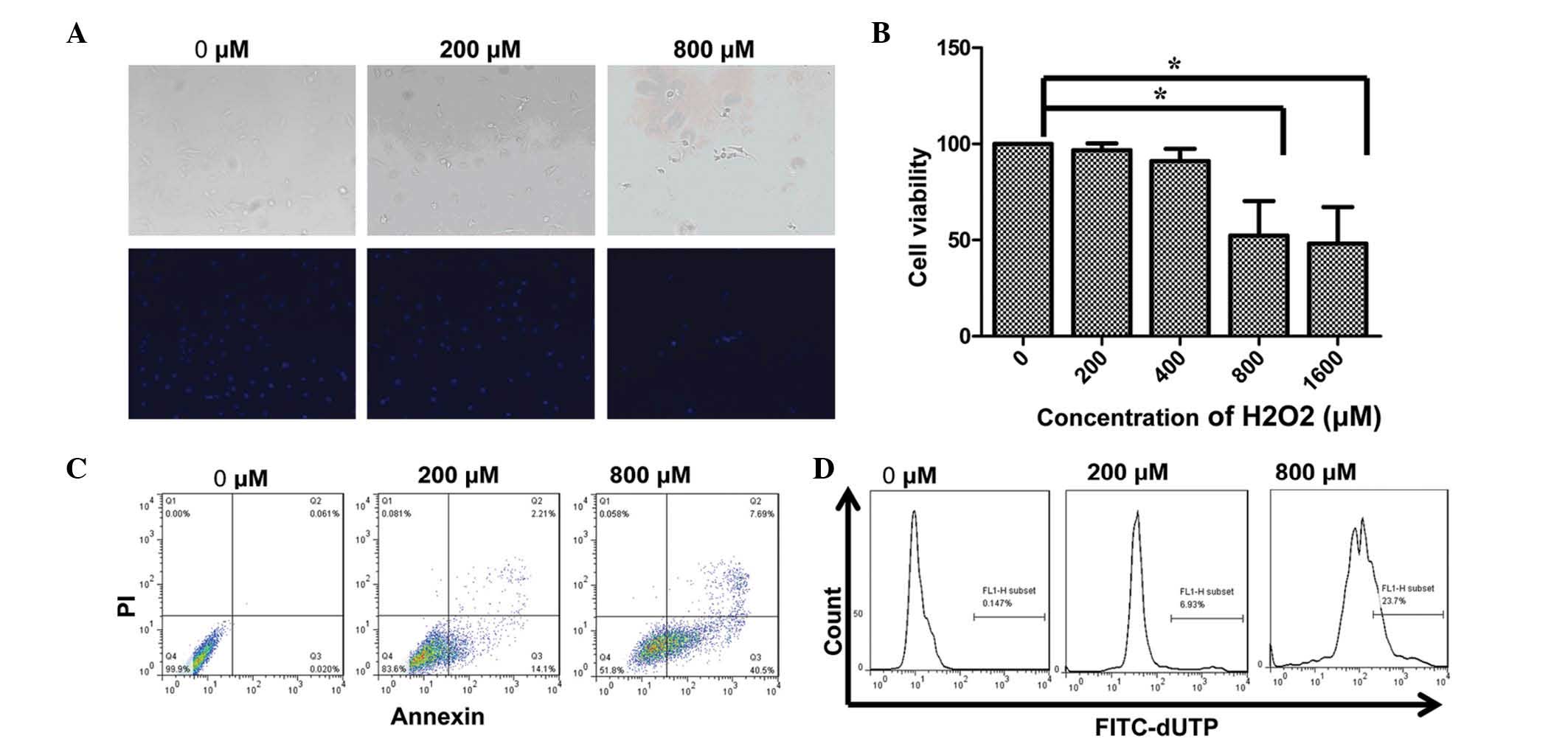
H2O2 negatively
affects HUVEC viability
The effect of H2O2 on HUVECs
is shown in Fig. 3. HUVECs were
exposed to different doses of H2O2 (0, 200,
400, 800 or 1,600 µM) for 4 h, and the cell viability was then
detected using CCK-8 assay. As shown in Fig. 3B, cell viability was not changed at
low H2O2 concentrations of 200 and 400 µM. At
higher concentrations of H2O2, cell viability
was significantly decreased. At H2O2
concentrations of 800 and 1,600 µM, the cell viability was reduced
to 52.34±31.06 and 48.15±32.92%, respectively; therefore, a
concentration of 800 µM was used to induce cell injury in
subsequent experiments. The results of the DAPI staining reflected
the findings of the cell viability assay (Fig. 3A). In cells not treated with
H2O2, no signs of morphological nuclear
damage were observed; by contrast, the
H2O2-treated cells exhibited changes in
nuclear and actin structure. At a concentration of 800 µM, nuclear
breakdown was evident from small fragments of nuclei inside the
cells.
H2O2 induces
apoptosis in HUVECs
It is well known that H2O2 can
cause lipid peroxidation and DNA damage, which can induce
apoptosis. In the present study, two assays were applied to examine
the changes in the levels of apoptosis following
H2O2-induced HUVEC injury. As shown by the
annexin V/PI staining in Fig. 3C,
the percentage of early apoptotic (annexin
V+/PI−) and late apoptotic (annexin
V+/PI+) untreated HUVECs was 0.020 and
0.061%, respectively. By contrast, 4 h of treatment with different
concentrations of H2O2 largely increased the
percentage of early and late apoptotic cells. At a concentration of
200 µM, the percentage of early and late apoptotic cells increased
to 14.4 and 2.21%, respectively, and at the concentration of 800
µM, these percentages increased to 40.5 and 7.69%. As shown by
TUNEL assay in Fig. 3D, comparisons
with untreated HUVECs revealed the percentage of TUNEL-positive
cells in the HUVECs treated with 200 and 800 µM
H2O2 to be 6.97 and 23.7%, respectively;
however, the combined percentages of annexin
V+/PI− and annexin
V+/PI+ cells were 16.31 and 48.19%,
respectively. These values were approximately two-fold higher than
the values determined by TUNEL assay. This indicated that annexin
V/PI staining was more sensitive than TUNEL assay, as reported by
Shen et al (26).
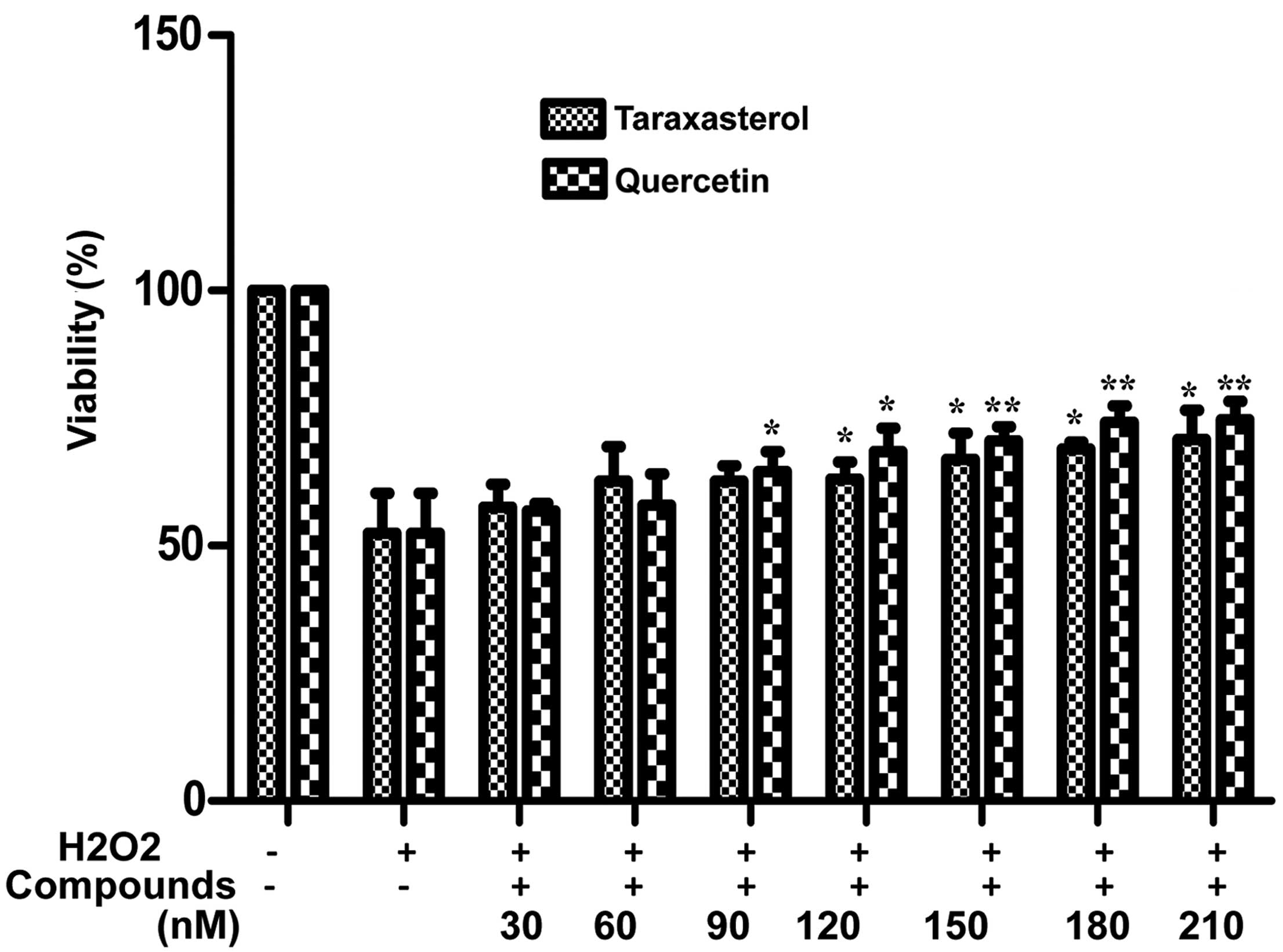
Quercetin and taraxasterol protect
HUVECs against H2O2-induced cytotoxicity
Following the apoptosis experiments, the optimal
concentrations of quercetin and taraxasterol for inducing the
proliferation of HUVECs without altering cell morphology were
determined. The protective effects of the two compounds against
H2O2-induced HUVECs injury were then
compared. The effects of quercetin and taraxasterol on the
viability of H2O2-treated HUVECs were
analyzed by CCK-8 assay. As shown in Fig. 4, treatment with 800 µM
H2O2 for 4 h caused significantly decreased
cell viability (~50%); however, pretreatment with different
concentrations of taraxasterol (30, 60, 90, 120, 150, 180 or 210
µM) for 12 h significantly improved the viability of the HUVECs (up
to 70.78±5.72% viability) and pretreatment with quercetin
significantly increased the viability of the HUVECs to a maximum of
74.74±6.12% at 210 µM (P<0.01). The inhibitory effect of
quercetin on H2O2-induced HUVEC injury was
stronger than that of taraxasterol at concentrations of 90–210 µM,
which is consistent with previous research (27–29). The
data also showed that the vascular protective effects of quercetin
and taraxasterol against oxidative damage occurred in a
dose-dependent manner.
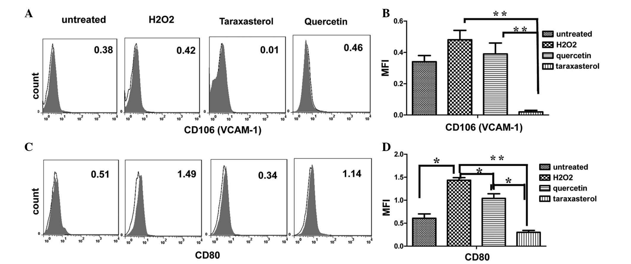
VCAM-1 and CD80 are involved in the
protection of endothelial cells
Based on the above observation and the
anti-inflammatory effects of the two compounds (26–30), we
speculated whether quercetin and taraxasterol would affect the
expression of pro-inflammatory CAMs and co-stimulatory molecules.
Previous research has indicated that selectins mediate the initial
rolling of leukocytes along the endothelium and that VCAM-1 and
ICAM-1 play important roles in the firm attachment and
transendothelial migration of the leukocytes (31). The next focus of the study was
therefore on the surface expression of the adhesive molecule VCAM-1
and the co-stimulatory molecule CD80.
Although VCAM-1 was not constitutively expressed by
HUVECs, it was markedly induced following the stimulation of the
HUVECs by 800 µM H2O2 for 4 h. The MFI
increased from 0.34±0.057 to 0.48±0.085. Pretreatment of the HUVECs
with quercetin or taraxasterol for 12 h prior to the stimulation
with 800 µM H2O2 for 4 h decreased the
expression of VCAM-1. Pretreatment with 210 µM quercetin for 12 h
decreased the expression of VCAM-1 to normal levels (0.39±0.099) or
lower, and pretreatment with taraxasterol significantly reduced the
MFI from 0.48±0.085 to 0.02±0.014 (P<0.01) (Fig. 5A and B).
Having observed that taraxasterol and quercetin
reduced the expression of VCAM-1, it was further investigated
whether taraxasterol and quercetin could change the expression of
co-stimulatory molecules. Due to the fact that deficiencies in the
co-stimulatory molecules CD80 and CD86 have been shown to reduce
atherosclerosis in mice (32), the
surface expression of CD80 molecule on HUVECs was evaluated in the
present study by assessing the MFI. As shown in Fig. 5C and D, the expression of CD80 was
low in resting HUVECs (0.60±0.13); however, when the HUVECs were
activated by H2O2, the expression of CD80
increased significantly by three-fold. Although taraxasterol and
quercetin both reduced the CD80 expression on activated HUVECs, the
inhibitory effect exerted by taraxasterol on the enhancement of
CD80 expression was more marked (from 1.44±0.77 to 0.30±0.056).
Discussion
Oxidative stress can result from a variety of
external stimuli, including toxins, cytokines, neurohormones,
ischemia or mechanical stress (33).
It has previously been suggested that an increased formation of
reactive oxygen species (ROS) (known as oxidative stress) promotes
smooth muscle cell proliferation and matrix formation, and
participants in long-term changes in the cardiac cellular
phenotype, as observed in hypertrophy, heart failure and apoptosis
(34). It is therefore believed that
increased ROS production may be partly responsible for a variety of
different diseases, such as hypertension (35), restenosis and arteriosclerosis
(36). Initially, redox processes
were suggested to be mediated predominantly by
H2O2. H2O2 can modify
LDL into oxidized-LDL, which may promote the development of
atherosclerosis (37). In the
present study, the construction of an
H2O2-induced cell injury model was used to
demonstrate that oxidative damage could alter the morphology and
function of endothelial cells.
At concentrations of 30–210 µM, both taraxasterol
and quercetin increased the viability and proliferation of the
HUVECs. At concentrations of 90–210 µM, the inhibitory effect of
quercetin on H2O2-induced HUVEC injury was
stronger than that of taraxasterol, and a dose-dependent effect was
observed. There are several mechanisms by which taraxasterol and
quercetin can protect against oxidative stress-induced epithelial
and endothelial cell dysfunction (38,39). A
previous study reported that quercetin inhibited the upregulation
of caveolin-1 to result in an increase in antioxidative capacity
(39). Furthermore, the chronic
administration of quercetin has been shown to significantly
attenuate elevations in lipid peroxidation and restore depletions
in reduced glutathione levels, acetylcholinesterase activity and
nitrite activity (27). Taraxasterol
has been suggested to exert its anti-inflammatory effect by
blocking the nuclear factor-κB (NF-κB) pathway (24).
It is well known that H2O2
induces apoptosis in HUVECs (40).
The findings of the present study also confirmed that
H2O2 could increase the level of HUVEC
apoptosis. Comparisons with untreated HUVECs revealed that the
percentage of TUNEL-positive cells among the cells treated with 200
and 800 µM H2O2 was 6.97 and 23.7%,
respectively. Furthermore, annexin V/PI staining was found to be a
more sensitive method of assessment than TUNEL assay.
In addition to inducing apoptosis,
H2O2 is a powerful pro-inflammatory factor
acting in endothelial cells. In a previous study, 2 h incubation
with H2O2 increased the release of tumor
necrosis factor-α and IL-6 to 2.67- and 1.67-fold that of control
levels, respectively (41). To
further confirm the ability of taraxasterol and quercetin to
prevent atherogenesis, the current study investigated the effect of
taraxasterol and quercetin on the expression of ICAM-1 and VCAM-1
in H2O2-induced HUVECs. Leukocyte adherence
and transmigration across the vascular endothelium are mediated by
CAMs (42). Increased levels of
ICAM-1 and VCAM-1 have been associated with the early events of
atherosclerosis. Quercetin, a bioflavonoid commonly present in a
variety of plants, is known to have several biological effects and
exerts anti-inflammatory, antioxidant (39), antiviral and antitumor activities.
The anti-inflammatory effect of quercetin has received particular
focus. In a previous study involving human atheroma, high levels of
ICAM-1 expression were found in endothelial cells and macrophages,
whereas VCAM-1 expression was observed in fewer than one-third of
lesions and its expression was predominantly restricted to
endothelial cells (43).
One of the main findings of the present study was
that short-term exposure (4 h) of HUVECs to
H2O2 resulted in the induction of the
expression of the adhesive molecule VCAM-1 and the co-stimulatory
molecule CD80 on HUVECs in vitro. Quercetin reduced the
expression of VCAM-1 on H2O2-injured HUVECs
in a non-significant manner (P=0.522). This finding was not
consistent with that of Rendig et al (44), which documented that quercetin
inhibited H2O2-induced
endothelium-independent coronary vasorelaxation and significantly
downregulated NF-κB, activator protein-1, IL-6 and VCAM-1.
Taraxasterol, a pentacyclic-triterpene isolated from Taraxacum
officinale, Cichorium glandulosum, Cynara
cardunculus L., Arnica montana L., Arctium and
Cichorium intybus (45), has
various biological activities and exerts antimicrobial (46), antiviral (47,48),
anti-inflammatory (24) and
chemopreventive (38) effects. In
the present study, we found that taraxasterol significantly reduced
the expression of VCAM-1 on H2O2-injured
HUVECs. Furthermore, both taraxasterol and quercetin significantly
reduced the expression of CD80 on
H2O2-injured HUVECs. A combination of these
two compounds may attenuate the inflammatory state in
atherosclerosis. These findings demonstrated the beneficial effect
of taraxasterol and quercetin in reducing VCAM-1 and CD80
expression on endothelial cells and thus in reducing the risk of
atherosclerosis.
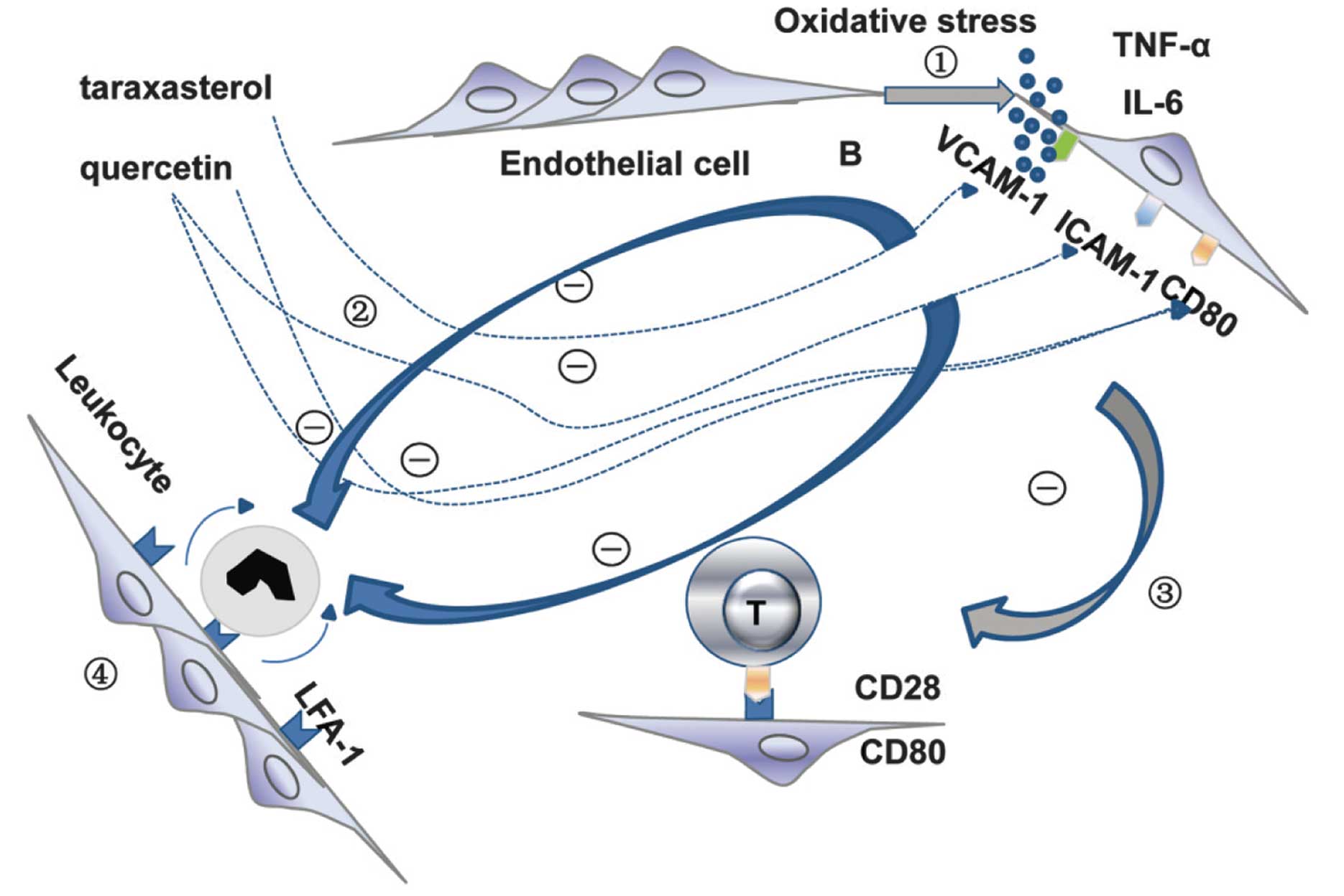
In conclusion, as a powerful pro-inflammatory
factor, H2O2 is known to increase the release
of TNF-α and IL-6 from HUVECs, on which the adhesive molecule
VCAM-1 and the co-stimulatory molecule CD80 are expressed (49). Pretreatment of the HUVECs with
taraxasterol or quercetin inhibited the expression of VCAM-1 and
CD80 on the activated HUVECs. Decreased expression of CD80 has been
found to result in the inactivation of T cells (50), while decreased expression of ICAM-1
results in a reduction in leukocyte rolling (51); both are associated with the
progression of atherosclerosis (Fig.
6). The results of the present study showed the protective
effects of quercetin and taraxasterol against cell injury and in
downregulating the expression of VCAM-1 and CD80 on HUVECs. This
study may provide preliminary data for further investigations into
the mechanism underlying the anti-atherosclerotic and
cardiovascular protective effects of quercetin and taraxasterol as
dietary supplements.
Acknowledgements
This study was supported by the Zhengzhou Doctor
Venture Fund Project (no. 2011-11).
References
|
1
|
Versari D, Daghini E, Virdis A, Ghiadoni L
and Taddei S: Endothelial dysfunction as a target for prevention of
cardiovascular disease. Diabetes Care 32 Suppl. 2:S314–S321. 2009.
View Article : Google Scholar
|
|
2
|
Savoia C, Sada L, Zezza L, et al: Vascular
inflammation and endothelial dysfunction in experimental
hypertension. Int J Hypertens. 2011:2812402011. View Article : Google Scholar : PubMed/NCBI
|
|
3
|
Thompson AM, Zhang Y, Tong W, et al:
Association of inflammation and endothelial dysfunction with
metabolic syndrome, prediabetes and diabetes in adults from Inner
Mongolia, China. BMC Endocr Disord. 11:162011. View Article : Google Scholar : PubMed/NCBI
|
|
4
|
Wannamethee SG, Tchernova J, Whincup P, et
al: Plasma leptin: associations with metabolic, inflammatory and
haemostatic risk factors for cardiovascular disease.
Atherosclerosis. 191:418–426. 2007. View Article : Google Scholar : PubMed/NCBI
|
|
5
|
Spagnoli LG, Bonanno E, Sangiorgi G and
Mauriello A: Role of inflammation in atherosclerosis. J Nucl Med.
48:1800–1815. 2007. View Article : Google Scholar : PubMed/NCBI
|
|
6
|
Lawson C and Wolf S: ICAM-1 signaling in
endothelial cells. Pharmacol Rep. 61:22–32. 2009. View Article : Google Scholar : PubMed/NCBI
|
|
7
|
Hwang SJ, Ballantyne CM, Sharrett AR, et
al: Circulating adhesion molecules VCAM-1, ICAM-1, and E-selectin
in carotid atherosclerosis and incident coronary heart disease
cases: the Atherosclerosis Risk In Communities (ARIC) study.
Circulation. 96:4219–4225. 1997. View Article : Google Scholar : PubMed/NCBI
|
|
8
|
Rubio Guerra, Vargas-Robles H, Serrano AM,
et al: Correlation between the levels of circulating adhesion
molecules and atherosclerosis in hypertensive type-2 diabetic
patients. Clin Exp Hypertens. 32:308–310. 2010. View Article : Google Scholar : PubMed/NCBI
|
|
9
|
Ellison S, Gabunia K, Kelemen SE, et al:
Attenuation of experimental atherosclerosis by interleukin-19.
Arterioscler Thromb Vasc Biol. 33:2316–2324. 2013. View Article : Google Scholar : PubMed/NCBI
|
|
10
|
McLaren JE, Michael DR, Salter RC, et al:
IL-33 reduces macrophage foam cell formation. J Immunol.
185:1222–1229. 2010. View Article : Google Scholar : PubMed/NCBI
|
|
11
|
Miller AM, Xu D, Asquith DL, et al: IL-33
reduces the development of atherosclerosis. J Exp Med. 205:339–346.
2008. View Article : Google Scholar : PubMed/NCBI
|
|
12
|
Ridker PM, Rifai N, Clearfield M, et al:
Air Force/Texas Coronary Atherosclerosis Prevention Study
Investigators: Measurement of C-reactive protein for the
targeting of statin therapy in the primary prevention of acute
coronary events. N Engl J Med. 344:1959–1965. 2001. View Article : Google Scholar : PubMed/NCBI
|
|
13
|
Kleemann R, Verschuren L, de Rooij BJ, et
al: Evidence for anti-inflammatory activity of statins and
PPARalpha activators in human C-reactive protein transgenic mice in
vivo and in cultured human hepatocytes in vitro. Blood.
103:4188–4194. 2004. View Article : Google Scholar : PubMed/NCBI
|
|
14
|
Mirjanic Azaric, Rizzo M, Sormaz L, et al:
Atorvastatin in stable angina patients lowers CCL2 and ICAM1
expression: pleiotropic evidence from plasma mRNA analyses. Clin
Biochem. 46:1526–1531. 2013. View Article : Google Scholar : PubMed/NCBI
|
|
15
|
Colucci R, Fornai M, Duranti E, et al:
Rosuvastatin prevents angiotensin II-induced vascular changes by
inhibition of NAD(P)H oxidase and COX-1. Br J Pharmacol.
169:554–566. 2013. View Article : Google Scholar : PubMed/NCBI
|
|
16
|
Nie P, Li D, Hu L, et al: Atorvastatin
improves plaque stability in ApoE-knockout mice by regulating
chemokines and chemokine receptors. PLoS One. 9:e970092014.
View Article : Google Scholar : PubMed/NCBI
|
|
17
|
Berman JP, Farkouh ME and Rosenson RS:
Emerging anti-inflammatory drugs for atherosclerosis. Expert Opin
Emerg Drugs. 18:193–205. 2013. View Article : Google Scholar : PubMed/NCBI
|
|
18
|
Larson AJ, Symons JD and Jalili T:
Therapeutic potential of quercetin to decrease blood pressure:
review of efficacy and mechanisms. Adv Nutr. 3:39–46. 2012.
View Article : Google Scholar : PubMed/NCBI
|
|
19
|
Egert S, Bosy-Westphal A, Seiberl J, et
al: Quercetin reduces systolic blood pressure and plasma oxidised
low-density lipoprotein concentrations in overweight subjects with
a high-cardiovascular disease risk phenotype: a double-blinded,
placebo-controlled cross-over study. Br J Nutr. 102:1065–1074.
2009. View Article : Google Scholar : PubMed/NCBI
|
|
20
|
Galindo P, Rodriguez-Gómez I,
González-Manzano S, et al: Glucuronidated quercetin lowers blood
pressure in spontaneously hypertensive rats via deconjugation. PLoS
One. 7:e326732012. View Article : Google Scholar : PubMed/NCBI
|
|
21
|
Chopra M, Fitzsimons PE, Strain JJ,
Thurnham DI and Howard AN: Nonalcoholic red wine extract and
quercetin inhibit LDL oxidation without affecting plasma
antioxidant vitamin and carotenoid concentrations. Clin Chem.
46:1162–1170. 2000.PubMed/NCBI
|
|
22
|
Galindo P, González-Manzano S, Zarzuelo
MJ, et al: Different cardiovascular protective effects of quercetin
administered orally or intraperitoneally in spontaneously
hypertensive rats. Food Funct. 3:643–650. 2012. View Article : Google Scholar : PubMed/NCBI
|
|
23
|
Hubbard GP, Wolffram S, de Vos R, et al:
Ingestion of onion soup high in quercetin inhibits platelet
aggregation and essential components of the collagen-stimulated
platelet activation pathway in man: a pilot study. Br J Nutr.
96:482–488. 2006.PubMed/NCBI
|
|
24
|
Zhang X, Xiong H and Liu L: Effects of
taraxasterol on inflammatory responses in
lipopolysaccharide-induced RAW 264.7 macrophages. J Ethnopharmacol.
141:206–211. 2012. View Article : Google Scholar : PubMed/NCBI
|
|
25
|
Liu J, Xiong H, Cheng Y, et al: Effects of
taraxasterol on ovalbumin-induced allergic asthma in mice. J
Ethnopharmacol. 148:787–793. 2013. View Article : Google Scholar : PubMed/NCBI
|
|
26
|
Shen HM, Dai J, Chia SE, Lim A and Ong CN:
Detection of apoptotic alterations in sperm in subfertile patients
and their correlations with sperm quality. Hum Reprod.
17:1266–1273. 2002. View Article : Google Scholar : PubMed/NCBI
|
|
27
|
Kumar A, Sehgal N, Kumar P, Padi SS and
Naidu PS: Protective effect of quercetin against ICV
colchicine-induced cognitive dysfunctions and oxidative damage in
rats. Phytother Res. 22:1563–1569. 2008. View Article : Google Scholar : PubMed/NCBI
|
|
28
|
Ishizawa K, Yoshizumi M, Kawai Y, et al:
Pharmacology in health food: metabolism of quercetin in vivo and
its protective effect against arteriosclerosis. J Pharmacol Sci.
115:466–470. 2011. View Article : Google Scholar : PubMed/NCBI
|
|
29
|
Nabavi SF, Nabavi SM, Mirzaei M and
Moghaddam AH: Protective effect of quercetin against sodium
fluoride induced oxidative stress in rat's heart. Food Funct.
3:437–441. 2012. View Article : Google Scholar : PubMed/NCBI
|
|
30
|
Matouk AI, Taye A, Heeba GH and El-Moselhy
MA: Quercetin augments the protective effect of losartan against
chronic doxorubicin cardiotoxicity in rats. Environ Toxicol
Pharmacol. 36:443–450. 2013. View Article : Google Scholar : PubMed/NCBI
|
|
31
|
Chi Z and Melendez AJ: Role of cell
adhesion molecules and immune-cell migration in the initiation,
onset and development of atherosclerosis. Cell Adh Migr. 1:171–175.
2007. View Article : Google Scholar : PubMed/NCBI
|
|
32
|
Buono C, Pang H, Uchida Y, et al:
B7-1/B7-2 costimulation regulates plaque antigen-specific T-cell
responses and atherogenesis in low-density lipoprotein
receptor-deficient mice. Circulation. 109:2009–2015. 2004.
View Article : Google Scholar : PubMed/NCBI
|
|
33
|
Prasad V, Lorenz JN, Miller ML, et al:
Loss of NHE1 activity leads to reduced oxidative stress in heart
and mitigates high-fat diet-induced myocardial stress. J Mol Cell
Cardiol. 65:33–42. 2013. View Article : Google Scholar : PubMed/NCBI
|
|
34
|
Cooper MP: Interplay of mitochondrial
biogenesis and oxidative stress in heart failure. Circulation.
127:1932–1934. 2013. View Article : Google Scholar : PubMed/NCBI
|
|
35
|
Paravicini TM and Touyz RM: NADPH
oxidases, reactive oxygen species, and hypertension: clinical
implications and therapeutic possibilities. Diabetes Care 31 Suppl.
2:S170–S180. 2008. View Article : Google Scholar
|
|
36
|
Moukdar F, Robidoux J, Lyght O, et al:
Reduced antioxidant capacity and diet-induced atherosclerosis in
uncoupling protein-2-deficient mice. J Lipid Res. 50:59–70. 2009.
View Article : Google Scholar : PubMed/NCBI
|
|
37
|
Watanabe T, Pakala R, Katagiri T and
Benedict CR: Oxidized low-density lipoproteins potentiate the
mitogenic effect of 5-hydroxytryptamine on vascular smooth muscle
cells. Jpn Heart J. 43:35–42. 2002. View Article : Google Scholar : PubMed/NCBI
|
|
38
|
Takasaki M, Konoshima T, Tokuda H, et al:
Anti-carcinogenic activity of Taraxacum plant I. Biol Pharm Bull.
22:602–605. 1999. View Article : Google Scholar : PubMed/NCBI
|
|
39
|
Kook D, Wolf AH, Yu AL, et al: The
protective effect of quercetin against oxidative stress in the
human RPE in vitro. Invest Ophthalmol Vis Sci. 49:1712–1720. 2008.
View Article : Google Scholar : PubMed/NCBI
|
|
40
|
Liu CL, Xie LX, Li M, et al: Salvianolic
acid B inhibits hydrogen peroxide-induced endothelial cell
apoptosis through regulating PI3K/Akt signaling. PLoS One.
2:e13212007. View Article : Google Scholar : PubMed/NCBI
|
|
41
|
Qian J, Jiang F, Wang B, et al:
Ophiopogonin D prevents H2O2-induced injury
in primary human umbilical vein endothelial cells. J
Ethnopharmacol. 128:438–445. 2010. View Article : Google Scholar : PubMed/NCBI
|
|
42
|
Alvarez A, Cerdá-Nicolas M, Naim Abu Nabah
Y, et al: Direct evidence of leukocyte adhesion in arterioles by
angiotensin II. Blood. 104:402–408. 2004. View Article : Google Scholar : PubMed/NCBI
|
|
43
|
Davies MJ, Gordon JL, Gearing AJ, et al:
The expression of the adhesion molecules ICAM-1, VCAM-1, PECAM, and
E-selectin in human atherosclerosis. J Pathol. 171:223–229. 1993.
View Article : Google Scholar : PubMed/NCBI
|
|
44
|
Rendig SV, Symons JD, Longhurst JC and
Amsterdam EA: Effects of red wine, alcohol, and quercetin on
coronary resistance and conductance arteries. J Cardiovasc
Pharmacol. 38:219–227. 2001. View Article : Google Scholar : PubMed/NCBI
|
|
45
|
Ovesná Z, Vachálková A and Horváthová K:
Taraxasterol and beta-sitosterol: new naturally compounds with
chemoprotective/chemopreventive effects. Neoplasma. 51:407–414.
2004.PubMed/NCBI
|
|
46
|
Villarreal ML, Alvarez L, Alonso D, et al:
Cytotoxic and antimicrobial screening of selected terpenoids from
Asteraceae species. J Ethnopharmacol. 42:25–29. 1994. View Article : Google Scholar : PubMed/NCBI
|
|
47
|
He W, Han H, Wang W and Gao B:
Anti-influenza virus effect of aqueous extracts from dandelion.
Virol J. 8:5382011. View Article : Google Scholar : PubMed/NCBI
|
|
48
|
Han H, He W, Wang W and Gao B: Inhibitory
effect of aqueous Dandelion extract on HIV-1 replication and
reverse transcriptase activity. BMC Complement Altern Med.
11:1122011. View Article : Google Scholar : PubMed/NCBI
|
|
49
|
Lakshminarayanan V, Beno DW, Costa RH and
Roebuck KA: Differential regulation of interleukin-8 and
intercellular adhesion molecule-1 by H2O2 and tumor necrosis
factor-alpha in endothelial and epithelial cells. J Biol Chem.
272:32910–32918. 1997. View Article : Google Scholar : PubMed/NCBI
|
|
50
|
McCoy KD and Le Gros G: The role of CTLA-4
in the regulation of T cell immune responses. Immunol Cell Biol.
77:1–10. 1999. View Article : Google Scholar : PubMed/NCBI
|
|
51
|
Kadono T, Venturi GM, Steeber DA and
Tedder TF: Leukocyte rolling velocities and migration are optimized
by cooperative L-selectin and intercellular adhesion molecule-1
functions. J Immunol. 169:4542–4550. 2002. View Article : Google Scholar : PubMed/NCBI
|