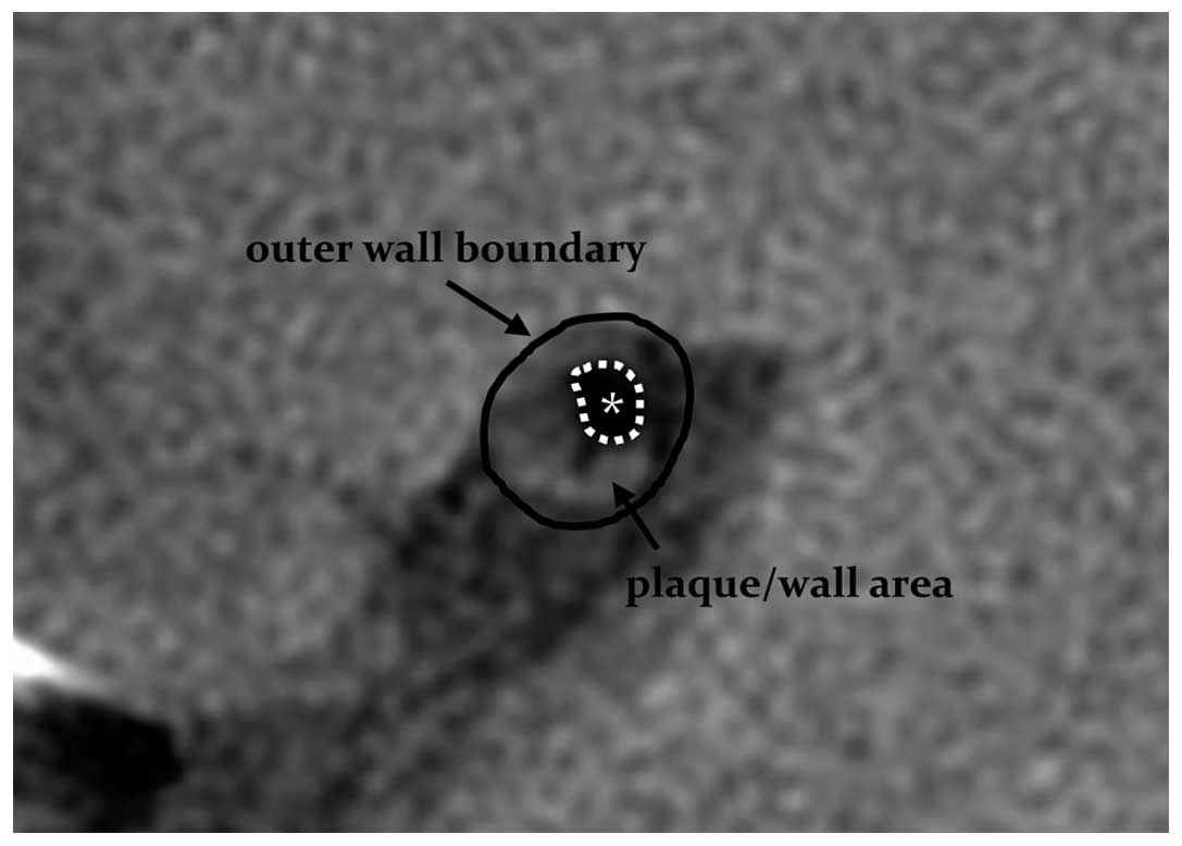
|
1
|
Wityk RJ, Lehman D, Klag M, Coresh J, Ahn
H and Litt B: Race and sex differences in the distribution of
cerebral atherosclerosis. Stroke. 27:1974–1980. 1996. View Article : Google Scholar : PubMed/NCBI
|
|
2
|
Wong KS, Gao S, Chan YL, Hansberg T, Lam
WW, Droste DW, Kay R and Ringelstein EB: Mechanisms of acute
cerebral infarctions in patients with middle cerebral artery
stenosis: A diffusion-weighted imaging and microemboli monitoring
study. Ann Neurol. 52:74–81. 2002. View Article : Google Scholar : PubMed/NCBI
|
|
3
|
Ryu CW, Jahng GH, Kim EJ, Choi WS and Yang
DM: High resolution wall and lumen MRI of the middle cerebral
arteries at 3 tesla. Cerebrovasc Dis. 27:433–442. 2009. View Article : Google Scholar : PubMed/NCBI
|
|
4
|
Xu WH, Li ML, Gao S, Ni J, Zhou LX, Yao M,
Peng B, Feng F, Jin ZY and Cui LY: In vivo high-resolution MR
imaging of symptomatic and asymptomatic middle cerebral artery
atherosclerotic stenosis. Atherosclerosis. 212:507–511. 2010.
View Article : Google Scholar : PubMed/NCBI
|
|
5
|
Yuan C, Mitsumori LM, Ferguson MS,
Polissar NL, Echelard D, Ortiz G, Small R, Davies JW, Kerwin WS and
Hatsukami TS: In vivo accuracy of multispectral magnetic resonance
imaging for identifying lipid-rich necrotic cores and intraplaque
hemorrhage in advanced human carotid plaques. Circulation.
104:2051–2056. 2001. View Article : Google Scholar : PubMed/NCBI
|
|
6
|
Ma N, Jiang WJ, Lou X, Ma L, Du B, Cai JF
and Zhao TQ: Arterial remodeling of advanced basilar
atherosclerosis: a 3-tesla MRI study. Neurology. 75:253–258. 2010.
View Article : Google Scholar : PubMed/NCBI
|
|
7
|
Yuan C, Beach KW, Smith LH Jr and
Hatsukami TS: Measurement of atherosclerotic carotid plaque size in
vivo using high resolution magnetic resonance imaging. Circulation.
98:2666–2671. 1998. View Article : Google Scholar : PubMed/NCBI
|
|
8
|
Niizuma K, Shimizu H, Takada S and
Tominaga T: Middle cerebral artery plaque imaging using 3-Tesla
high-resolution MRI. J Clin Neurosci. 15:1137–1141. 2008.
View Article : Google Scholar : PubMed/NCBI
|
|
9
|
Klein IF, Lavallée PC, Mazighi M,
Schouman-Claeys E, Labreuche J and Amarenco P: Basilar artery
atherosclerotic plaques in paramedian and lacunar pontine
infarctions: A high-resolution MRI study. Stroke. 41:1405–1409.
2010. View Article : Google Scholar : PubMed/NCBI
|
|
10
|
Schoenhagen P, Ziada KM, Kapadia SR, Crowe
TD, Nissen SE and Tuzcu EM: Extent and direction of arterial
remodeling in stable versus unstable coronary syndromes: An
intravascular ultrasound study. Circulation. 101:598–603. 2000.
View Article : Google Scholar : PubMed/NCBI
|
|
11
|
Li ML, Xu WH, Song L, Feng F, You H, Ni J,
Gao S, Cui LY and Jin ZY: Atherosclerosis of middle cerebral
artery: Evaluation with high-resolution MR imaging at 3T.
Atherosclerosis. 204:447–452. 2009. View Article : Google Scholar : PubMed/NCBI
|
|
12
|
Yamagishi M, Terashima M, Awano K, Kijima
M, Nakatani S, Daikoku S, Ito K, Yasumura Y and Miyatake K:
Morphology of vulnerable coronary plaque: Insights from follow-up
of patients examined by intravascular ultrasound before an acute
coronary syndrome. J Am Coll Cardiol. 35:106–111. 2000. View Article : Google Scholar : PubMed/NCBI
|
|
13
|
Jiang WJ, Yu W, Ma N, Du B, Lou X and
Rasmussen PA: High resolution MRI guided endovascular intervention
of basilar artery disease. J Neurointerv Surg. 3:375–378. 2011.
View Article : Google Scholar : PubMed/NCBI
|
|
14
|
Swartz RH, Bhuta SS, Farb RI, Agid R,
Willinsky RA, Terbrugge KG, Butany J, Wasserman BA, Johnstone DM,
Silver FL and Mikulis DJ: Intracranial arterial wall imaging using
high-resolution 3-tesla contrast-enhanced MRI. Neurology.
72:627–634. 2009. View Article : Google Scholar : PubMed/NCBI
|
|
15
|
Fayad ZA and Fuster V: Characterization of
atherosclerotic plaques by magnetic resonance imaging. Ann NY Acad
Sci. 902:173–186. 2000. View Article : Google Scholar : PubMed/NCBI
|
|
16
|
Saam T, Cai J, Ma L, Cai YQ, Ferguson MS,
Polissar NL, Hatsukami TS and Yuan C: Comparison of symptomatic and
asymptomatic atherosclerotic carotid plaque features with in vivo
MR imaging. Radiology. 240:464–472. 2006. View Article : Google Scholar : PubMed/NCBI
|
|
17
|
Saam T, Yuan C, Chu B, Takaya N, Underhill
H, Cai J, Tran N, Polissar NL, Neradilek B, Jarvik GP, et al:
Predictors of carotid atherosclerotic plaque progression as
measured by noninvasive magnetic resonance imaging.
Atherosclerosis. 194:e34–e42. 2007. View Article : Google Scholar : PubMed/NCBI
|
|
18
|
Kim JM, Jung KH, Sohn CH, Moon J, Han MH
and Roh JK: Middle cerebral artery plaque and prediction of the
infarction pattern. Arch Neurol. 69:1470–1475. 2012. View Article : Google Scholar : PubMed/NCBI
|
|
19
|
Demarco JK, Ota H, Underhill HR, Zhu DC,
Reeves MJ, Potchen MJ, Majid A, Collar A, Talsma JA, Potru S, et
al: MR carotid plaque imaging and contrast-enhanced MR angiography
identifies lesions associated with recent ipsilateral
thromboembolic symptoms: An in vivo study at 3T. AJNR Am J
Neuroradiol. 31:1395–1402. 2010. View Article : Google Scholar : PubMed/NCBI
|
|
20
|
Chen XY, Wong KS, Lam WW, Zhao HL and Ng
HK: Middle cerebral artery atherosclerosis: Histological comparison
between plaques associated with and not associated with infarct in
a postmortem study. Cerebrovasc Dis. 25:74–80. 2008. View Article : Google Scholar : PubMed/NCBI
|