Introduction
Corneal neovascularization (CNV) is typically
associated with inflammatory, infectious, traumatic, toxic,
degenerative or immunological disorders of the ocular surface and
cornea (1,2). CNV may result in significant visual
impairment and blindness, due to edema, scar formation or lipid
deposition (3,4). The regulation of corneal angiogenesis
is known to be a complex multistep process controlled by
stimulatory and inhibitory factors (5). Among various other proangiogenic
factors, the vascular endothelial growth factor (VEGF) family
serves a crucial function in stimulating the multiplication of
endothelial cells and the formation of new blood vessels (6). Furthermore, VEGF inhibitors have
exhibited potential for the treatment of CNV through the direct
inhibition of angiogenesis at a molecular level (3). In addition, numerous pharmacological
agents, including glucocorticosteroids, interleukin-1 receptor
antagonist, cyclosporine A, plasminogen fragments, doxycycline and
triamcinolone acetonide, appear to exhibit anti-angiogenic activity
(7–12). However, there is currently no clear
consensus regarding the most effective treatment option for CNV,
which underlines the requirement for novel therapies for the
treatment of CNV.
90Sr-90Y β-irradiation has
been widely used for the treatment of various diseases, including
coronary artery in-stent restenosis, post-operative scar
hyperplasia and skin hemangioma (13–15).
Notably, a previous study demonstrated that the inhibition of the
budding process of CNV pathogenesis via β-irradiation may limit the
formation and development of new corneal blood vessels (16); however, the effect of
90Sr-90Y β-irradiation in the cornea, and
specifically in animal models of CNV, has not yet been described.
The aim of the present study was to evaluate the safety and
efficacy of 90Sr-90Y β-irradiation in an
experimental rat model of alkali burn-induced CNV.
Materials and methods
Materials
The 90Sr-90Y ophthalmic
applicator (SSR9013) used in the present study was provided by the
China Institute of Atomic Energy (Beijing, China). The slit-lamp
microscope was purchased from Topcon Corporation (Tokyo, Japan) and
the BX41-72H02 binocular optical microscope was obtained from
Olympus Corporation (Tokyo, Japan).
Animals
A total of 40 female Wistar rats (age, 55–60 weeks;
weight, 200–250 g) obtained from the Experimental Animal Center of
Changchun Institute for Biological Sciences (Changchun, China) were
used in the present study. Approval of the experimental protocol
was obtained from the Jilin University Medical School Research
Committee (Changchun, China). The rats were treated and maintained
in accordance with the guidelines of the Statement for the Use of
Animals in Ophthalmic and Visual Research by the Association for
Research in Vision and Ophthalmology (17). The present study was approved by the
Ethics Committee of Jilin University. The rats were stored in a
specific pathogen-free facility, within filter-topped cages, under
a 12 h light/dark cycle at room temperature and 50–60% humidity.
The rats had ad libitum access to standard rodent chow and
water throughout the study.
Alkali-induced corneal injury model
and drug treatment protocol
A study population of 30 female Wistar rats were
anesthetized with an intraperitoneal injection of ketamine
hydrochloride (25 mg/kg) and xylazine hydrochloride (5 mg/kg; both
Sigma-Aldrich, St. Louis, MO, USA). All eyes were examined under a
binocular microscope to exclude corneal scaring, opacity and NV
prior to the study. Corneal injury was induced by placing a
monolayer filter saturated with 1 mol/l NaOH onto the right eye of
the rat for 2 min, as previously described (18–20).
Following the establishment of the alkali burn corneal injury, the
30 alkali-injured rats were allocated at random into three groups:
Alkali burn control group, which received, 3 drops of balanced salt
solution (Sigma-Aldrich) 3 times a day for 7 days in the
alkali-treated eyes; group 1, which received 1% cyclosporine
(Sigma-Aldrich) from day 1 following alkali injury, 3 drops 3 times
a day for 7 days in the alkali-treated eyes; and group 2, which
received 90Sr-90Y β-irradiation from day 1
following alkali injury, 1 Gy once a day for 7 days in the
alkali-treated eyes. In addition, 10 Wistar rats which did not
receive any treatment were selected as the alkali burn control
group, receiving 3 drops of the balanced salt solution, 3 times a
day for 7 days).
Evaluation of CNV
The CNV and edema formation in each group under
anesthesia was observed using the slit-lamp microscope on days 2, 5
and 7 following the experiment. The average NV length (VL), corneal
radius (r) and corneal hours (CH) were calculated. The NV area was
measured according to the following formula (21): Area (mm2) = CH/12 ×
3.14[r2-(r-VL)2].
Photographic analysis
All rats were sacrificed by exsanguination on day 7
immediately followed by observation using the slit-lamp microscope.
Briefly, the eyes were enucleated and the globes were fixed in
freshly prepared 4% paraformaldehyde. Following fixation for 24 h,
corneal samples were prepared by macroscopic incisions from limbus
to limbus passing through the central cornea to include the region
with the highest NV intensity. Subsequently, fixed tissues were
sectioned serially in the horizontal plane at 4 µm. In the majority
of sections, the NV density was obtained from the central region of
the cornea. The sections were stained with hematoxylin and eosin
(H&E; Sigma-Aldrich). The degree of CNV was evaluated
histomorphometrically using the optical microscope, as described in
a previous study (22). In addition,
the inflammatory index was evaluated using slit-lamp biomicroscopy,
and inflammatory cells that had infiltrated into the cornea tissue
were detected by histological analysis at days 1, 7 and 14
following the alkali burn, as previously described (23).
Western blot analysis
The rats were sacrificed by exsanguination and the
corneas harvested from the treated eyes were dissected and frozen
at −70°C, then homogenized in ice-cold RIPA lysis buffer solution
(Santa Cruz Biotechnology Inc., Santa Cruz, CA, USA). Following
centrifugation for 5 min at 12,000 × g, the supernatants were
collected and the protein concentrations were determined using the
Bradford reagent (Sigma-Aldrich Chemie GmbH, Steinheim, Germany).
Equal quantities of protein (15 µg/lane) from the cell lysates were
separated using 8–15% sodium dodecyl sulfate polyacrylamide gel
electrophoresis (SDS-PAGE) and transferred onto nitrocellulose
membranes (Santa Cruz Biotechnology, Inc.). Membranes were
incubated for 2 h in phosphate-buffered saline plus 0.1% Tween-20
and 5% non-fat skim milk to block non-specific binding. The
membranes were then incubated overnight at 4°C with the following
antibodies: Goat monoclonal anti-mouse VEGF (1:2,000; sc-7269;
Santa Cruz Biotechnology, Inc.), goat monoclonal anti-mouse VEGF
receptor (VEGFR)-1 (1:3,000; 4762; Sigma-Aldrich), goat monoclonal
anti-mouse VEGFR-2 (1:1,000; 3817; Cell Signaling Technology,
Danvers, MA, USA.), goat monoclonal anti-mouse matrix
metalloproteinase (MMP)-9 (1:2,000; sc-21733; Santa Cruz
Biotechnology, Inc.), and goat monoclonal anti-mouse β-actin
(1:10,000; sc-47778; Santa Cruz Biotechnology, Inc.), which was
used as a loading control. Subsequently, the membranes were
incubated with the anti-mouse horseradish peroxidase-conjugated
immunoglobulin G (1:10,000; sc-2005; Santa Cruz Biotechnology,
Inc.), and protein bands were visualized using a SuperSignal
chemiluminescence detection kit (Thermo Fisher Scientific, Inc.,
Rockford, Illinois, USA).
Statistical analysis
Data are presented as the mean ± standard deviation.
The statistical comparison among >2 groups was performed using
one-way analysis of variance followed by the Tukey's post-hoc test.
Statistical analysis was performed using SPSS software, version
16.0 (SPSS Inc., Chicago, IL, USA) and GraphPad Prism, version 5.01
(GraphPad Software Inc., San Diego, CA, USA) for Windows. P<0.05
was considered to indicate a statistically significant
difference.
Results
Anti-NV effects of
90Sr-90Y β-irradiation in the cornea
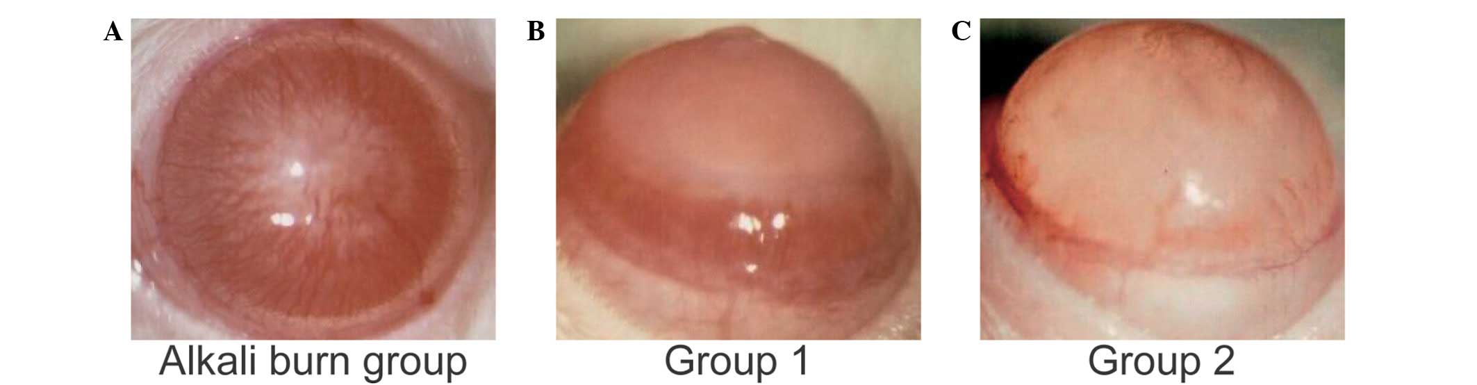
The clinical indication of CNV was examined first.
The results showed that numerous new vessels had invaded the suture
area from the limbal region in the alkali burn control group on day
7 (Fig. 1A). The rats treated with
angiogenesis inhibitors (group 1) and
90Sr-90Y β-irradiation (group 2) exhibited
reduced CNV (Fig. 1B and C), as
compared with the alkali burn control group (Fig. 1A). The results for the NV length and
area revealed that angiogenesis inhibitors (group 1) and
90Sr-90Y β-irradiation (group 2) treatment
significantly reduced the CNV at the various time points, compared
with the alkali burn control group (Tables I and II); however, the CNV length and area in
group 1 were significantly decreased, as compared with group 2 on
day 2 (P<0.05; Tables I and
II), and increased on days 5 and 7
(P<0.05; Tables I and II).
 | Table I.Average length of CNV among the
different groups at each time-point (n=10 per group). |
Table I.
Average length of CNV among the
different groups at each time-point (n=10 per group).
|
| Average length of
CNV (mm) |
|---|
|
|
|
|---|
| Group | Day 2 | Day 5 | Day 7 |
|---|
| Alkali burn
group | 0.410±0.024 | 1.980±0.015 | 2.580±0.037 |
| Group 1 |
0.278±0.025a |
1.678±0.017a |
2.178±0.032a |
| Group 2 |
0.352±0.021ab |
1.482±0.030ab |
1.882±0.033ab |
| Table II.Average area of CNV among the
different groups at each time-point (n=10 per group). |
Table II.
Average area of CNV among the
different groups at each time-point (n=10 per group).
|
| Average area of CNV
(mm2) |
|---|
|
|
|
|---|
| Group | Day 2 | Day 5 | Day 7 |
|---|
| Alkali burn
group | 4.596±0.184 | 14.516±0.112 | 21.739±0.209 |
| Group 1 |
2.167±0.181a |
12.465±0.132a |
17.851±0.199a |
| Group 2 |
3.471±0.178ab |
10.499±0.202ab |
14.471±0.211ab |
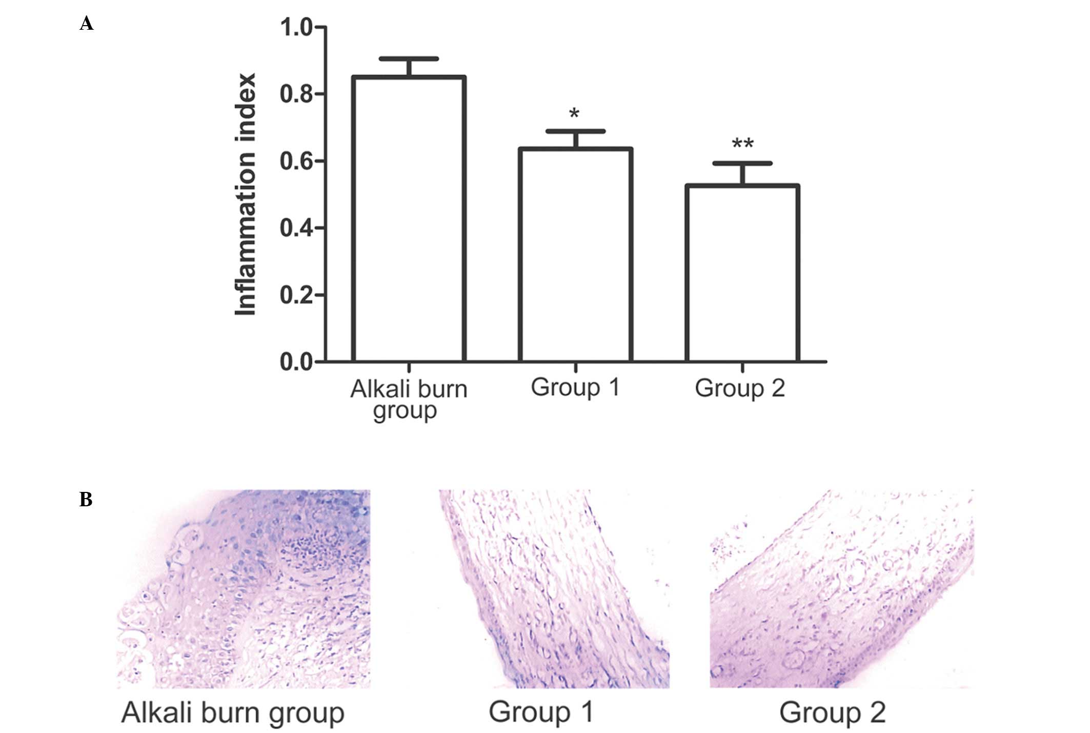
Anti-inflammatory effects of
90Sr-90Y β-irradiation in the cornea
Corneal inflammation was detected and analyzed
simultaneously. The inflammatory index results demonstrated that
the treatments administered in groups 1 and 2 significantly reduced
inflammation on day 7 compared with the alkali burn control group
(Fig. 2A). The the most marked
reduction in the number of inflammatory cells and the degree of
edema was observed in group 2. Histological examination of H&E
staining showed that the alkali burn control group exhibited
increased inflammatory cell infiltration in the corneal stroma
compared with the other groups (Fig.
2B), while the treatments administered in groups 1 and 2
decreased inflammatory cell infiltration (Fig. 2B). In addition, the angiogenesis
inhibitors and 90Sr-90Y β-irradiation
treatment resulted in reduced corneal edema compared with the
alkali burn control group.
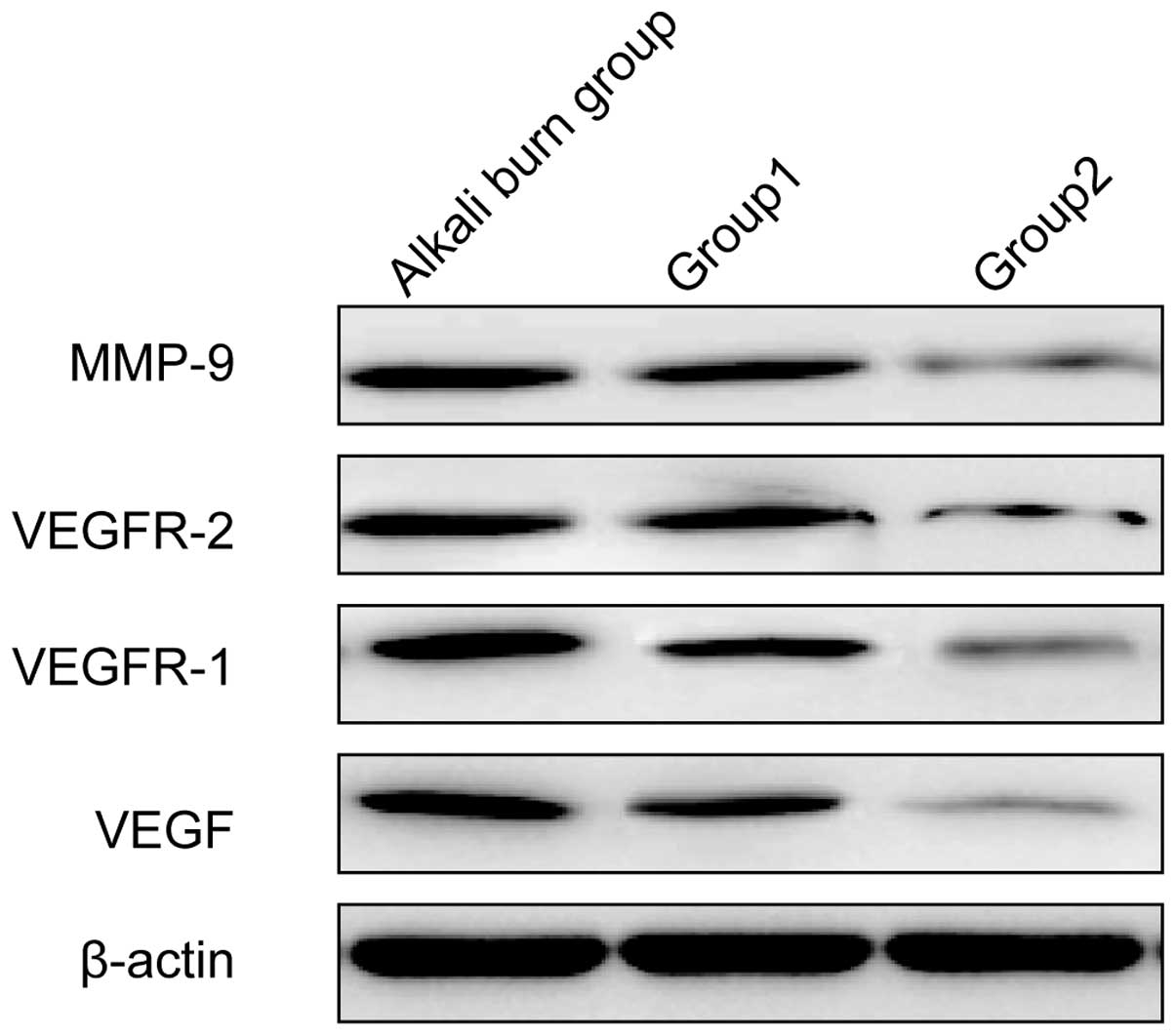
Inhibition of angiogenic factor
expression in alkali-injured corneas by
90Sr-90Y β-irradiation
The balance of proangiogenic factors is crucial for
angiogenesis. Therefore, the effect of
90Sr-90Y β-irradiation on the expression
levels of a number of proangiogenic factors in alkali-burn corneal
wounds was examined. The expression of MMP-9, VEGF, VEGFR-1 and
VEGFR-2 was measured using western blot analysis. The angiogenesis
inhibitors (group 1) and 90Sr-90Y
β-irradiation (group 2) appeared to produce marked reductions in
the expression levels of MMP-9, VEGF, VEGFR-1 and VEGFR-2 compared
with the alkali burn control group (Fig.
3). Compared with the treatment in group 1, the treatment in
group 2 appeared to produce reductions in the protein expression
levels of MMP-9, VEGF, VEGFR-1 and VEGFR-2.
Discussion
CNV is able to cause edema, scar formation or lipid
deposition, resulting in significant visual impairment and
blindness (3). Therefore, treating
this potentially blinding condition is of clinical significance
(24). Previous studies have shown
that thalidomide, photodynamic therapy, steroids, cyclosporine,
conjunctival limbal allograft and fine needle diathermy may exert
inhibitory effects against CNV (25–29);
however, these drugs are not always effective and may result in
complications (30). Radiation
therapy is widely used to treat post-operative scar hyperplasia and
skin hemangioma (14,15), which suggests that it may exert an
inhibitory effect against CNV. The aim of the present study was to
compare the treatment efficacy of 90Sr-90Y
β-irradiation with that of angiogenesis inhibitors in a rat model
of alkali burn-induced CNV, and to evaluate the inhibitory effects
of 90Sr-90Y β-irradiation on CNV. The present
results showed that 90Sr-90Y β-irradiation
exhibits inhibitory effects on alkali burn-induced CNV, which were
found to be superior to those of the angiogenesis inhibitor
cyclosporine.
Alkali burn may result in corneal endothelial cell
division and angiogenesis (31). The
growth trend of NV in the angiogenesis inhibitors and
90Sr-90Y β-irradiation groups were similar to
that of the alkali burn control group, but with a limited degree of
inhibition at different time points (P<0.05). The curative
effect of angiogenesis inhibitors was more marked compared with
that of 90Sr-90Y β-irradiation in the early
stage of treatment (P<0.05), while the opposite occurred in the
later stage (P<0.05). These results suggested that
90Sr-90Y β-irradiation was more effective
compared with angiogenesis inhibitors in inhibiting alkali
burn-induced CNV. The phenomenon in the early stage of treatment
could be explained by the interference of 1% cyclosporine with
certain cytokines associated with angiogenesis, thus achieving a
rapid biological effect (32).
The corneal of rats in the alkali burn control group
exhibited marked NV and numerous inflammatory cells, and presented
obvious edema. These pathological manifestations were consistent
with the results of a previous study (33). In the 90Sr-90Y
β-irradiation group, the degree of NV and edema, in addition to the
number of inflammatory cells, were reduced compared with the
angiogenesis inhibitors group. These results implied that
90Sr-90Y β-irradiation was more effective in
inhibiting CNV compared with angiogenesis inhibitors. This may be
explained by the biological effects of ionizing radiation, which is
able to damage DNA in endothelial cells of cornea angiogenesis,
inhibit cell proliferation and accelerate apoptosis, which caused
the vascular permeability to decrease and capillaries to atrophy.
Furthermore, the superiority of 90Sr-90Y
β-irradiation may be a result of the ability of ionizing radiation
to reduce plasma leakage and corneal inflammation, thus reducing
cytokine expression and leading to a subsequent reduction in CNV
(34).
To investigate the effects of the
90Sr-90Y β-irradiation on mediators
responsible for angiogenic activity, the expression levels of VEGF,
VEGFR-1, VEGFR-2 and MMP-9 were investigated in the present study.
It is generally acknowledged that an upregulation of angiogenic
factors occurs during CNV (35).
VEGF is a major mediator of the process of angiogenesis and is
crucially involved in the development of NV (36). Two high-affinity receptor tyrosine
kinases, soluble VEGFR-1 and VEGFR-2 have been shown to regulate
the angiogenic activity of VEGF (37). Therapeutic strategies involving the
targeting of VEGF, in order to inhibit the cascade of neovascular
formation, have recently been investigated (3). Abnormal MMPs are potent proangiogenic
factors and are known to be involved in CNV progression (38). The present study demonstrated that
90Sr-90Y β-irradiation suppressed the
expression of VEGF, VEGFR-1, VEGFR-2 and MMP-9, suggesting that
90Sr-90Y β-irradiation may inhibit CNV by
downregulating the expression of angiogenic factors.
In conclusion, the results of the present study
indicate that 90Sr-90Y β-irradiation and
angiogenesis inhibitors have inhibitory effects on CNV induced by
alkali burn, with the former being more effective, suggesting that
90Sr-90Y β-irradiation possesses therapeutic
potential for CNV. Although the short-term inhibiting effect of
90Sr-90Y β-irradiation in Wistar rats was
promising, its long-term effect and mechanism remains to be
elucidated.
Acknowledgements
The authors thank the National Natural Science
Foundation of China projects (grant no. 81271606) and Research Fund
of the Science and Technology Department of Jilin Province (grant
no. 201015185 and 201201041) for the financial support.
References
|
1
|
Siemerink MJ, Augustin AJ and Schlingemann
RO: Mechanisms of ocular angiogenesis and its molecular mediators.
Dev Ophthalmol. 46:4–20. 2010. View Article : Google Scholar : PubMed/NCBI
|
|
2
|
Maddula S, Davis DK, Maddula S, Burrow MK
and Ambati BK: Horizons in therapy for corneal angiogenesis.
Ophthalmology. 118:591–599. 2011. View Article : Google Scholar : PubMed/NCBI
|
|
3
|
Chang JH, Garg NK, Lunde E, Han KY, Jain S
and Azar DT: Corneal neovascularization, An anti-VEGF therapy
review. Surv Ophthalmol. 57:415–429. 2012. View Article : Google Scholar : PubMed/NCBI
|
|
4
|
Chang JH, Gabison EE, Kato T and Azar DT:
Corneal neovascularization. Curr Opin Ophthalmol. 12:242–249. 2001.
View Article : Google Scholar : PubMed/NCBI
|
|
5
|
Hsu CC, Chang HM, Lin TC, Hung KH, Chien
KH, Chen SY, Chen SN and Chen YT: Corneal neovascularization and
contemporary antiangiogenic therapeutics. J Chin Med Assoc.
78:323–330. 2015. View Article : Google Scholar : PubMed/NCBI
|
|
6
|
Morabito A, De Maio E, Di Maio M, Normanno
N and Perrone F: Tyrosine kinase inhibitors of vascular endothelial
growth factor receptors in clinical trials: Current status and
future directions. Oncologist. 11:753–764. 2006. View Article : Google Scholar : PubMed/NCBI
|
|
7
|
Phillips K, Arffa R, Cintron C, Rose J,
Miller D, Kublin CL and Kenyon KR: Effects of prednisolone and
medroxyprogesterone on corneal wound healing, ulceration, and
neovascularization. Arch Ophthalmol. 101:640–643. 1983. View Article : Google Scholar : PubMed/NCBI
|
|
8
|
Dana R: Comparison of topical
interleukin-1 vs tumor necrosis factor-alpha blockade with
corticosteroid therapy on murine corneal inflammation
neovascularization, and transplant survival (an American
Ophthalmological Society thesis). Trans Am Ophthalmol Soc.
105:330–343. 2007.PubMed/NCBI
|
|
9
|
Lipman RM, Epstein RJ and Hendricks RL:
Suppression of corneal neovascularization with cyclosporine. Arch
Ophthalmol. 110:405–407. 1992. View Article : Google Scholar : PubMed/NCBI
|
|
10
|
Li C, Li L, Cheng R, Dai Z, Li C, Yao Y,
Zhou T, Yang Z, Gao G and Yang X: Acidic/neutral amino acid
residues substitution in NH2 terminal of plasminogen kringle 5
exerts enhanced effects on corneal neovascularization. Cornea.
32:680–688. 2013. View Article : Google Scholar : PubMed/NCBI
|
|
11
|
Jovanovic V and Nikolic L: The effect of
topical doxycycline on corneal neovascularization. Curr Eye Res.
39:142–148. 2014. View Article : Google Scholar : PubMed/NCBI
|
|
12
|
Mehrjardi HZ, Ghaffari R, Mahbod M and
Hashemi H: Triamcinolone acetonide as an adjunct to bevacizumab for
prevention of corneal neovascularization in a rat model. J
Ophthalmic Vis Res. 9:162–168. 2014.PubMed/NCBI
|
|
13
|
Schiele TM, Herbst J, Pӧllinger B, Rieber
J, Kӧnig A, Sohn HY, Krötz F, Leibig M, Belka C and Klauss V: Late
and very late catch-up after 90Sr/90Y
beta-irradiation for the treatment of coronary in-stent restenosis.
Acute Card Care. 13:9–13. 2011. View Article : Google Scholar : PubMed/NCBI
|
|
14
|
Shi CB, Yuan B, Lu JR, Xu JL, Yang WD,
Deng JL and Wang J: Continuous low-dose-rate radiation of
radionuclide phosphorus-32 for hemangiomas. Cancer Biother
Radiopharm. 27:198–203. 2012. View Article : Google Scholar : PubMed/NCBI
|
|
15
|
Huang CM, Lee KW and Huang CJ: Radiation
therapy for life-threatening huge laryngeal hemangioma involving
pharynx and parapharyngeal space. Head Neck. 35:E98–E101. 2013.
View Article : Google Scholar : PubMed/NCBI
|
|
16
|
Feng K, Yang J and Li B: An investigation
on morphology of experimental corneal neovascularization. Zhonghua
Yan Ke Za Zhi. 37:384–386. 2001.(In Chinese). PubMed/NCBI
|
|
17
|
British Photobiology Society; Association
for Research in Vision and Ophthalmology. Proceedings of a meeting
on visual sensitivity and adaptation. Sponsored by the British
Photobiology Society and the Association for Research in Vision and
Ophthalmology Inc. held at the University of Surrey Guildford,
19-23 September, 1978. Vision research. 19:351–440. 1979.
|
|
18
|
Lu P, Li L, Mukaida N and Zhang X:
Alkali-induced corneal neovascularization is independent of
CXCR2-mediated neutrophil infiltration. Cornea. 26:199–206. 2007.
View Article : Google Scholar : PubMed/NCBI
|
|
19
|
Zhou WJ, Liu GQ, Li LB, Zhang XG and Lu
PR: Inhibitory effect of CCR3 signal on alkali-induced corneal
neovascularization. Int J Ophthalmol. 5:251–257. 2012.PubMed/NCBI
|
|
20
|
Zhou Q, Yang L, Qu M, Wang Y, Chen P, Wang
Y and Shi W: Role of senescent fibroblasts on alkali-induced
corneal neovascularization. J Cell Physiol. 227:1148–1156. 2012.
View Article : Google Scholar : PubMed/NCBI
|
|
21
|
Needleman P, Turk J, Jakschik BA, Morrison
AR and Lefkowith JB: Arachidonic acid metabolism. Annu Rev Biochem.
55:69–102. 1986. View Article : Google Scholar : PubMed/NCBI
|
|
22
|
Nakayama M, Iejima D, Akahori M, Kamei J,
Goto A and Iwata T: Overexpression of HtrA1 and exposure to
mainstream cigarette smoke leads to choroidal neovascularization
and subretinal deposits in aged mice. Invest Ophthalmol Vis Sci.
55:6514–6523. 2014. View Article : Google Scholar : PubMed/NCBI
|
|
23
|
Peng LH, Shen W, Yong W, Lu L and Liu L:
Effects of AMD3100 subconjunctival injection on alkali burn induced
corneal neovascularization in mice. Int J Ophthalmol. 4:44–48.
2011.PubMed/NCBI
|
|
24
|
Bachmann B, Taylor RS and Cursiefen C: The
association between corneal neovascularization and visual acuity, A
systematic review. Acta Ophthalmol. 91:12–19. 2013. View Article : Google Scholar : PubMed/NCBI
|
|
25
|
Kruse FE, Joussen AM, Rohrschneider K,
Becker MD and Vӧlcker HE: Thalidomide inhibits corneal angiogenesis
induced by vascular endothelial growth factor. Graefes Arch Clin
Exp Ophthalmol. 236:461–466. 1998. View Article : Google Scholar : PubMed/NCBI
|
|
26
|
Gohto Y, Obana A, Kanai M, Nagata S,
Nakajima S and Miki T: Treatment parameters for selective occlusion
of experimental corneal neovascularization by photodynamic therapy
using a water soluble photosensitizer, ATX-S10(Na). Exp Eye Res.
72:13–22. 2001. View Article : Google Scholar : PubMed/NCBI
|
|
27
|
Haynes WL, Proia AD and Klintworth GK:
Effect of inhibitors of arachidonic acid metabolism on corneal
neovascularization in the rat. Invest Ophthalmol Vis Sci.
30:1588–1593. 1989.PubMed/NCBI
|
|
28
|
Daya SM and Ilari FA: Living related
conjunctival limbal allograft for the treatment of stem cell
deficiency. Ophthalmology. 108:126–134. 2001. View Article : Google Scholar : PubMed/NCBI
|
|
29
|
Faraj LA, Elalfy MS, Said DG and Dua HS:
Fine needle diathermy occlusion of corneal vessels. Br J
Ophthalmol. 98:1287–1290. 2014. View Article : Google Scholar : PubMed/NCBI
|
|
30
|
Williams KA, Irani YD and Klebe S: Novel
therapeutic approaches for corneal disease. Discov Med. 15:291–299.
2013.PubMed/NCBI
|
|
31
|
Giacomini C, Ferrari G, Bignami F and Rama
P: Alkali burn versus suture-induced corneal neovascularization in
C57BL/6 mice, An overview of two common animal models of corneal
neovascularization. Exp Eye Res. 121:1–4. 2014. View Article : Google Scholar : PubMed/NCBI
|
|
32
|
Navarro-Antolín J, Redondo-Horcajo M,
Zaragoza C, Alvarez-Barrientos A, Fernández AP, León-Gómez E,
Rodrigo J and Lamas S: Role of peroxynitrite in endothelial damage
mediated by Cyclosporine A. Free Radic Biol Med. 42:394–403. 2007.
View Article : Google Scholar : PubMed/NCBI
|
|
33
|
Li X, Li NC, Wang G and Na YQ: Effect and
safety of transrectal 137 CS gamma-rays in the treatment of benign
prostatic hyperplasia. Zhonghua Nan Ke Xue. 12:525–527. 2006.(In
Chinese). PubMed/NCBI
|
|
34
|
Onoda JM, Kantak SS and Diglio CA:
Radiation induced endothelial cell retraction in vitro, Correlation
with acute pulmonary edema. Pathol Oncol Res. 5:49–55. 1999.
View Article : Google Scholar : PubMed/NCBI
|
|
35
|
Martin G, Schlunck G, Hansen LL and
Agostini HT: Differential expression of angioregulatory factors in
normal and CNV-derived human retinal pigment epithelium. Graefes
Arch Clin Exp Ophthalmol. 242:321–326. 2004. View Article : Google Scholar : PubMed/NCBI
|
|
36
|
Carmeliet P and Jain RK: Angiogenesis in
cancer and other diseases. Nature. 407:249–257. 2000. View Article : Google Scholar : PubMed/NCBI
|
|
37
|
Philipp W, Speicher L and Humpel C:
Expression of vascular endothelial growth factor and its receptors
in inflamed and vascularized human corneas. Invest Ophthalmol Vis
Sci. 41:2514–2522. 2000.PubMed/NCBI
|
|
38
|
Lee CM, Jung WK, Na G, Lee DS, Park SG,
Seo SK, Yang JW, Yea SS, Lee YM, Park WS and Choi IW: Inhibitory
effects of the platelet-activating factor receptor antagonists,
CV-3988 and Ginkgolide B, on alkali burn-induced corneal
neovascularization. Cutan Ocul Toxicol. 34:53–60. 2015. View Article : Google Scholar : PubMed/NCBI
|