Introduction
Osteoarthritis is one of the most common joint
disorders affecting human health, with no evident racial and
regional differences (1).
Osteoarthritis is a cause of long-term disability second only to
cardiovascular disease in China (2).
On the basis of a review, osteoarthritis giving rise to damage
affects 2–6% of the population (3).
Osteoarthritis has a greater influence on elderly patients than
other diseases, affecting, for example, the ability to go up and
down the stairs and other lower limb functions (4). Therefore, osteoarthritis is one of the
major diseases leading to functional disability in people aged
>50 years, causing loss to the economy and affecting social
development (5).
Osteoarthritis is characterized by bone
regeneration, and involves the degeneration and loss of articular
cartilage with chronic arthritis of the joint edge and subchondral
bone (6). The initial site of the
disease is in the cartilage. The etiology and pathogenesis of
osteoarthritis have not been fully elucidated; however, it is
generally considered that systemic factors are involved, such as
age, gender and family susceptibility, as well as local
biomechanics, cartilage cell apoptosis, cell factors and the
effects of degradative enzymes; it is a disease involving multiple
linked factors (7). According to the
literature, cartilage cell apoptosis may be important in the
pathogenesis of osteoarthritis (8).
At present, the signaling pathways involved in the induction of
cartilage cell apoptosis, the regulation of signaling factors in
such apoptosis, and indicators of chondrocyte apoptosis signal
stimulation are topics of particular interest for experimental
study. Numerous studies have confirmed that the pathogenic
mechanism of osteoarthritis involves interleukin-1 (IL)-1, tumor
necrosis factor (TNF)-α and matrix metalloproteinases (9). Through the determination of these
indicators, the evolution process of osteoarthritis can be directly
understood, particularly the association between osteoarthritis
pathogenesis and cartilage cell apoptosis, which may be useful in
the evaluation of targets and mechanisms for osteoarthritis
intervention measures (10).
Lithospermum is the dry root of the borage perennial
herbaceous plant Arnebia euchroma (Royle) Johnst. or
Arnebia guttata Bunge (11).
In China, it has a long medical history, and has been used
clinically as a traditional Chinese medicine mainly for the
treatment of wet macula, purpura, hematuria, dehydration, heat,
constipation, burns, eczema and erysipelas, and for the promotion
of circulation and removal of stasis (12). Shikonin is a naphthalene dione
compound; studies have shown that shikonin plays an important role
in the pharmacological effects of lithospermum, having
anti-inflammatory and antibacterial effects, and inhibiting tumor
proliferation and angiogenesis, thereby limiting tumor development
(13). In the present study, whether
shikonin exhibits a protective effect by inhibiting inflammation
and chondrocyte apoptosis in a rat model of osteoarthritis was
explored. In addition, the molecular mechanism of shikonin on
osteoarthritis was investigated by researching changes in the
phosphoinositide 3-kinase (PI3K)/Akt and mitochondrial signaling
pathways.
Materials and methods
Materials
Shikonin was obtained from Sigma-Aldrich (St. Louis,
MO, USA). IL-1β, TNF-α and inducible nitric oxide synthase (iNOS)
enzyme-linked immunosorbent assay (ELISA) kits were obtained from
Beijing 4A Biotech Co., Ltd.(Beijing, China). This study was
approved by the ethics committee of Anhui Provincial Hospital
(Hefei, China).
Osteoarthritis animal model
Healthy male Sprague-Dawley rats (n=30; 8–10-weeks
old, 250–300 g), obtained from the Animal Science Laboratory of
Anhui Provincial Hospital were anesthetized with 50 mg/kg
pentobarbital intraperitoneally (i.p.) and shaved in a sterile
state. Under sterile conditions, the right knee joint of the
anesthetized rat was exposed through a medial parapatellar
approach. Following anterior cruciate ligament transection and
medial meniscus resection using micro-scissors, the patella was
dislocated laterally and the knee was placed in full flexion. The
rats were maintained under a 12-h light/dark cycle at 22±2°C with
55±5% humidity, and were allowed free access to food and water.
Experimental groups and treatment
The rats were randomly assigned to three groups:
Sham-operated group (n=10), osteoarthritis model group (n=10) and
shikonin-treated group (n=10). In the sham-operated group, the
right knee joint of the anesthetized rat was only exposed under
sterile conditions, and the rats were treated with 0.1 ml/100 g
physiological saline (i.p.). In the osteoarthritis model group,
osteoarthritis model rats were treated with 0.1 ml/100 g
physiological saline (i.p.). In the shikonin-treated group,
osteoarthritis model rats were treated with 10 mg/kg shikonin
(i.p.) once daily for 4 days after osteoarthritis modeling
(14,15).
ELISA analysis
Following treatment with 10 mg/kg shikonin,
peripheral blood was collected from the abdominal aorta of rats in
each group (n=10). The blood was centrifuged at 12,000 × g for 10
min at 4°C and the supernatant was analyzed for IL-1β, TNF-α and
iNOS using ELISA assay kits according to the manufacturer's
protocol (Beijing 4A Biotech Co., Ltd.).
Western blot analysis
Following the treatment with 10 mg/kg shikonin, rats
were anesthetized with 50 mg/kg pentobarbital intraperitoneally
(i.p.), sacrificed by decapitation, and samples of arthrotic tissue
were collected (n=10 per group). The samples were homogenized with
radioimmunoprecipitation assay (RIPA) lysis buffer (Beijing 4A
Biotech Co., Ltd.). The homogenate was centrifuged at 12,000 × g
for 10 min at 4°C and analyzed using a bicinchoninic acid (BCA)
assay kit (Beijing 4A Biotech Co., Ltd.). Approximately 50 µg
protein was separated by electrophoresis on a 12% sodium dodecyl
sulfate (SDS)-polyacrylamide gel and then transferred onto a
nitrocellulose filter membrane. Proteins were detected using mouse
anti-nuclear factor (NF)-κB p65 (sc-29311; 1:500),
anti-cyclooxygenase (COX)-2 (sc-23984; 1:300), anti-Akt (sc-8312;
1:500) and anti-phosphorylated-Akt (anti-p-Akt; sc-135650;
1:1,000), all from Santa Cruz Biotechnology, Inc. (Dallas, TX, USA)
and anti-β-actin (BB-2101-1; 1:5,000; BestBio Inc., Shanghai,
China) followed by horseradish peroxidase-conjugated goat antimouse
antibody (sc-2777; 1:5,000; Santa Cruz Biotechnology, Inc.). The
relative quantities of protein expression were measured using
AlphaEase FC (FluorChem FC2) software (ProteinSimple, Inc., San
Jose, CA, USA).
Caspase-3 activity analysis
Following the 4-day treatment with 10 mg/kg
shikonin, rats were sacrificed and osteoarthritis samples were
collected. The samples were homogenized with RIPA lysis buffer. The
homogenate was centrifuged at 12,000 × g for 10 min at 4°C and
analyzed using a BCA assay kit. Protein (20 µg) was mixed with the
substrate Ac-DEVD-pNA (BB-4106-1; BestBio Inc.) in reaction buffer,
and incubated at 37°C for 2 h in the dark. The absorption was then
detected at a wavelength of 405 nm.
Statistical analysis
All data are expressed as the mean ± standard
deviation. Data from each group were statistically analyzed by
one-way analysis of variance followed by Tukey's tests. P<0.05
was considered to indicate a statistically significant
difference.
Results
Effect of shikonin on inflammation in
the rat model of osteoarthritis
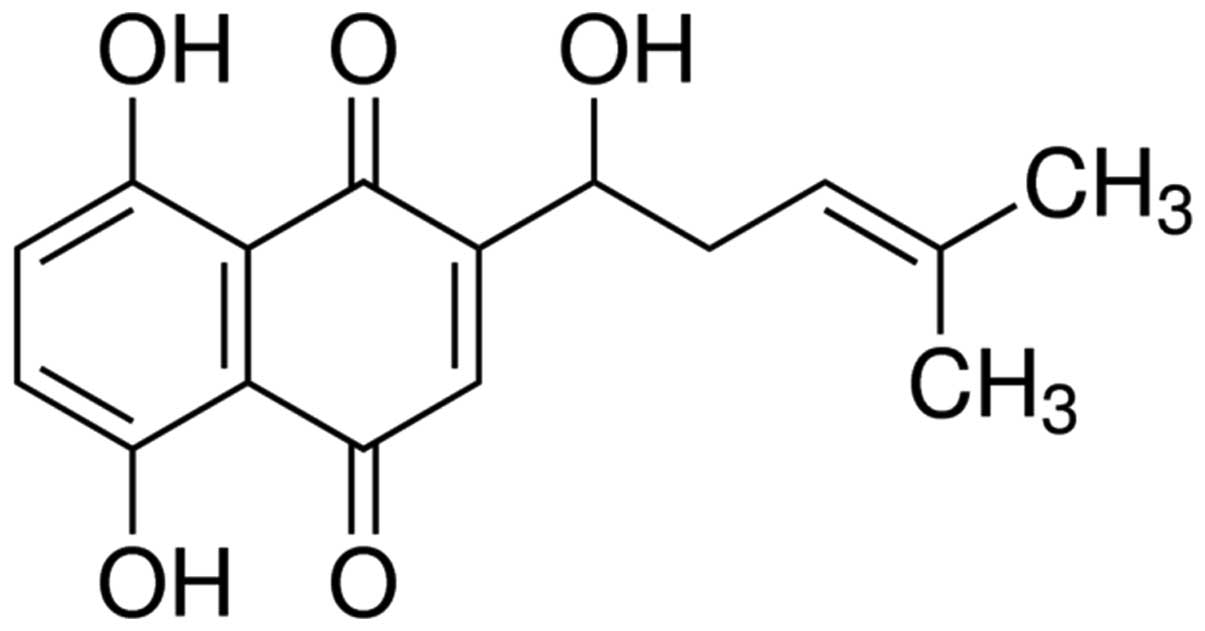
The chemical structure of shikonin is displayed in
Fig. 1. To evaluate the
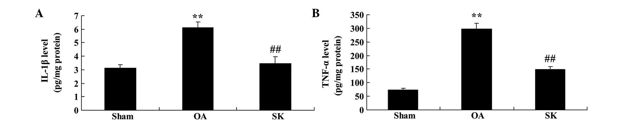
anti-inflammatory effect of shikonin in a rat model of
osteoarthritis, IL-1β and TNF-α levels were measured. As shown in
Fig. 2, the expression levels of
IL-1β and TNF-α were significantly increased in the rat model of
osteoarthritis, compared with those in the sham group (P<0.01;
Fig. 2). However, shikonin
significantly inhibited the increase in IL-1β and TNF-α expression
levels in the rat model of osteoarthritis, compared with those in
the osteoarthritis group (P<0.01; Fig. 2).
Effect of shikonin on NF-κB in the rat
model of osteoarthritis
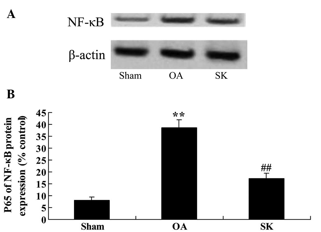
To appraise the anti-inflammatory mechanisms of
shikonin in the rat model of osteoarthritis, the protein expression
levels of NF-κB were examined using western blot analysis. There
was a significant increase of the NF-κB protein expression level in
the rat model of osteoarthritis, compared with that in the sham
group (P<0.01; Fig. 3). However,
the NF-κB protein expression level was significantly suppressed by
shikonin in the rat model of osteoarthritis, compared with that in
the osteoarthritis group (P<0.01; Fig. 3).
Effect of shikonin on iNOS level in
the rat model of osteoarthritis
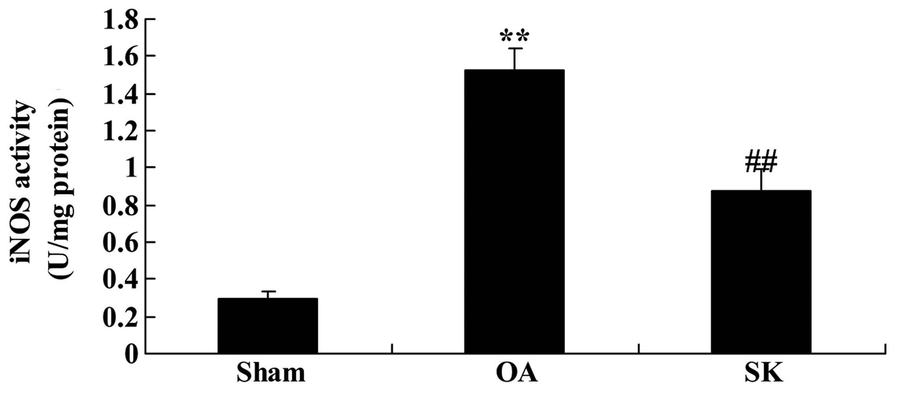
The effects of shikonin on the iNOS level in the rat
model of osteoarthritis were investigated. The iNOS level in the
rat model of osteoarthritis was significantly increased compared
with that of the sham group (P<0.01; Fig. 4). The induction of the iNOS level was
suppressed by treatment with shikonin in the rat model of
osteoarthritis, compared with that in the osteoarthritis group
(P<0.01; Fig. 4).
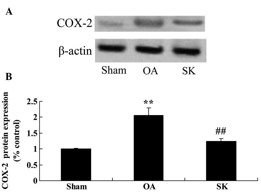
Effect of shikonin on COX-2 expression
in the rat model of osteoarthritis
To investigate the effects of shikonin on COX-2 in
the rat model of osteoarthritis, COX-2 protein expression levels
were measured by western blot analysis. The results showed that the
protein expression level of COX-2 was markedly promoted in the rat
model of osteoarthritis, as compared with that in the sham group
(P<0.01; Fig. 5). Notably, the
administration of shikonin markedly weakened the upregulation of
COX-2 protein expression in the rat model of osteoarthritis, as
compared with that in the osteoarthritis group (P<0.01; Fig. 5).
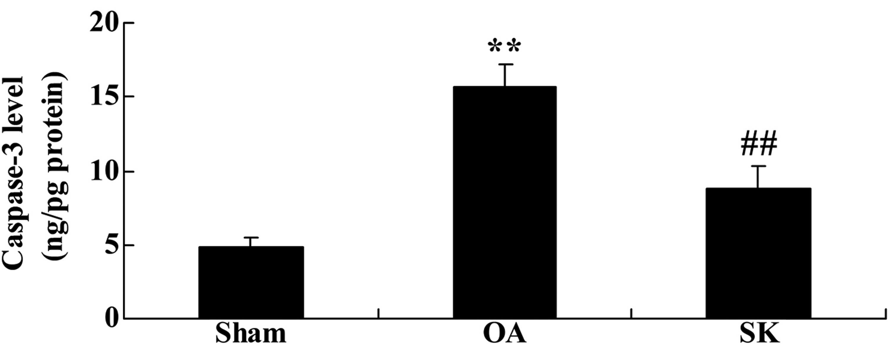
Effect of shikonin on caspase-3
activity in the rat model of osteoarthritis
In order to investigate the anti-apoptotic effect of
shikonin on osteoarthritis, caspase-3 activity was measured. There
was a significant increase in caspase-3 activity in the rat model
of osteoarthritis, as compared with the caspase-3 activity in the
sham group (P<0.01; Fig. 6).
However, the elevation of caspase-3 activity was significantly
reduced by shikonin treatment in the rat model of osteoarthritis,
compared with that in the osteoarthritis group (P<0.01; Fig. 6).
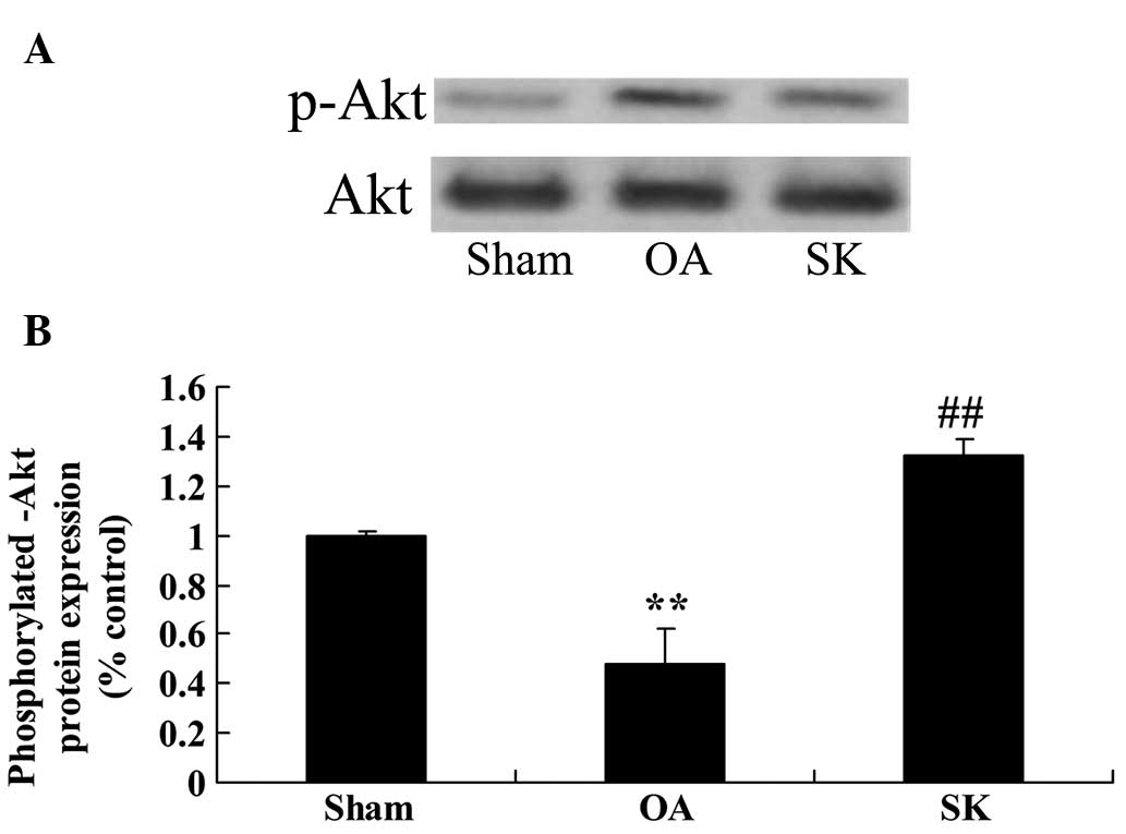
Effect of shikonin on the
phosphorylation of Akt in the rat model of osteoarthritis
The effects of shikonin on the phosphorylation of
Akt in the rat model of osteoarthritis were examined; p-Akt
expression levels were determined using western blot analysis.
Notably, p-Akt protein expression of the osteoarthritic model group
was lower than that of the sham group (P<0.01; Fig. 7). The downregulation of Akt
phosphorylation was significantly recovered by treatment with
shikonin in the rat model of osteoarthritis, compared with that in
the osteoarthritis group (P<0.01; Fig. 7).
Discussion
Osteoarthritis is the most common chronic joint
degenerative disease, and is also known as degenerative joint
disease, hypertrophic arthritis and senile osteoarthropathy
(16). The main pathological feature
of osteoarthritis is degeneration of the articular cartilage, which
results in articular cartilage injury, damage, joint edge and
subchondral bone reactive hyperplasia and osteophyte formation
(17). The incidence of
osteoarthritis is 3.0–8.3% in China, and in people aged 55–64 years
is ~40% (18). However, the specific
pathogenesis of osteoarthritis is unclear, and no reliable method
to cure osteoarthritis clinically is available (19). The results of the present study
revealed that shikonin treatment effectively inhibited the
expression of IL-1β and TNF-α in a rat model of osteoarthritis.
Furthermore, a previous study has indicated that shikonin exerts an
anti-inflammatory effect via proteasome inhibition (14). Andújar et al (20) reported that shikonin exerts
anti-inflammatory effects by inhibiting the activation of NF-κB.
These findings suggest that shikonin has an anti-inflammatory
effect in rats with osteoarthritis.
NF-κB is a nuclear transcription factor, widely
present in eukaryotes, which plays a role in the central control of
cell processes including inflammation, immune response,
differentiation, proliferation, apoptosis and tumorigenesis
(21). In the synthesis of various
cytokines in the body, NF-κB has a regulatory role in
transcription. Under normal circumstances NF-κB and inhibitor of κB
(Iκb) combine and exist in the cytoplasm in an inactive form. When
an activating signal is received, Iκb is phosphorylated, which
releases NF-κB; at this point NF-κB is in an activated state
(22). Following activation, NF-κB
translocates to the nucleus and induces target gene transcription,
so as to regulate the synthesis of cytokines and other inflammatory
factors (23). Studies have
confirmed that NF-κB has extensive biological effects, plays a
significant role in the regulation of inflammation, and is a key
link in the complex network of inflammatory cytokines (24,25).
Moreover, it modulates the occurrence and development of
inflammation. For instance, NF-κB is able to increase the
transcription levels of TNF-α and IL-1, causing the secretion of
these two cytokines to increase (26). Animal experiments have shown that in
arthritis development, the activation of the NF-κB precedes the
clinical manifestations of arthritis (27). In the present study, the
anti-inflammatory action of shikonin suppressed NF-κB protein
expression in a rat model of osteoarthritis. Yang et al
(28) reported that shikonin
inhibited inflammation in RAW264.7 cells through NF-κB signaling
pathways. Andújar et al (20)
reported that shikonin exerted anti-inflammatory effects by
inhibiting NF-κB activation. These results indicate that the
anti-inflammatory activity of shikonin may be mediated via the
downregulation of NF-κB signaling pathways in rats with
osteoarthritis.
Apoptosis is a genetically regulated process of
programmed cell death occurring in multicellular organisms; the
regulatory mechanism of cell apoptosis-related gene expression is
complex (29). The removal of cells
by apoptosis is necessary for multicellular organisms to sustain
life. There are numerous studies in which tumor cell proliferation
is inhibited through apoptosis induction, or cell apoptosis is
inhibited to repress chronic inflammation and potentially achieve
anti-aging effects (30). In normal
circumstances, the proliferation and apoptosis of articular
cartilage cells are in dynamic equilibrium, which keeps the cell
number, morphology and function of the articular cartilage
generally stable; excessive apoptosis is pathological and harmful
(31). Chondrocyte apoptosis has
been confirmed to occur in articular cartilage affected by
osteoarthritis (32). In addition,
the excessive apoptosis of cartilage cells is considered to be one
of the pathological factors in the degenerative changes of
articular cartilage; thus cartilage cell apoptosis may be an
important factor in the pathogenesis of osteoarthritis (33). Therefore, when considering how to
reduce the excessive apoptosis of cartilage cells in order to
prevent and treat osteoarthritis, maintaining a balance of
cartilage cell proliferation and apoptosis is key (34). The present study showed that shikonin
treatment significantly suppressed iNOS level elevation, COX-2
protein expression upregulation and increased caspase-3 activity in
a rat model of osteoarthritis. Prasad et al (35) indicated that in
lipopolysaccharide-stimulated BV2 microglial cells, shikonin
downregulated the gene expression of proinflammatory NOS and COX-2.
Yang et al (28) reported
that shikonin protects against interleukin-1β-induced apoptosis in
chondrocytes via the inhibition of caspase-3 activity. Thus, the
aforementioned results suggest that iNOS, COX-2 and caspase-3 are
important molecular targets of shikonin in the treatment of
osteoarthritis.
The mechanism of cartilage cell apoptosis is complex
and has not been fully elucidated. However, it may be associated
with various signal transduction pathways, among which the PI3K/Akt
signal transduction pathway is regarded as an important pathway of
cartilage cell apoptosis (36). As a
membrane protein, PI3K receives incoming signals from tyrosine
kinase receptors, cytokine receptors, CD19, B-cell receptors and
G-protein-coupled receptors, which directly or indirectly activates
Akt and downstream factors (37).
Akt has functions in protein synthesis, cell apoptosis, cell cycle
regulation, glucose metabolism and nerve degeneration (38). The current study demonstrates that
treatment with shikonin significantly activated the downregulation
of Akt activation in a rat model of osteoarthritis. Huang et
al (39) found that shikonin
inhibited oxidized low-density lipoprotein-induced monocyte
adhesion through the upregulation of PI3K/Akt and the suppression
of NF-κB activation. Kamei et al (40) suggested that in 3T3-L1 adipocytes,
shikonin stimulated glucose uptake via a mechanism involving Akt
phosphorylation. The results of the present study indicate that
shikonin has anti-inflammatory effects in rats with osteoarthritis
and one of the underlying mechanisms may be the upregulation of
PI3K/Akt signaling pathways.
In conclusion, the results of the present study
confirmed that shikonin inhibits inflammation and chondrocyte
apoptosis by regulating the PI3K/Akt signaling pathway in a rat
model of osteoarthritis. These findings suggest that shikonin has
therapeutic potential for osteoarthritis.
References
|
1
|
Wu C, Tian B, Qu X, Liu F, Tang T, Qin A,
Zhu Z and Dai K: MicroRNAs play a role in chondrogenesis and
osteoarthritis (review). Int J Mol Med. 34:13–23. 2014.PubMed/NCBI
|
|
2
|
Yu H, Wang Y, Guo Y, Wang H, Chen B and
Zhao X: Quality assessment of randomized controlled trials
reporting on knee osteoarthritis treated with warming needle
moxibustion. J Tradit Chin Med. 34:621–626. 2014. View Article : Google Scholar
|
|
3
|
Wang X, Wei S, Liu T, Pang J, Gao N, Ding
D, Duan T, Cao Y, Zheng Y and Zhan H: Effectiveness, medication
patterns and adverse events of traditional Chinese herbal patches
for osteoarthritis: A systematic review. Evid Based Complement
Alternat Med. 2014:3431762014. View Article : Google Scholar : PubMed/NCBI
|
|
4
|
Baker KR, Xu L, Zhang Y, Nevitt M, Niu J,
Aliabadi P, Yu W and Felson D: Quadriceps weakness and its
relationship to tibiofemoral and patellofemoral knee osteoarthritis
in Chinese: The Beijing osteoarthritis study. Arthritis Rheum.
50:1815–1821. 2004. View Article : Google Scholar : PubMed/NCBI
|
|
5
|
Robertson C, Archibald D, Avenell A,
Douglas F, Hoddinott P, van Teijlingen E, Boyers D, Stewart F,
Boachie C, Fioratou E, et al: Systematic reviews of and integrated
report on the quantitative, qualitative and economic evidence base
for the management of obesity in men. Health Technol Assess.
18(v-vi): xxiii–xxix, 1-424. 2014.
|
|
6
|
Lu H, Hou G, Zhang Y, Dai Y and Zhao H:
C-Jun transactivates Puma gene expression to promote
osteoarthritis. Mol Med Rep. 9:1606–1612. 2014.PubMed/NCBI
|
|
7
|
Patel DV, Sawant MG and Kaur G: Evaluation
of anti-osteoarthritic activity of Vigna mungo in papain induced
osteoarthritis model. Indian J Pharmacol. 47:59–64. 2015.
View Article : Google Scholar : PubMed/NCBI
|
|
8
|
Wang F, Wu L, Li L and Chen S: Monotropein
exerts protective effects against IL-1β-induced apoptosis and
catabolic responses on osteoarthritis chondrocytes. Int
Immunopharmacol. 23:575–580. 2014. View Article : Google Scholar : PubMed/NCBI
|
|
9
|
Zangerle PF, De Groote D, Lopez M,
Meuleman RJ, Vrindts Y, Fauchet F, Dehart I, Jadoul M, Radoux D and
Franchimont P: Direct stimulation of cytokines (IL-1 beta,
TNF-alpha, IL-6, IL-2, IFN-gamma and GM-CSF) in whole blood: II.
Application to rheumatoid arthritis and osteoarthritis. Cytokine.
4:568–575. 1992.
|
|
10
|
Zamli Z, Brown K Robson, Tarlton JF, Adams
MA, Torlot GE, Cartwright C, Cook WA, Vassilevskaja K and Sharif M:
Subchondral bone plate thickening precedes chondrocyte apoptosis
and cartilage degradation in spontaneous animal models of
osteoarthritis. Biomed Res Int. 2014:6068702014. View Article : Google Scholar : PubMed/NCBI
|
|
11
|
Wang X, Hayashi S, Umezaki M, Yamamoto T,
Kageyama-Yahara N, Kondo T and Kadowaki M: Shikonin, a constituent
of Lithospermum erythrorhizon exhibits anti-allergic effects by
suppressing orphan nuclear receptor Nr4a family gene expression as
a new prototype of calcineurin inhibitors in mast cells. Chem Biol
Interact. 224C:117–127. 2014. View Article : Google Scholar
|
|
12
|
Wada N, Kawano Y, Fujiwara S, Kikukawa Y,
Okuno Y, Tasaki M, Ueda M, Ando Y, Yoshinaga K, Ri M, et al:
Shikonin, dually functions as a proteasome inhibitor and a
necroptosis inducer in multiple myeloma cells. Int J Oncol.
46:963–972. 2015.PubMed/NCBI
|
|
13
|
Wang R, Yin R, Zhou W, Xu D and Li S:
Shikonin and its derivatives: A patent review. Expert Opin Ther
Pat. 22:977–997. 2012. View Article : Google Scholar : PubMed/NCBI
|
|
14
|
Lu L, Qin A, Huang H, Zhou P, Zhang C, Liu
N, Li S, Wen G, Zhang C, Dong W, et al: Shikonin extracted from
medicinal Chinese herbs exerts anti-inflammatory effect via
proteasome inhibition. Eur J Pharmacol. 658:242–247. 2011.
View Article : Google Scholar : PubMed/NCBI
|
|
15
|
Öberg AI, Yassin K, Csikasz RI, Dehvari N,
Shabalina IG, Hutchinson DS, Wilcke M, Östenson CG and Bengtsson T:
Shikonin increases glucose uptake in skeletal muscle cells and
improves plasma glucose levels in diabetic Goto-Kakizaki rats. PLoS
One. 6:e225102011. View Article : Google Scholar : PubMed/NCBI
|
|
16
|
Fox BA and Stephens MM:
Glucosamine/chondroitin/primorine combination therapy for
osteoarthritis. Drugs Today (Barc). 45:21–31. 2009. View Article : Google Scholar : PubMed/NCBI
|
|
17
|
Cicero AF and Laghi L: Activity and
potential role of licofelone in the management of osteoarthritis.
Clin Interv Aging. 2:73–79. 2007. View Article : Google Scholar : PubMed/NCBI
|
|
18
|
Guo D, Cao XW, Liu JW, Niu W, Ma ZW, Lin
DK, Chen JY, Lian WD, Ouyang WW and Liu J: Clinical effectiveness
and micro-perfusion alteration of Jingui external lotion in
patients with knee osteoarthritis: Study protocol for a randomized
controlled trial. Trials. 16:1242015. View Article : Google Scholar : PubMed/NCBI
|
|
19
|
Li Y, Zhang H, Zhang J, Li X, Song G and
Feng H: Clinical outcome of simultaneous high tibial osteotomy and
anterior cruciate ligament reconstruction for medial compartment
osteoarthritis in young patients with anterior cruciate
ligament-deficient knees: A systematic review. Arthroscopy.
31:507–519. 2015. View Article : Google Scholar : PubMed/NCBI
|
|
20
|
Andújar I, Recio MC, Bacelli T, Giner RM
and Ríos JL: Shikonin reduces oedema induced by phorbol ester by
interfering with IkappaBalpha degradation thus inhibiting
translocation of NF-kappaB to the nucleus. Br J Pharmacol.
160:376–388. 2010. View Article : Google Scholar : PubMed/NCBI
|
|
21
|
Hilgendorff A, Muth H, Parviz B, Staubitz
A, Haberbosch W, Tillmanns H and Hölschermann H: Statins differ in
their ability to block NF-kappaB activation in human blood
monocytes. Int J Clin Pharmacol Ther. 41:397–401. 2003. View Article : Google Scholar : PubMed/NCBI
|
|
22
|
Kwak SC, Lee C, Kim JY, Oh HM, So HS, Lee
MS, Rho MC and Oh J: Chlorogenic acid inhibits osteoclast
differentiation and bone resorption by down-regulation of receptor
activator of nuclear factor kappa-B ligand-induced nuclear factor
of activated T cells c1 expression. Biol Pharm Bull. 36:1779–1786.
2013. View Article : Google Scholar : PubMed/NCBI
|
|
23
|
Bowles RD, Mata BA, Bell RD, Mwangi TK,
Huebner JL, Kraus VB and Setton LA: In vivo luminescence imaging of
NF-κB activity and serum cytokine levels predict pain sensitivities
in a rodent model of osteoarthritis. Arthritis Rheumatol.
66:637–646. 2014. View Article : Google Scholar : PubMed/NCBI
|
|
24
|
Cho HJ, Lee KW and Park JH: Erucin exerts
anti-inflammatory properties in murine macrophages and mouse skin:
Possible mediation through the inhibition of NFκB signaling. Int J
Mol Sci. 14:20564–20577. 2013. View Article : Google Scholar : PubMed/NCBI
|
|
25
|
Toegel S, Weinmann D, André S, Walzer SM,
Bilban M, Schmidt S, Chiari C, Windhager R, Krall C, Bennani-Baiti
IM and Gabius HJ: Galectin-1 couples glycobiology to inflammation
in osteoarthritis through the activation of an NF-κB-regulated gene
network. J Immunol. 196:1910–1921. 2016. View Article : Google Scholar : PubMed/NCBI
|
|
26
|
Ni S, Miao K, Zhou X, Xu NW, Li CK, Zhu
RX, Sun RB and Wang YJ: The involvement of follistatin-like protein
1 in osteoarthritis by elevating NF-κB-mediated inflammatory
cytokines and enhancing fibroblast like synoviocyte proliferation.
Arthritis Res Ther. 17:912015. View Article : Google Scholar : PubMed/NCBI
|
|
27
|
Chan DD, Xiao WF, Li J, de la Motte CA,
Sandy JD and Plaas A: Deficiency of hyaluronan synthase 1 (Has1)
results in chronic joint inflammation and widespread
intra-articular fibrosis in a murine model of knee joint cartilage
damage. Osteoarthritis Cartilage. 23:1879–1889. 2015. View Article : Google Scholar : PubMed/NCBI
|
|
28
|
Yang Y, Wang J, Yang Q, Wu S, Yang Z, Zhu
H, Zheng M, Liu W, Wu W, He J and Chen Z: Shikonin inhibits the
lipopolysaccharide-induced release of HMGB1 in RAW264.7 cells via
IFN and NF-κB signaling pathways. Int Immunopharmacol. 19:81–87.
2014. View Article : Google Scholar : PubMed/NCBI
|
|
29
|
Fang HY, Chen CY, Hung MF, Hsiao YT,
Chiang TC, Lin TY, Chang HW, Chow KC and Ko WJ: Caspase-14 is an
anti-apoptotic protein targeting apoptosis-inducing factor in lung
adenocarcinomas. Oncol Rep. 26:359–369. 2011.PubMed/NCBI
|
|
30
|
Chan DD, Xiao WF, Li J, de la Motte CA,
Sandy JD and Plaas A: Deficiency of hyaluronan synthase 1 (Has1)
results in chronic joint inflammation and widespread
intra-articular fibrosis in a murine model of knee joint cartilage
damage. Osteoarthritis Cartilage. 23:1879–1889. 2015. View Article : Google Scholar : PubMed/NCBI
|
|
31
|
Machner A, Baier A, Wille A, Drynda S, Pap
G, Drynda A, Mawrin C, Bühling F, Gay S, Neumann W and Pap T:
Higher susceptibility to Fas ligand induced apoptosis and altered
modulation of cell death by tumor necrosis factor-alpha in
periarticular tenocytes from patients with knee joint
osteoarthritis. Arthritis Res Ther. 5:R253–R261. 2003. View Article : Google Scholar : PubMed/NCBI
|
|
32
|
Sezgin M, Barlas İÖ, Yıldır S, Türköz G,
Ankaralı HÇ, Şahin G and Erdal ME: Apoptosis-related Fas and FasL
gene polymorphisms' associations with knee osteoarthritis.
Rheumatol Int. 33:2039–2043. 2013. View Article : Google Scholar : PubMed/NCBI
|
|
33
|
López-Armada MJ, Caramés B, Cillero-Pastor
B, Lires-Deán M, Maneiro E, Fuentes I, Ruíz C, Galdo F and Blanco
FJ: Phosphatase-1 and-2A inhibition modulates apoptosis in human
osteoarthritis chondrocytes independently of nitric oxide
production. Ann Rheum Dis. 64:1079–1082. 2005. View Article : Google Scholar : PubMed/NCBI
|
|
34
|
Takács-Buia L, Iordachel C, Efimov N,
Caloianu M, Montreuil J and Bratosin D: Pathogenesis of
osteoarthritis: Chondrocyte replicative senescence or apoptosis?
Cytometry B Clin Cytom. 74:356–362. 2008.PubMed/NCBI
|
|
35
|
Prasad RG, Choi YH and Kim GY: Shikonin
isolated from lithospermum erythrorhizon downregulates
proinflammatory mediators in lipopolysaccharide-stimulated BV2
microglial cells by suppressing crosstalk between reactive oxygen
species and NF-κB. Biomol Ther (Seoul). 23:110–118. 2015.
View Article : Google Scholar : PubMed/NCBI
|
|
36
|
Yu SM and Kim SJ: Withaferin A-caused
production of intracellular reactive oxygen species modulates
apoptosis via PI3K/Akt and JNKinase in rabbit articular
chondrocytes. J Korean Med Sci. 29:1042–1053. 2014. View Article : Google Scholar : PubMed/NCBI
|
|
37
|
Sugimori K, Matsui K, Motomura H, Tokoro
T, Wang J, Higa S, Kimura T and Kitajima I: BMP-2 prevents
apoptosis of the N1511 chondrocytic cell line through
PI3K/Akt-mediated NF-kappaB activation. J Bone Miner Metab.
23:411–419. 2005. View Article : Google Scholar : PubMed/NCBI
|
|
38
|
Lee KN, Seo MC, Bae IH, Oh SH, Jang WG,
Jeong BC, Oh WM, Kim SH, Lee SE, Shim KM, et al: COMP-Ang1, a
variant of angiopoietin 1, inhibits serum-deprived apoptosis of
mesenchymal cells via PI3K/Akt and mitogen-activated protein kinase
pathways. Pharmacology. 86:327–335. 2010. View Article : Google Scholar : PubMed/NCBI
|
|
39
|
Huang CS, Lin AH, Yang TC, Liu KL, Chen HW
and Lii CK: Shikonin inhibits oxidized LDL-induced monocyte
adhesion by suppressing NFκB activation via up-regulation of
PI3K/Akt/Nrf2-dependent antioxidation in EA hy926 endothelial
cells. Biochem Pharmacol. 93:352–361. 2015. View Article : Google Scholar : PubMed/NCBI
|
|
40
|
Kamei R, Kitagawa Y, Kadokura M, Hattori
F, Hazeki O, Ebina Y, Nishihara T and Oikawa S: Shikonin stimulates
glucose uptake in 3T3-L1 adipocytes via an insulin-independent
tyrosine kinase pathway. Biochem Biophys Res Commun. 292:642–651.
2002. View Article : Google Scholar : PubMed/NCBI
|