Introduction
Asthma, one of the most frequent diseases worldwide,
is a chronic inflammatory disease of the airways characterized by
hyperresponsiveness, reversible airway obstruction and airway
inflammation (1). The pathological
features of allergic asthma include edema, inflammatory cell
infiltration, denudation of the airway epithelium, mast cell
activation and collagen deposition (2). Moreover, asthma is associated with an
imbalance of T helper (Th)1/Th2 cells and their different cytokine
profiles (3,4). Th2-specific cytokines, including
interleukin (IL)-4, 5 and 13, are important in the mediation of
humoral immune responses and immunoglobulin E (IgE) production by
affecting eosinophils in the airway. Furthermore, the release of
Th2 cytokines, including histamine and leukotriene, is associated
with hyperresponsiveness (5,6). Th1 cytokines, including interferon
(IFN)-γ and IL-12, mediate cellular immune reactions, the
antagonism of Th2 immune responses, and IgE synthesis in order to
restrain asthma development (7). If
the dynamic balance of Th1/Th2 cells is disturbed, the subject is
more likely to experience a disease (8). Therefore, researchers have examined
ways of inhibiting the activation of Th2 cells or the modulation of
the Th1/Th2 balance as a way of preventing and treating asthma
(9).
Nuclear factor-kappa B (NF-κB), a well-studied
transcriptional factor that is important in signaling pathways, is
also important in airway remodeling in asthma (10,11).
Cytokines and other factors triggered as part of inflammatory
processes activate NF-κB via several signaling pathways, leading to
a signaling cascade that amplifies inflammation (10). Several studies associate NF-κB
overactivation with airway remodeling (12,13). The
p38 mitogen-activated protein kinases (MAPKs) represent a point of
convergence for multiple signaling processes that are activated in
inflammation, as well as a diverse range of events that are
important in inflammation (14,15).
Various proinflammatory transcription factors, including activator
protein 1 (AP-1) and NF-κB (16–18) are
regulated by the p38 MAPK pathway.
Lignans from Sesamum indicum L. seeds are
potent antioxidants. Sesamin, a type of lignan, is the most
abundant in sesame seed oil (19).
Previous studies reveal that sesamin reduces the frequency of
chemically-induced mammary tumors, enhances hepatic detoxification
and protects against oxidative stress (20,21).
Moreover, previous studies indicate that sesamin has other
potential pathways for bioactivity, including the inhibition of
delta-5 desaturase activity in the fatty acid metabolism, which
results in the accumulation of dihomo-γ-linolenic acid.
Dihomo-γ-linolenic acid is capable of displacing arachidonic acid
and decreasing the formation of proinflammatory mediators, such as
prostaglandin E2 and leukotriene B4 (22,23).
Moreover, another study of ours demonstrated that sesamin inhibits
lipopolysaccharide-induced IL-6 production by suppressing NF-κB and
p38 MAPK activation (24). Sesamin
also partially reverses IL-1β signaling in human
astrocyte-cerebellar cultures, leading to the inhibition of matrix
metallopeptidase-1, −3 and −13 expression (25). The study further demonstrated that
this inhibition occurred through the p38 and c-Jun N-terminal
kinase signaling pathways, but not through extracellular regulated
protein kinases 1/2 (25). The
present study examines the anti-inflammatory mechanism of sesamin
in a mouse asthma model.
Materials and methods
Animals
A total of 35 specific pathogen-free inbred female
BALB/c mice (age, 7 weeks) were purchased from Yanbian University
Health Science Center (Yanji, China). The mice were kept in an
animal facility under standard laboratory conditions for 1 week
prior to the experiments, with water and standard food ad libitum.
All animal experiments were conducted according to the guidelines
approved by the Institutional Animal Care and Use Committee of
Yanbian University School of Medical Sciences (Yanji, China).
The mice were intraperitoneally immunized with 10 µg
chicken egg ovalbumin (OVA; Sigma-Aldrich; Merck KGaA, Darmstadt,
Germany) and 1.0 mg aluminum hydroxide adjuvant (Imject Alum;
Pierce Protein Biology; Thermo Fisher Scientific, Inc., Rockford,
IL, USA). The mice received a booster injection with 10 µg OVA and
1.0 mg aluminum hydroxide adjuvant 10 days later. In addition,
between days 17 and 19, the immunized mice were exposed to a 1% OVA
aerosol in phosphate-buffered saline (PBS) for 20 min. The
bronchoprovocation procedure was performed in vented plastic
chambers (18×14×8 cm) that were adapted for mice. Aerosol particles
of 3–5 µm diameter were created using an ultrasonic nebulizer
(NE-U12; Omron Corporation, Kyoto, Japan), then vented into a fume
hood. Each test group consisted of seven mice. Mice in the control
group were treated with aerosol of saline. Sesamin dissolved in
vehicle composed of 0.5% (w/v) carboxy methylcellulose and 0.025%
Tween 20 in distilled water (200 mg/kg body weight; Sigma-Aldrich),
or the reference drug dexamethasone (DXM; 0.5 mg/kg body weight;
Sigma-Aldrich) was administered by oral gavage to each animal at
24-h intervals on days 17–19, beginning 1 h before the first
provocation.
Immediately after the assessment of airway
responsiveness, the mice were anesthetized by intraperitoneal
injection of pentobarbital (50 mg/kg; Wuhan Boster Biological
Technology, Ltd., Wuhan, China), and their tracheas cannulated
while gently massaging their thoraxes. Moreover, the lungs were
lavaged with 0.7 ml PBS. Bronchoalveolar lavage (BAL) fluid samples
were collected and the number of total cells in a 0.05-ml aliquot
were counted using a hemocytometer. The remaining samples were
centrifuged at 1,200 × g for 10 min at 4°C (model 5424R;
Eppendorf Instrumente GmbH, Hamburg, Germany), and the supernatants
stored at −70°C before use. Cell pellets were resuspended in PBS
and cytospin (Cytospin 3; Shandon Scientific Limited, Astmoor, UK)
preparations of BAL cells were stained with Diff-Quik solution
(International Reagents Corporation, Kobe, Japan). A total of two
independent, double-blinded investigators counted the cells using a
microscope (CX41; Olympus Corporation, Tokyo, Japan). In total,
~400 cells were counted in each of four different random locations.
The inter-investigator variation was <5%, and the mean number
from the two investigators was used to estimate cell
differentials.
Enzyme-linked immunosorbent assay
(ELISA)
IL-4 (M4000B), IL-5 (M5000), IL-13 (M1300CB) and
IFN-γ (MIF00) levels in BAL were determined using mouse ELISA kits
(R&D Systems, Inc., Minneapolis, MN, USA) according to the
manufacturer's instructions. The sensitivity was 2.0 pg/ml for
IL-4, IL-5, IL-13 and IFN-γ.
Hematoxylin and eosin (HE)
staining
Following BAL, murine lungs were resected, fixed
with 4% paraformaldehyde and embedded in paraffin. Specimens were
cut into 4-µm sections using a rotary microtome (2165; Leica
Biosystems Nussloch Gmbh, Nussloch, Germany), and the microsections
were stained with HE (Richard-Allan Scientific, Kalamazoo, MI,
USA). Furthermore, they were examined under a magnification of
×100.
Nuclear protein extraction
Freshly isolated lung tissues were washed and lysed
in 2 volumes of lysis buffer A containing 50 mM Tris-HCl, pH 7.5, 1
mM EDTA, 10% glycerol, 0.5 mM dithiothreitol, 5 mM
MgCl2, 1 mM phenylmethylsulfonyl fluoride (PMSF) and
protease inhibitor cocktails for 5 min at 4°C (all lysis buffer
reagents purchased from Sigma-Aldrich). The suspension was
centrifuged at 1,000 × g for 15 min at 4°C. Cytosolic
proteins were extracted from the supernatant by incubating on ice
for 10 min and centrifuging at 100,000 × g for 1 h at 4°C.
The pelleted nuclei were resuspended in buffer B containing 1.3 M
sucrose, 1.0 mM MgCl2 and 10 mM potassium phosphate
buffer (pH 6.8), and centrifuged at 1,000 × g for 15 min.
The pellets were suspended in buffer B to a final sucrose
concentration of 2.2 M and centrifuged at 100,000 × g for 1
h. The nuclear pellets were washed once with a solution containing
0.25 M sucrose, 0.5 mM MgCl2 and 20 mM Tris-HCl (pH
7.2), and centrifuged again at 1,000 × g for 10 min. The new
pellets were then solubilized with a solution containing 50 mM
Tris-HCl (pH 7.2), 0.3 M sucrose, 150 mM NaCl, 2 mM EDTA, 20%
glycerol, 2% Triton X-100, 2 mM PMSF and protease inhibitor
cocktails. Furthermore, the mixture was kept on ice for 1 h with
gentle stirring and centrifuged at 12,000 × g for 30 min.
The resulting supernatant was used as the soluble nuclear protein
sample for western blot analysis.
Western blot analysis
Lung tissue samples were homogenized and lysed in
two volumes of lysis buffer containing 50 mM Tris-HCl (pH 7.5), 150
mM NaCl, 1% Nonidet-P40, 1% sodium deoxycholate, 0.1 mM
dithiothreitol, 0.5 mM EDTA, 1 mM sodium vanadate, 2 mM
phenylmethylsulfonyl fluoride, 1 µg/ml aprotinin, 1 µg/ml leupeptin
and 1 µg/ml pepstatin. The mixture was kept on ice for 1 h with
gentle vortexing and centrifuged at 8,000 × g for 15 min.
The resulting supernatant was stored at −80°C (total extracts).
Protein samples (30 µg) from the lung homogenates were separated
using 12% SDS-PAGE, and the proteins were then transferred onto a
nitrocellulose membranes. Nonspecific sites were blocked with 5%
non-fat dry milk in TBST buffer [25 mM Tris (pH 7.5), 150 mM NaCl,
0.1% Tween 20] for 1 h, and washed with TBST for four times of 10
min. Western blot analysis was performed using polyclonal
antibodies against IL-5 (sc-7887; 1:1,000), p38, phosphorylated p38
(p-p38; sc-535; 1:1,000), poly ADP ribose polymerase (sc-25780;
1:1,000), NF-κB p65 (sc-109; 1:1,000), IκB-α (sc-847; 1:1,000),
β-actin (sc-130656; 1:1,000; Santa Cruz Biotechnology, Inc., Santa
Cruz, CA, USA), IL-13 (AF-413-NA; 1:1,000; R&D Systems, Inc.),
IFN-γ (sc-9344; 1:1,000; Santa Cruz Biotechnology, Inc.) or IL-4
(AAM36; 1:1,000; Bio-Rad Laboratories, Inc.). Membranes were probed
with primary antibody (1:1,000) overnight on a shaker at 4°C. After
washing with TBST for four times of 10 min, membranes were
incubated with secondary antibody (1:2,500) for 2 h. Binding of the
antibodies was detected using an enhanced chemiluminescence
detection system (Amersham Life Science, Arlington Heights, IL,
USA) according to the manufacturer's instructions. The band
intensity was quantified using Quantity One software (Bio-Rad
Laboratories, Inc.). All experiments were repeated for at least
three times.
Statistical analysis
All immunoreactive and phosphorylation signals were
analyzed by densitometric scanning (Gel Doc XR; Bio-Rad
Laboratories, Inc.). The results were analyzed using SPSS 17.0
(SPSS, Inc., Chicago, IL, USA). The data were expressed as the mean
± standard error of the mean. Statistical evaluation of the data
was performed using analysis of variance followed by Dunnett's
post-hoc test. P<0.05 was used to indicate a statistically
significant difference.
Results
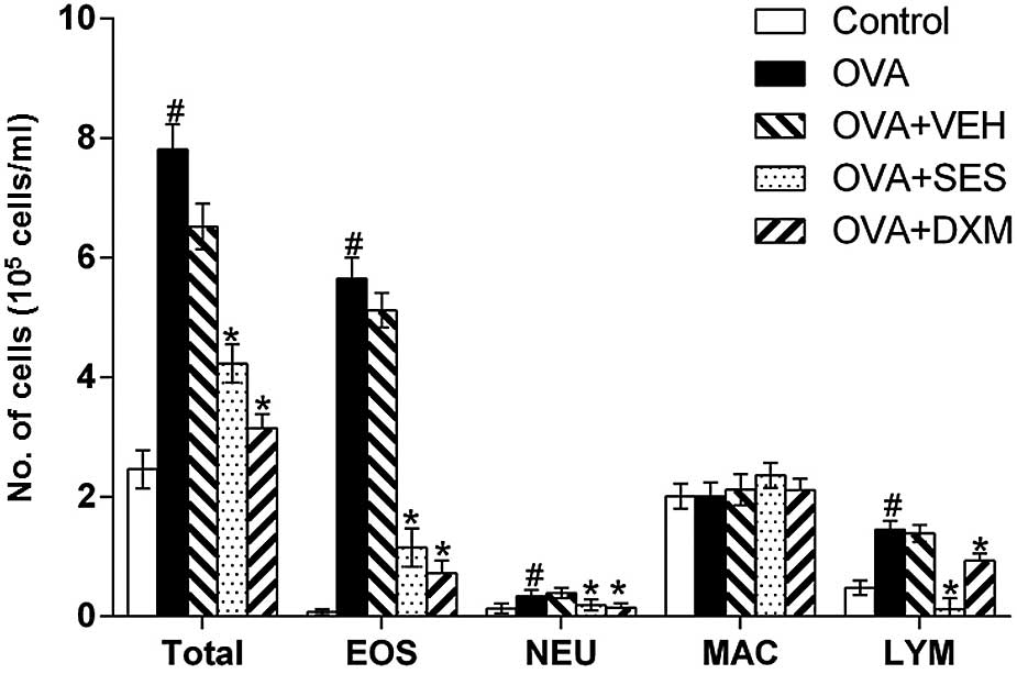
Sesamin reduces the number of cells in
BAL fluids of OVA-induced asthma
In order to examine the effect of sesamin on
cellular changes in BAL fluids of OVA-induced asthma, the number of
various cells were counted. The data revealed that the numbers
lymphocytes, eosinophils and neutrophils in BAL fluids were
significantly increased at 48 h after OVA inhalation compared with
those following saline inhalation (Fig.
1). In addition, administration of sesamin or DXM significantly
reduced the number of these cells. These results indicate that
sesamin reduces the number of cells in BAL fluids of OVA-induced
asthma.
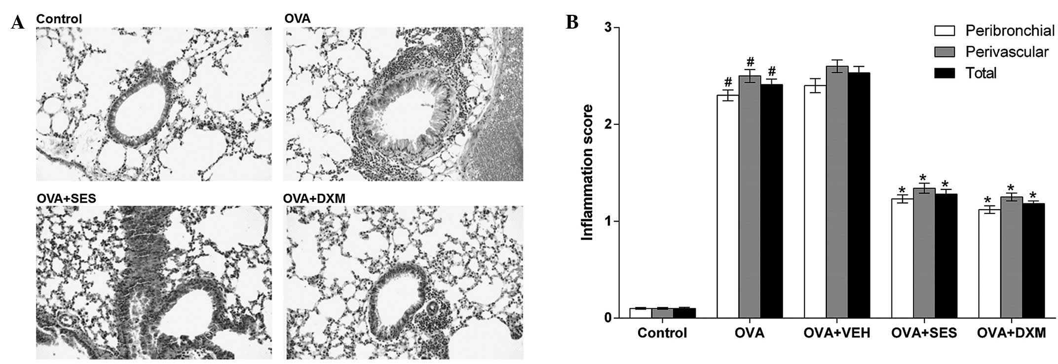
Sesamin inhibits the infiltration of
inflammatory cells and attenuates antigen-induced airway
inflammation
In order to study the effect of sesamin on the
pathological changes of OVA-induced asthma, histological
examinations were undertaken. The examinations revealed typical
pathological features of asthma in OVA-challenged mice through
widespread perivascular and peribronchiolar inflammatory cell
infiltrates, as compared to the control. Moreover, the mice treated
with sesamin or DXM demonstrated significantly reduced inflammatory
cell infiltration in the peribronchiolar and perivascular regions,
compared with the OVA group (Fig.
2). These results indicate that sesamin attenuates
antigen-induced airway inflammation and inhibits the infiltration
of inflammatory cells.
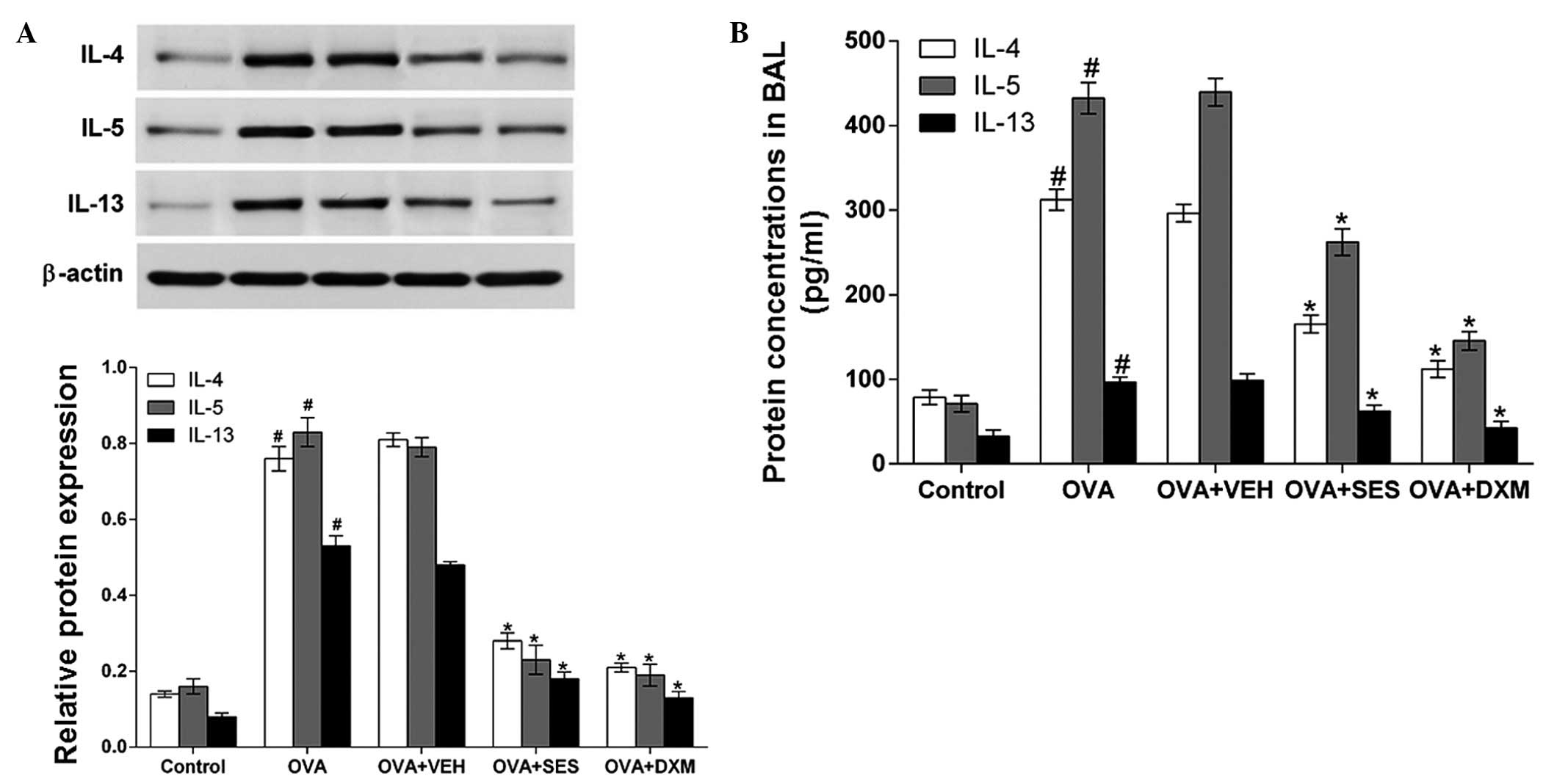
Treatment with sesamin suppresses the
increase in IL-4, IL-5 and IL-13 levels
Western blot analysis and ELISA were used to
investigate the effect of sesamin on IL-4, IL-5 and IL-13 protein
levels in lung tissues and in BAL fluids of OVA-induced asthma,
respectively. Western blotting demonstrated that IL-4, IL-5 and
IL-13 protein levels in lung tissues were significantly increased
48 h after OVA inhalation compared with the levels following saline
inhalation. Moreover, the increased IL-4, IL-5 and IL-13 levels
were significantly reduced by the administration of sesamin or DXM
(Fig. 3A). Consistent with these
results, ELISA revealed that the levels of IL-4, IL-5 and IL-13 in
BAL fluids were also significantly increased at 48 h after OVA
inhalation compared with the control group (Fig. 3B). These results indicate that
treatment with sesamin suppresses the increase in the IL-4, IL-5
and IL-13 levels.
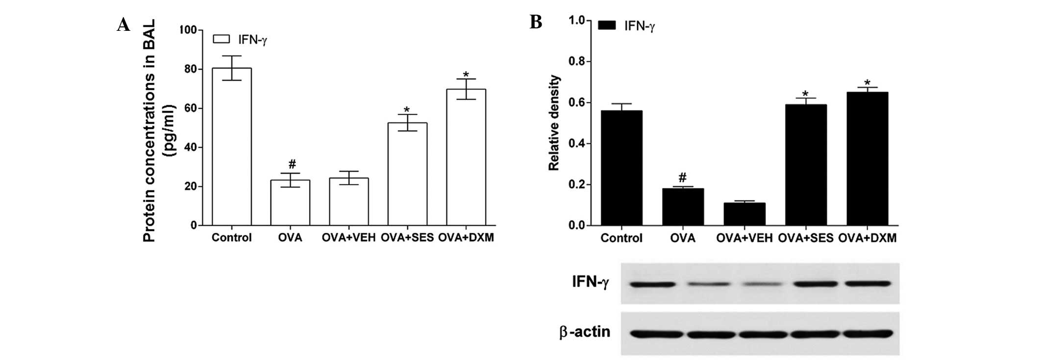
Treatment with sesamin increases the
decreased IFN-γ level in allergic mice
In order to determine the effect of sesamin on the
Th1 response, IFN-γ levels in lung tissues and BAL fluids were
measured using western blotting and ELISA, respectively. The IFN-γ
level in OVA-challenged mice was lower than that in the control
mice (Fig. 4A). However,
administration of DXM or sesamin increased the levels of IFN-γ in
OVA-challenged mice. In agreement with this result, western
blotting demonstrated that the IFN-γ protein level in the lung
tissue was significantly decreased in allergic mice compared with
that in the control mice (Fig. 4B).
It is noteworthy that the decreased IFN-γ level was increased after
pretreatment with sesamin or DXM. These results indicate that
treatment with sesamin increases the decreased IFN-γ level in the
lung tissue of allergic mice.
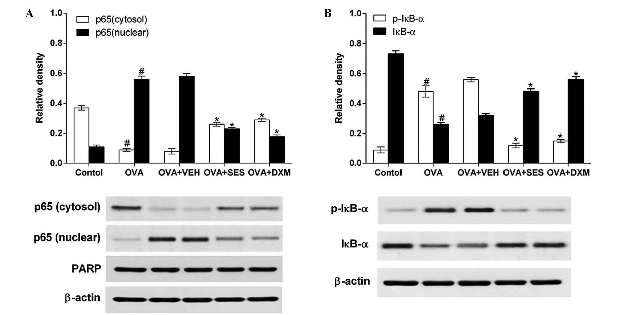
Sesamin prevents the translocation of
NF-κB by blocking phosphorylation and degradation of IκB-α in
OVA-challenged mice
Western blotting was used to determine the effect of
sesamin on NF-κB p65 and phosphorylation levels of IκB-α protein in
the lung tissues of mice with OVA-induced asthma. The data
demonstrated that the levels of NF-κB p65 in nuclear protein
extracts from lung tissues were increased at 48 h after OVA was
inhaled compared with the levels in mice used as the control. The
increased NF-κB p65 levels at 48 h after OVA inhalation were
decreased after sesamin or DXM was administered. By contrast, the
levels of NF-κB p65 in cytosolic protein extracts from lung tissues
were decreased at 48 h after OVA inhalation in comparison to the
control (Fig. 5A). The decreased
NF-κB p65 levels in the cytosolic preparations were increased after
sesamin or DXM was administered. Moreover, the effect of sesamin on
degradation of IκB-α and on OVA-induced phosphorylation was studied
in order to investigate the molecular mechanisms by which sesamin
inhibits NF-κB transcriptional activity. Sesamin was observed to
significantly block OVA-induced phosphorylation and degradation of
IκB-α (Fig. 5B). These results
indicate that sesamin prevents NF-κB translocation by blocking the
phosphorylation and degradation of IκB-α in OVA-challenged
mice.
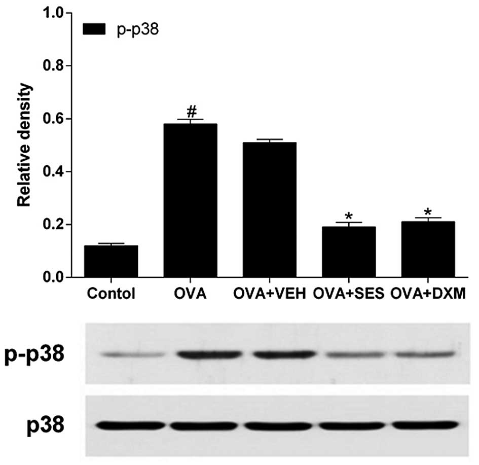
Treatment with sesamin reduces the
phosphorylation levels of p38 MAPK protein in lung tissue of mice
with OVA-induced asthma
In order to test the effect of sesamin on the
phosphorylation levels of p38 MAPK protein in the lung tissue of
mice with OVA-induced asthma, western blotting was used to
determine the activation of p38 MAPK, which is an upstream
signaling mediator of NF-κB. The data revealed that the levels of
p-p38 protein in lung tissues were increased at 48 h after OVA
inhalation compared with the levels in the control mice (Fig. 6). However, no significant changes
were observed in the p38 protein levels in any of the groups
tested. The administration of sesamin or DMX significantly reduced
the levels of p-p38 at Thr 180 and Tyr 182 (but not the total p38
protein levels) in lung tissues at 48 h after OVA inhalation. This
result indicates that treatment with sesamin reduces the
phosphorylation levels of the p38 MAPK protein in the lung tissue
of mice with OVA-induced asthma.
Discussion
Allergic asthma is a chronic inflammatory illness of
the airways characterized by airway inflammatory cell infiltration
and hyperresponsiveness to inhaled allergens and nonspecific
stimuli (26). Inflammatory cells,
including Th2, mast cells, macrophages, lymphocytes, eosinophils
and neutrophils, as well as the release of cytokines (particularly
IL-4, IL-5 and IL-13) are important in asthma (27,28). The
results of the present study reveal that sesamin reverses or
prevents some of these effects in mice suffering from asthmatic
symptoms. The number of total cells, eosinophils, neutrophils and
lymphocytes in BAL fluid were much lower in OVA-induced mice
treated with sesamin compared to those in the control group. The
expression of Th2 cytokines, including IL-4, IL-5 and IL-13 was
increased, and the expression of Th1 cytokines such as IFN-γ was
decreased during the induction of asthma. Sesamin significantly
reduced OVA-induced enhancement of IL-4, IL-5 and IL-13 expression,
and increased OVA-induced attenuation of IFN-γ expression in lung
tissues and BAL fluids. Consistent with these biochemical
observations, HE staining revealed that sesamin treatments improved
airway inflammation.
Airway inflammation is connected by complex
signaling networks. As a result, we still do not fully understand
the molecular mechanisms of this disorder. NF-κB is present in the
majority of cell types and is known to be important in immune and
inflammatory responses, including asthma (29–32). In
unstimulated cells, IκBα proteins sequester NF-κB into the
cytoplasm. However, phosphorylation and degradation of IkBα allows
the translocation of NF-κB into the nucleus where it regulates
transcription of target genes, which encode numerous inflammatory
proteins (33) such as cytokines
IL-4, IL-5 and IL-13, all of which are closely implicated in the
pathogenesis of asthma (34). It is
consistent with the observations of the present study that NF-κB
levels in nuclear protein extracts from lung tissues are
substantially increased in the OVA-induced model of allergic airway
disease. Sesamin significantly reduced the degradation of IκBα, the
translocation of the p65 subunit of NF-κB into the nucleus, and the
levels of Th2 cytokines (IL-4, IL-5 and IL-13) in the lungs of
OVA-induced mice. These results suggest that sesamin alleviates
asthmatic symptoms through the modulation of NF-κB activation.
MAPKs belong to the serine/threonine kinase family
that includes extracellular signal-regulated kinases, c-Jun
N-terminal kinases (JNK) and p38 MAPK. MAPKs relay signals
generated by endogenous and exogenous stimuli to the intracellular
space through the phosphorylation of proteins (35–37). The
activities and/or phosphorylation states of all three MAPK members
are upregulated in asthma animal models (38,39). In
addition, a potent and selective p38 MAPK inhibitor named SB239063
promotes the apoptosis of eosinophils in BAL fluid and
significantly inhibits antigen-induced eosinophilia (40). In addition, p38 MAPK inhibitors
reduce levels of IL-4, IL-5 and IL-13, mucus hypersecretion,
antigen-induced airway inflammatory cell infiltration and airway
hyperresponsiveness (41).
Furthermore, sesamin inhibits IL-1β-induced phosphorylation of JNK
and p38 MAPK in human articular chondrocytes (25). Consistent with this data, the present
study reveals that phosphorylation of p38 MAPK is evidently
increased following OVA inhalation, and that sesamin significantly
decreases the levels of p-p38 MAPK in lung tissues of OVA-inhaled
mice. In addition, it was revealed that sesamin does not only
reduce allergen-induced airway inflammation, but also interrupts
the translocation of the p65 subunit of NF-κB and degrades IκBα.
These observations indicate that the modulation of the p38 MAPK
signaling pathway, involving IκB and NF-κB, may be one of the
molecular bases for the beneficial effects of sesamin on allergic
airway disease.
In summary, the present study investigated the
effects of sesamin on allergen-induced airway inflammation. At the
same time, the roles of p38 MAPK and NF-κB in this process were
clarified. Following treatment of OVA-inhaled mice with sesamin,
p38 MAPK phosphorylation and NF-κB activation was reduced. The
level of Th2 cytokine airway inflammation was also attenuated.
According to the results of the present study, the inhibitory
effects of sesamin on OVA-induced allergic airway inflammation were
partly mediated by regulating the p38 MAPK/NF-κB pathways. The
present study also provided a crucial molecular basis for the
preventive and/or therapeutic capability of sesamin for allergic
airway diseases.
Acknowledgements
The present study was supported by the Natural
Science Foundation of China (grant nos. 81260665 and 81160176) and
the Project of Research & Innovation of Jilin Youth Leader and
Team (grant no. 20140519013JH).
References
|
1
|
Pascual RM and Peters SP: Airway
remodeling contributes to the progressive loss of lung function in
asthma: an overview. J Allergy Clin Immunol. 116:477–486. 2005.
View Article : Google Scholar : PubMed/NCBI
|
|
2
|
Kay AB: Asthma and inflammation. J Allergy
Clin Immunol. 87:893–910. 1991. View Article : Google Scholar : PubMed/NCBI
|
|
3
|
Janssen EM, van Oosterhout AJ, van Rensen
AJ, van Eden W, Nijkamp FP and Wauben MH: Modulation of Th2
responses by peptide analogues in a murine model of allergic
asthma: Amelioration or deterioration of the disease process
depends on the Th1 or Th2 skewing characteristics of the
therapeutic peptide. J Immunol. 164:580–588. 2000. View Article : Google Scholar : PubMed/NCBI
|
|
4
|
Mazzarella G, Bianco A, Catena E, De Palma
R and Abbate GF: Th1/Th2 lymphocyte polarization in asthma.
Allergy. 55:(Suppl 61). S6–S9. 2000. View Article : Google Scholar
|
|
5
|
Primhak RA and Powell CV: AHR in asthma.
Thorax. 57:186author reply 186. 2002. View Article : Google Scholar : PubMed/NCBI
|
|
6
|
Robinson DS, Hamid Q, Ying S, Tsicopoulos
A, Barkans J, Bentley AM, Corrigan C, Durham SR and Kay AB:
Predominant TH2-like bronchoalveolar T-lymphocyte population in
atopic asthma. N Engl J Med. 326:298–304. 1992. View Article : Google Scholar : PubMed/NCBI
|
|
7
|
Tournoy KG, Kips JC and Pauwels RA: Is Th1
the solution for Th2 in asthma? Clin Exp Allergy. 32:17–29. 2002.
View Article : Google Scholar : PubMed/NCBI
|
|
8
|
Kidd P: Th1/Th2 balance: The hypothesis,
its limitations, and implications for health and disease. Altern
Med Rev. 8:223–246. 2003.PubMed/NCBI
|
|
9
|
Umetsu DT, McIntire JJ, Akbari O, Macaubas
C and DeKruyff RH: Asthma: An epidemic of dysregulated immunity.
Nat Immunol. 3:715–720. 2002. View Article : Google Scholar : PubMed/NCBI
|
|
10
|
Bao Z, Guan S, Cheng C, Wu S, Wong SH,
Kemeny DM, Leung BP and Wong WS: A novel antiinflammatory role for
andrographolide in asthma via inhibition of the nuclear
factor-kappaB pathway. Am J Respir Crit Care Med. 179:657–665.
2009. View Article : Google Scholar : PubMed/NCBI
|
|
11
|
Janssen-Heininger YM, Poynter ME, Aesif
SW, Pantano C, Ather JL, Reynaert NL, Ckless K, Anathy V, van der
Velden J, Irvin CG and van der Vliet A: Nuclear factor kappaB,
airway epithelium and asthma: Avenues for redox control. Proc Am
Thorac Soc. 6:249–255. 2009. View Article : Google Scholar : PubMed/NCBI
|
|
12
|
Tully JE, Hoffman SM, Lahue KG, Nolin JD,
Anathy V, Lundblad LK, Daphtary N, Aliyeva M, Black KE, Dixon AE,
et al: Epithelial NF-κB orchestrates house dust mite-induced airway
inflammation, hyperresponsiveness, and fibrotic remodeling. J
Immunol. 191:5811–5821. 2013. View Article : Google Scholar : PubMed/NCBI
|
|
13
|
Ichikawa T, Sugiura H, Koarai A, Kikuchi
T, Hiramatsu M, Kawabata H, Akamatsu K, Hirano T, Nakanishi M,
Matsunaga K, et al: 25-hydroxycholesterol promotes
fibroblast-mediated tissue remodeling through NF-κB dependent
pathway. Exp Cell Res. 319:1176–1186. 2013. View Article : Google Scholar : PubMed/NCBI
|
|
14
|
Schieven GL: The biology of p38 kinase: A
central role in inflammation. Curr Top Med Chem. 5:921–928. 2005.
View Article : Google Scholar : PubMed/NCBI
|
|
15
|
Fu J, Meng X, He J and Gu J: Inhibition of
inflammation by a p38 MAP kinase targeted cell permeable peptide.
Med Chem. 4:597–604. 2008. View Article : Google Scholar : PubMed/NCBI
|
|
16
|
Saatian B, Zhao Y, He D, Georas SN,
Watkins T, Spannhake EW and Natarajan V: Transcriptional regulation
of lysophosphatidic acid-induced interleukin-8 expression and
secretion by p38 MAPK and JNK in human bronchial epithelial cells.
Biochem J. 393:657–668. 2006. View Article : Google Scholar : PubMed/NCBI
|
|
17
|
Saccani S, Pantano S and Natoli G:
p38-Dependent marking of inflammatory genes for increased NF-kappa
B recruitment. Nat Immunol. 3:69–75. 2002. View Article : Google Scholar : PubMed/NCBI
|
|
18
|
Woo CH, Lim JH and Kim JH:
Lipopolysaccharide induces matrix metalloproteinase-9 expression
via a mitochondrial reactive oxygen species-p38 kinase-activator
protein-1 pathway in Raw 264.7 cells. J Immunol. 173:6973–6980.
2004. View Article : Google Scholar : PubMed/NCBI
|
|
19
|
Nasirullah and Latha RB: Storage stability
of sunflower oil with added natural antioxidant concentrate from
sesame seed oil. J Oleo Sci. 58:453–459. 2009. View Article : Google Scholar : PubMed/NCBI
|
|
20
|
Yokota T, Matsuzaki Y, Koyama M, Hitomi T,
Kawanaka M, Enoki-Konishi M, Okuyama Y, Takayasu J, Nishino H,
Nishikawa A, Osawa T and Sakai T: Sesamin, a lignan of sesame,
down-regulates cyclin D1 protein expression in human tumor cells.
Cancer Sci. 98:1447–1453. 2007. View Article : Google Scholar : PubMed/NCBI
|
|
21
|
Utsunomiya T, Shimada M, Rikimaru T,
Hasegawa H, Yamashita Y, Hamatsu T, Yamasaki M, Kaku S, Yamada K
and Sugimachi K: Antioxidant and anti-inflammatory effects of a
diet supplemented with sesamin on hepatic ischemia-reperfusion
injury in rats. Hepatogastroenterology. 50:1609–1613.
2003.PubMed/NCBI
|
|
22
|
Chavali SR, Zhong WW and Forse RA: Dietary
alpha-linolenic acid increases TNF-alpha, and decreases IL-6, IL-10
in response to LPS: Effects of sesamin on the delta-5 desaturation
of omega6 and omega3 fatty acids in mice. Prostaglandins Leukot
Essent Fatty Acids. 58:185–191. 1998. View Article : Google Scholar : PubMed/NCBI
|
|
23
|
Iversen L, Fogh K and Kragballe K: Effect
of dihomogammalinolenic acid and its 15-lipoxygenase metabolite on
eicosanoid metabolism by human mononuclear leukocytes in vitro:
Selective inhibition of the 5-lipoxygenase pathway. Arch Dermatol
Res. 284:222–226. 1992. View Article : Google Scholar : PubMed/NCBI
|
|
24
|
Jeng KC, Hou RC, Wang JC and Ping LI:
Sesamin inhibits lipopolysaccharide-induced cytokine production by
suppression of p38 mitogen-activated protein kinase and nuclear
factor-kappaB. Immunol Lett. 97:101–106. 2005. View Article : Google Scholar : PubMed/NCBI
|
|
25
|
Phitak T, Pothacharoen P, Settakorn J,
Poompimol W, Caterson B and Kongtawelert P: Chondroprotective and
anti-inflammatory effects of sesamin. Phytochemistry. 80:77–88.
2012. View Article : Google Scholar : PubMed/NCBI
|
|
26
|
Bousquet J, Jeffery PK, Busse WW, Johnson
M and Vignola AM: Asthma. From bronchoconstriction to airways
inflammation and remodeling. Am J Respir Crit Care Med.
161:1720–1745. 2000. View Article : Google Scholar : PubMed/NCBI
|
|
27
|
Wegmann M: Th2 cells as targets for
therapeutic intervention in allergic bronchial asthma. Expert Rev
Mol Diagn. 9:85–100. 2009. View Article : Google Scholar : PubMed/NCBI
|
|
28
|
Holt PG and Sly PD: Th2 cytokines in the
asthma late-phase response. Lancet. 370:1396–1398. 2007. View Article : Google Scholar : PubMed/NCBI
|
|
29
|
Siebenlist U, Franzoso G and Brown K:
Structure, regulation and function of NF-kappa B. Annu Rev Cell
Biol. 10:405–455. 1994. View Article : Google Scholar : PubMed/NCBI
|
|
30
|
Baeuerle PA and Baltimore D: NF-kappa B:
Ten years after. Cell. 87:13–20. 1996. View Article : Google Scholar : PubMed/NCBI
|
|
31
|
Baldwin AS Jr: The NF-kappa B and I kappa
B proteins: New discoveries and insights. Annu Rev Immunol.
14:649–683. 1996. View Article : Google Scholar : PubMed/NCBI
|
|
32
|
Barnes PJ: Nuclear factor-kappa B. Int J
Biochem Cell Biol. 29:867–870. 1997. View Article : Google Scholar : PubMed/NCBI
|
|
33
|
Ray A, Siegel MD, Prefontaine KE and Ray
P: Anti-inflammation: Direct physical association and functional
antagonism between transcription factor NF-KB and the
glucocorticoid receptor. Chest. 107:(Suppl 3). S1391995. View Article : Google Scholar
|
|
34
|
Imanifooladi AA, Yazdani S and Nourani MR:
The role of nuclear factor-kappaB in inflammatory lung disease.
Inflamm Allergy Drug Targets. 9:197–205. 2010. View Article : Google Scholar : PubMed/NCBI
|
|
35
|
Schaeffer HJ and Weber MJ:
Mitogen-activated protein kinases: Specific messages from
ubiquitous messengers. Mol Cell Biol. 19:2435–2444. 1999.
View Article : Google Scholar : PubMed/NCBI
|
|
36
|
Davis RJ: Signal transduction by the JNK
group of MAP kinases. Cell. 103:239–252. 2000. View Article : Google Scholar : PubMed/NCBI
|
|
37
|
Han J and Ulevitch RJ: Emerging targets
for anti-inflammatory therapy. Nat Cell Biol. 1:E39–E40. 1999.
View Article : Google Scholar : PubMed/NCBI
|
|
38
|
Kumar A, Lnu S, Malya R, Barron D, Moore
J, Corry DB and Boriek AM: Mechanical stretch activates nuclear
factor-kappaB, activator protein-1, and mitogen-activated protein
kinases in lung parenchyma: Implications in asthma. FASEB J.
17:1800–1811. 2003. View Article : Google Scholar : PubMed/NCBI
|
|
39
|
Taube C, Nick JA, Siegmund B, Duez C,
Takeda K, Rha YH, Park JW, Joetham A, Poch K, Dakhama A, Dinarello
CA and Gelfand EW: Inhibition of early airway neutrophilia does not
affect development of airway hyperresponsiveness. Am J Respir Cell
Mol Biol. 30:837–843. 2004. View Article : Google Scholar : PubMed/NCBI
|
|
40
|
Underwood DC, Osborn RR, Kotzer CJ, Adams
JL, Lee JC, Webb EF, Carpenter DC, Bochnowicz S, Thomas HC, Hay DW
and Griswold DE: SB 239063, a potent p38 MAP kinase inhibitor,
reduces inflammatory cytokine production, airways eosinophil
infiltration, and persistence. J Pharmacol Exp The. 293:281–288.
2000.
|
|
41
|
Duan W, Chan JH, McKay K, Crosby JR, Choo
HH, Leung BP, Karras JG and Wong WS: Inhaled p38alpha
mitogen-activated protein kinase antisense oligonucleotide
attenuates asthma in mice. Am J Respir Crit Care Med. 171:571–578.
2005. View Article : Google Scholar : PubMed/NCBI
|