Introduction
Previous findings showed that uveitis, as a common
eye disease in clinical practice, mainly occurs in young adults
(1). No fundamental solutions have
been found to its repeated attacks. Research results of Xu and Lv
(2) indicated that uveitis was a
type of auto-immunodeficiency disease caused by T lymphocyte which
was divided into three main types in clinical practice: Anterior,
intermediate and posterior uveitis. The pathogenesis of uveitis is
very complex.
In recent years, it has been found that Th1-type
immune response played an important role in uveitis attacks.
Research results of Trinh et al (3) suggested that Toll-like receptors (TLRs)
can play a role on the body cell surface as a kind of cell
signaling receptor. Thibault et al (4) suggested that TLRs played an important
role in identifying bacteria, fungi and other exogenous pathogens
and triggering body immune reaction signal, and Sun et al
(5) demonstrated that TLR9, as an
important member of TLRs family, was mainly involved in the body
for the recognition of invasion exogenous pathogen, whose main
mechanism is to activate the body's own immune system by methylated
CpG motif in invasion exogenous pathogen. Different to TLR9, the
main mechanism of TLR7 is to activate the body's own immune system
by the identification of single-stranded RNA virus, promoting the
elimination of exogenous pathogen with TLR9 combined action
(6), to maintain the stability of
the environment within body. Glucocorticoid is also termed
‘adrenocortical hormone’, which has extensive functions in
collectivity, such as biosynthesis and metabolism adjustment of
sugar, protein and fat, and obvious anti-inflammatory action found
in recent studies as well demonstrating (7) that glucocorticoid could be used for the
treatment on auto-immunodeficiency diseases (such as rheumatoid
arthritis and systemic lupus), at the same time, it also has a good
therapeutic effect for allergic diseases such as urticarial, and
hay fever.
In the present study, we provided certain reference
significance for the follow-up study of uveitis pathogenesis and
the development of specific medicine by conducting glucocorticoid
treatment on uveitis and detecting the cytokine content change of
TLR9 and TLR7 in peripheral blood to clarify the molecular
mechanism of glucocorticoid treatment on uveitis.
Subjects and methods
Subjects
Forty-six patients (25 males and 21 females) with
uveitis admitted to the Women and Infants Hospital of Zhengzhou
(Henan, China) from April, 2014 to April, 2015 were selected as the
research observational group. Their age averaged 32.4±8.64 years.
Thirty-five able-bodied individuals, including 18 males and 17
females with an average age of 34.1±9.72 years were enrolled at the
same period as members of the control group. There was no
statistical difference between the observation and control groups
regarding age and gender (χ2=0.318, t=0.523, P>0.05).
The diagnosis of patients with uveitis in observational group
refers to the International Uveitis Research Organization Diagnosis
and Classification Criteria (8).
Exclusion criteria: the patients with other eye diseases, such as
keratitis, cataracts, diabetic retinopathy and so on, were
excluded. The able-bodied individuals in the control group were
healthy and did not take any antibiotics, glucocorticoid hormone
and other related drugs in the recent 3 months that could affect
the experiment.
Methods
Blood (5 ml) was extracted from the patients vein in
early stage of uveitis attack and after glucocorticoid treatment
for 1–2 months, respectively. Blood samples were stored in heparin
anticoagulant tube and placed within liquid nitrogen and used for
the follow-up extraction of RNA and proteins.
Fluorogenic quantitative PCR
RNA extraction
RNA extraction plan was conducted according to the
Takara kit instructions and improved to some extent (9).
Fluorogenic quantitative PCR
Kit of fluorogenic quantitative PCR applied was from
Applied Biosystems (Beijing, China). Two-step method was used to
detect different gene expression quantity. Detailed plan was
preceded according to the instruction and improved to some extent,
and the primers was combined by Invitrogen-Life Technologies
(Carlsbad, CA, USA), the primer sequences are shown in Table I.
 | Table I.Fluorogenic quantitative PCR
primer. |
Table I.
Fluorogenic quantitative PCR
primer.
| Genes | Primer sequences |
|---|
| TLR9 | F:
5′-CTGCCTTCCTACCCTGTGAG-3′ |
|
| R:
5′-AGTTGCCGTCCATGAATACG-3′ |
| TLR7 | F:
5′-CTGGAGGTATTCCCACGAAC-3′ |
|
| R:
5′-AAACAGTAGGGACGGCTGTG-3′ |
| GAPDH | F:
5′-TCATGGGTGTGAACCATGAGAA-3′ |
|
| R:
5′-GGCAGGACTGTGGTCATGAG-3′ |
Enzyme-linked immunosorbent assay (ELISA)
In the experiment, the genetic expression quantity
was detected by double antibody sandwich method, and the detailed
method is as follows:
i) Coat: In this study, we made a proper dilution of
antibody protein by PBS buffer solution of pH 9.0, among which, the
concentration was about 1–10 µg/m. Then, 0.1 ml was added into a
96-well plate with overnight process under 4°C, the liquid in the
96-well plate was discarded the next day, the plate was cleaned 5
times by scrubbing solution, 2 min/time.
ii) Sample adding: 0.1 ml processed serum sample was
added into above 96-well plates, keeping static for 1 h under 37°C,
and cleaned 5 times by scrubbing in buffer solution, 2 min/time
(conducted with blank well, negative control and positive
control).
iii) Second antibody: 0.1 ml new second antibody
(Roche Diagnostics, Basel, Switzerland) was added to a 96-well
plate after cleaning for 0.5–1.2 h under 37°C, and cleaned 5 times
by scrubbing solution, 2 min/time.
iv) Chromogenic substrate: 0.1 ml new chromogenic
substrate (Roche Diagnostics) was added to a 96-well plate after
cleaning, which was incubated for 30 min under 37°C.
v) Stop solution: At the appropriate time to finish,
0.005 ml 0.2 M vitriol stop solution was added to the
above-mentioned 96-well plates.
vi) Indication; qualitative detection: The
above-mentioned 96-well plate was placed on plain paper to observe
the shade of color, that is to say, the deeper color, the stronger
the positive level, meaning the higher protein content of
TLR9/TLR7, negative control without any color; quantitative
detection: 96-well plate was placed on ELISA to conduct
quantitative detection, with a wavelength of 450 nm. Zero set was
carried out by blank well, in the experimental results, OD value
greater than negative control value was determined to be
positive.
Western blotting detection
In the present study, we used the combined protein
of animal cell protein extraction kit from Roche Diagnostics
(according to the instructions) with some improvements. Then
antibody dilution was carried out according to the instruction
provided by Roche Diagnostics, the final dilution ratio was
1:4,000, and the correlation operation referred to molecular clone
handbook.
Data processing
Software SPSS 20.0 (Chicago, IL, USA) was used for
statistical analysis. Data were presented as mean ± standard
deviation. The comparison of multi-sample mean adopted one-way
analysis of variance. The comparison between two groups was made
using t-test and pairwise comparison within group employed q-test
to check. P<0.05 was considered statistically significant.
Results
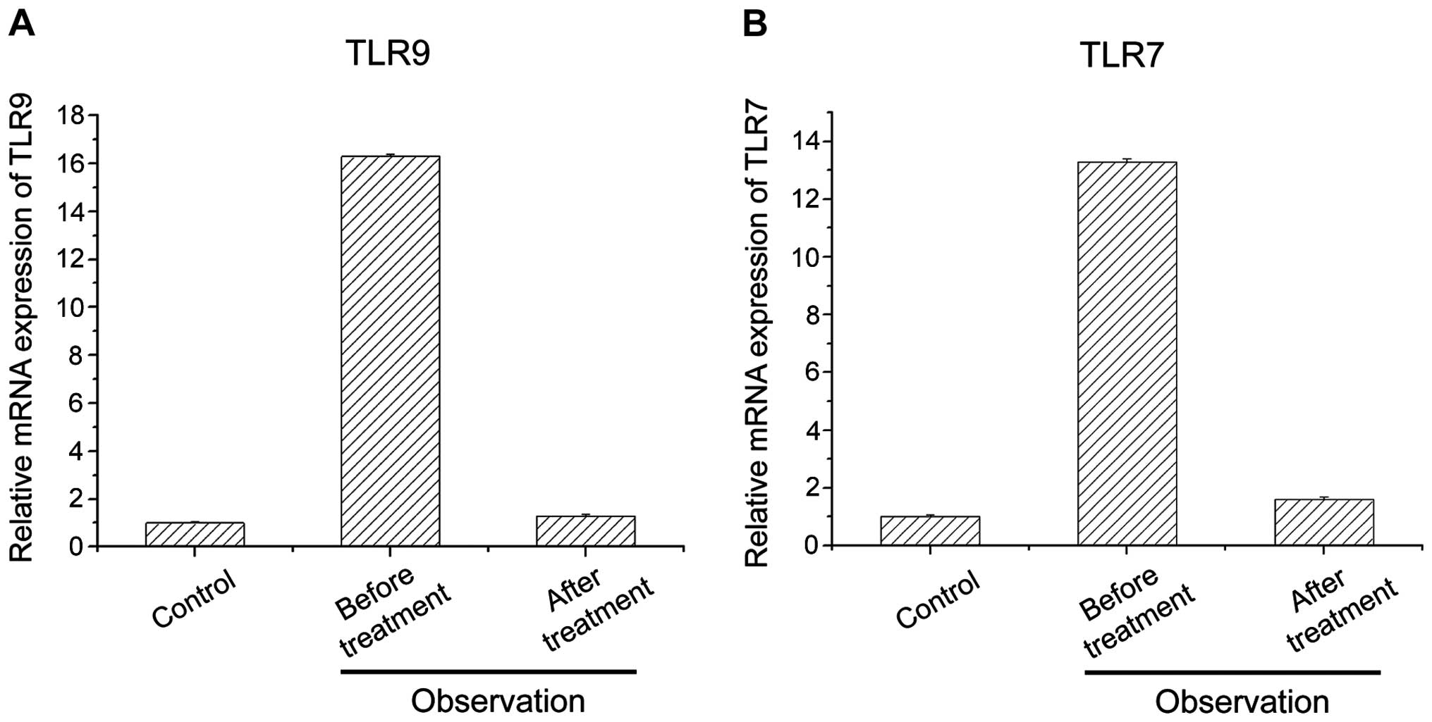
Detection of cytokine TLR9 and TLR7
mRNA content in peripheral blood of patients from observational
group before and after treatment
From the comparison between the relative quantity of
expression of cytokines TLR9 and TLR7 mRNA in peripheral blood of
patients from the observational group before and after treatment,
and those of patients from the control group, we determined that
the relative expression of the quantity of cytokines TLR9 and TLR7
mRNA in peripheral blood after glucocorticoid treatment decreased
significantly (P<0.05) (Fig.
1).
The relative expression of the quantity of cytokines
TLR9 and TLR7 mRNA in peripheral blood for the patients with
uveitis after glucocorticoid treatment had no significant
difference with those of control group. This suggested that
glucocorticoid could reduce the expression of cytokines TLR9 and
TLR7 mRNA in peripheral blood in the patients suffering uveitis.
This reduction may be associated with glucocorticoid treatment on
uveitis.
Detection of cytokine TLR9 and TLR7
protein expression quantity of observation group and control group
by ELISA
From the results of ELISA detection of the quantity
of expression of protein TLR9 and TLR7 in peripheral blood from
observational group before and after treatment and those of control
group, we can see that the TLR9 and TLR7 content in peripheral
blood of patients from observational group before treatment was
0.48±0.03 and 0.38±0.02 ng/l and the TLR9 and TLR7 content in
peripheral blood of patients from observation group after
glucocorticoid treatment was 0.21±0.01 and 0.19±0.01 ng/l (Table II). The comparison between them has
significant deference (P<0.05). As well, compared with control
group, the TLR9 and TLR7 content in peripheral blood of patients
from observation group before treatment was significantly higher
(P<0.05) than the TLR9 and TLR7 protein content of control group
patients. This indicated that glucocorticoid could reduce protein
content of cytokines TLR9 and TLR7 in peripheral blood for patients
with uveitis. The related articles showed that TLR9 and TLR7
content in peripheral blood had a positive correlation with uveitis
disease, which may relate to the therapeutic mechanism of
glucocorticoid on uveitis (10,11).
| Table II.Detection of TLR9 and TLR7 protein
content (ng/l) in peripheral blood fromthe observation group
patients after glucocorticoid treatment. |
Table II.
Detection of TLR9 and TLR7 protein
content (ng/l) in peripheral blood fromthe observation group
patients after glucocorticoid treatment.
| Groups | Cases | TLR9 cytokine | TLR7 cytokine |
|---|
| Observation |
|
|
|
| Before
treatment | 46 |
0.48±0.03a |
0.38±0.02a |
| After
treatment | 46 |
0.21±0.01a,b |
0.19±0.01a,b |
| Control | 35 | 0.19±0.01 | 0.17±0.01 |
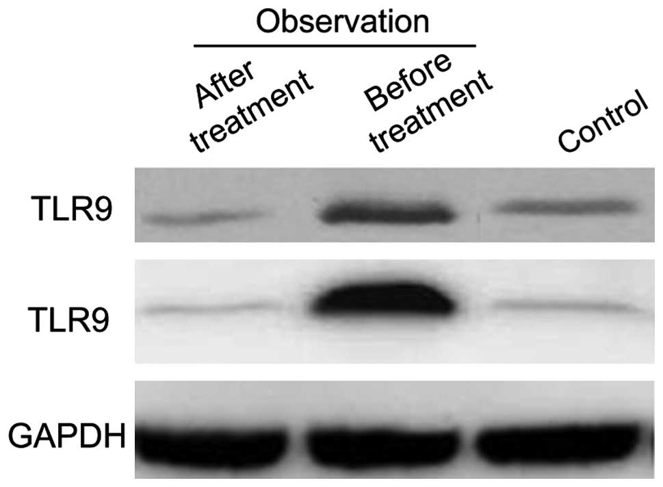
Detection of cytokine TLR9 and TLR7
protein expression quantity of the observation and control groups
by western blotting
The quantity of cytokine TLR9 and TLR7 protein
expression of observational group before and after treatment in
peripheral blood of observational group after glucocorticoid
treatment decreased insignificantly, whereas obviously compared
with cytokines TLR9 and TLR7 protein content in peripheral blood of
control group, which was accordant with ELISA result and suggested
that glucocorticoid could improve uveitis by reducing the protein
content of cytokines TLR9 and TLR7 in peripheral blood (Fig. 2).
Correlation detection of TLR9 and TLR7
content in peripheral blood before and after glucocorticoid
treatment
Through the detection of TLR9 and TLR7 content in
peripheral blood before and after glucocorticoid treatment by SPSS
20.0, we found that for the patients with uveitis, the content of
TLR9 and TLR7 in peripheral blood before and after treatment showed
a positive correlation (r=0.653, P=0.012). This showed that TLR9
and TLR7 content in peripheral blood had a positive correlation
with uveitis disease and glucocorticoid could improve uveitis by
reducing cytokines TLR9 and TLR7 content in peripheral blood. At
the same time, due to the correlation between TLR9 and TLR7, both
can be used as a new detection and recovery index for uveitis
disease.
Discussion
Uveitis, is a kind of autoimmune disease whose
pathogenesis is not clear, so that there is no specific effect
medicines for its treatment (12).
The results of Yu and Liu showed that uveitis, as a type of
auto-immunodeficiency disease, was caused by the deficiency of
immune response mechanism that involved T lymphocytes (12). Sun et al suggested that TLRs,
as a kind of natural immune receptor protein of human body, played
an important role of signal transduction in the body's immune
response (13). For example, the
expression of TLR can be detected in macrophages, B lymphocytes,
monocytes and T lymphocyte, which explained that TLRs widely
participated in collective immune response mechanism (14). Additionally, TLRs could selectively
identify the echogenic microbes and virus that entered the body by
their special spatial structure and triggered the response
mechanism of body's immune system to echogenic pathogenic
microorganism by other related signal transduction pathways
(15). Therefore, TLRs play a
significant role in identifying exogenous pathogenic factors and
starting body's immune response mechanism to clear pathogens. As a
type of autoimmune deficiency disease, the main cause of uveitis is
that the immune system starts mechanism for their own cells and
tissues incorrectly, leading to immune deficiency diseases. Thus,
as an important part of the body's own immune response mechanism,
the distribution and dysfunction of TLRs also can result in the
generation and deterioration of immunodeficiency diseases (16). Studies have shown that the main
expression of TLR9 and TLR7 was in the endoplasmic reticulum, TLR7
often participated in CpGDNA of TLR9 mediation as an auxiliary
receptor, with the function of immune cell activation (17). Prinz et al (18) found that the quantity of expression
of TLR9 and TLR7 mRNA had positive correlation with the state of
this illness by the detection of TLR9 and TLR7 mRNA expression in
experimental autoimmunity encephalomyelitis, which illustrated that
TLR9 and TLR7 are involved in the process of experimental
autoimmunity encephalomyelitis attack (19).
As a kind of autoimmunity deficiency disease, the
main pathogenesis of uveitis is also caused by body's autoimmunity
system deficiency (20,21). In the present study, we selected
glucocorticoid that has good effect on uveitis treatment. The
results showed that the content of TLR9 and TLR7 in peripheral
blood in the patients with uveitis decreased significantly compared
with the data before treatment, and the content of TLR9 and TLR7 in
peripheral blood was close to the content of normal people. This
suggested that glucocorticoids could reduce the content of TLR9 and
TLR7 in the body and the response of body's immune system to
external environment to reach our aim of cure. However, the present
study did not investigate the way by which glucocorticoid acts on
TLR9 and TLR7 to decrease their content. This is crucial to future
studies that are to be conducted.
References
|
1
|
Yang Y, Zhang M and Zhuang Z: Causes
investigation of uveitis recurrence. Medical J Peking Union Med
College Hospital. 4:150–153. 2013.(In Chinese).
|
|
2
|
Xu X and Jiahua Lv: Observation of T
lymphocyte change of rat with autoimmunity uveitis. Chinese
Ophthalmic Res. 26:841–844. 2008.(In Chinese).
|
|
3
|
Trinh L, Brignole-Baudouin F, Pauly A,
Liang H, Houssier M and Baudouin C: Th1- and Th2-related chemokine
and chemokine receptor expression on the ocular surface in
endotoxin-induced uveitis. Mol Vis. 14:2428–2434. 2008.PubMed/NCBI
|
|
4
|
Thibault DL, Chu AD, Graham KL, Balboni I,
Lee LY, Kohlmoos C, Landrigan A, Higgins JP, Tibshirani R and Utz
PJ: IRF9 and STAT1 are required for IgG autoantibody production and
B cell expression of TLR7 in mice. J Clin Invest. 118:1417–1426.
2008. View
Article : Google Scholar : PubMed/NCBI
|
|
5
|
Sun S, Rao NL, Venable J, Thurmond R and
Karlsson L: TLR7/9 antagonists as therapeutics for immune-mediated
inflammatory disorders. Inflamm Allergy Drug Targets. 6:223–235.
2007. View Article : Google Scholar : PubMed/NCBI
|
|
6
|
Takase H, Yu CR, Ham DI, Chan CC, Chen J,
Vistica BP, Wawrousek EF, Durum SK, Egwuagu CE and Gery I:
Inflammatory processes triggered by TCR engagement or by local
cytokine expression: differences in profiles of gene expression and
infiltrating cell populations. J Leukoc Biol. 80:538–545. 2006.
View Article : Google Scholar : PubMed/NCBI
|
|
7
|
Wu W: Observation and analysis of
glucocorticoid treatment on uveitis in clinical effect. World
Latest Medicine Information. 15:94–97. 2015.
|
|
8
|
Jabs DA, Nussenblatt RB and Rosenbaum JT:
Standardization of Uveitis Nomenclature (SUN) Working Group:
Standardization of uveitis nomenclature for reporting clinical
data. Results of the First International Workshop. Am J Ophthalmol.
140:509–516. 2005. View Article : Google Scholar : PubMed/NCBI
|
|
9
|
Sugita S, Kawazoe Y, Imai A, Yamada Y,
Horie S and Mochizuki M: Inhibition of Th17 differentiation by
anti-TNF-alpha therapy in uveitis patients with Behçet's disease.
Arthritis Res Ther. 14:R992012. View
Article : Google Scholar : PubMed/NCBI
|
|
10
|
Allensworth JJ, Planck SR, Rosenbaum JT
and Rosenzweig HL: Investigation of the differential potentials of
TLR agonists to elicit uveitis in mice. J Leukoc Biol.
90:1159–1166. 2011. View Article : Google Scholar : PubMed/NCBI
|
|
11
|
Chinnery HR, Leong CM, Chen W, Forrester
JV and McMenamin PG: TLR9 and TLR7/8 activation induces formation
of keratic precipitates and giant macrophages in the mouse cornea.
J Leukoc Biol. 97:103–110. 2015. View Article : Google Scholar : PubMed/NCBI
|
|
12
|
Yu Z and Liu J: Research summary of
related-uveitis cytokines. Heilongjiang Med J. 40:8–9. 2016.(In
Chinese).
|
|
13
|
Sun R, Sun L, Bao M, Zhang Y and Wang L,
Wu X, Hu D, Liu Y, Yu Y and Wang L: A human microsatellite
DNA-mimicking oligodeoxynucleotide with CCT repeats negatively
regulates TLR7/9-mediated innate immune responses via selected TLR
pathways. Clin Immunol. 134:262–276. 2010. View Article : Google Scholar : PubMed/NCBI
|
|
14
|
Sacco RE, Nicholson TL, Waters TE and
Brockmeier SL: Porcine TLR3 characterization and expression in
response to influenza virus and Bordetella bronchiseptica. Vet
Immunol Immunopathol. 142:57–63. 2011. View Article : Google Scholar : PubMed/NCBI
|
|
15
|
Beutler BA: TLRs and innate immunity.
Blood. 113:1399–1407. 2009. View Article : Google Scholar : PubMed/NCBI
|
|
16
|
Hsu LC, Park JM, Zhang K, Luo JL, Maeda S,
Kaufman RJ, Eckmann L, Guiney DG and Karin M: The protein kinase
PKR is required for macrophage apoptosis after activation of
Toll-like receptor 4. Nature. 428:341–345. 2004. View Article : Google Scholar : PubMed/NCBI
|
|
17
|
Krieg AM and Vollmer J: Toll-like
receptors 7, 8, and 9: linking innate immunity to autoimmunity.
Immunol Rev. 220:251–269. 2007. View Article : Google Scholar : PubMed/NCBI
|
|
18
|
Prinz M, Garbe F, Schmidt H, Mildner A,
Gutcher I, Wolter K, Piesche M, Schroers R, Weiss E, Kirschning CJ,
et al: Innate immunity mediated by TLR9 modulates pathogenicity in
an animal model of multiple sclerosis. J Clin Invest. 116:456–464.
2006. View
Article : Google Scholar : PubMed/NCBI
|
|
19
|
Forsbach A, Nemorin JG, Völp K, Samulowitz
U, Montino C, Müller C, Tluk S, Hamm S, Bauer S, Lipford GB, et al:
Characterization of conserved viral leader RNA sequences that
stimulate innate immunity through TLRs. Oligonucleotides.
17:405–417. 2007. View Article : Google Scholar : PubMed/NCBI
|
|
20
|
Hasimu A, Ge L, Li QZ, Zhang RP and Guo X:
Expressions of Toll-like receptors 3, 4, 7, and 9 in cervical
lesions and their correlation with HPV16 infection in Uighur women.
Chin J Cancer. 30:344–350. 2011. View Article : Google Scholar : PubMed/NCBI
|
|
21
|
De Trez C, Pajak B, Brait M, Glaichenhaus
N, Urbain J, Moser M, Lauvau G and Muraille E: TLR4 and Toll-IL-1
receptor domain-containing adapter-inducing IFN-beta, but not
MyD88, regulate Escherichia coli-induced dendritic cell maturation
and apoptosis in vivo. J Immunol. 175:839–846. 2005. View Article : Google Scholar : PubMed/NCBI
|