Introduction
During the process of orthodontic treatment, tooth
movement is closely related to alveolar bone remodeling, which
consists of bone resorption in direction of tooth movement and bone
formation on the opposite side. The use of an optimal force system
is important for an adequate biological response in the periodontal
system. Heavy forces could overcompress the periodontal ligament
and induce hyalinization, which could impede resorption of the
alveolar bone surface (1,2).
A previous study on the periodontal tissues, and
especially on the microstructure of the trabecular bone used to
adopt specimens destructive methods of histological techniques
which was capable of the two-dimensional microstructure, but was
hard to obtain and describe the parameters of the alveolar bone
internal microstructure (3).
Micro-computed tomography (micro-CT) is a new imaging examination
and analysis technique of high resolution and non-intrusive has
developed fast in recent years, which can provide three-dimensional
qualitative as well as quantitative data of the tested specimens,
to better understand and study the remolding of alveolar bone
microstructure (4,5).
The purpose of this study was to evaluate the
dynamic changes of the microstructure of alveolar bone during
orthodontic tooth movement under different force magnitudes in rats
employing micro-CT system, and to explore the pattern of changes of
the trabecular bone microstructure in the alveolar bone so as to
provide theoretical reference for clinical orthodontic
treatment.
Materials and methods
Grouping of the laboratory
animals
Seventy 11-week-old adult female Wistar rats
(approximate weight, 220–260 g) were used for this study. A total
of 10 rats were selected randomly as control, and the remaining 60
rats were divided into 25-g and 75-g groups of equal number. The
rats were raised under standard conditions: Room temperature at
25±1°C, relative humidity at 55±5%, and 12-h light and dark cycles.
All the experimental protocols followed were approved by the Ethics
Committee of Harbin Medical University, and the experiments were
carried out under the control of the University's Guidelines for
Animal Experimentation.
Installing orthodontic appliance
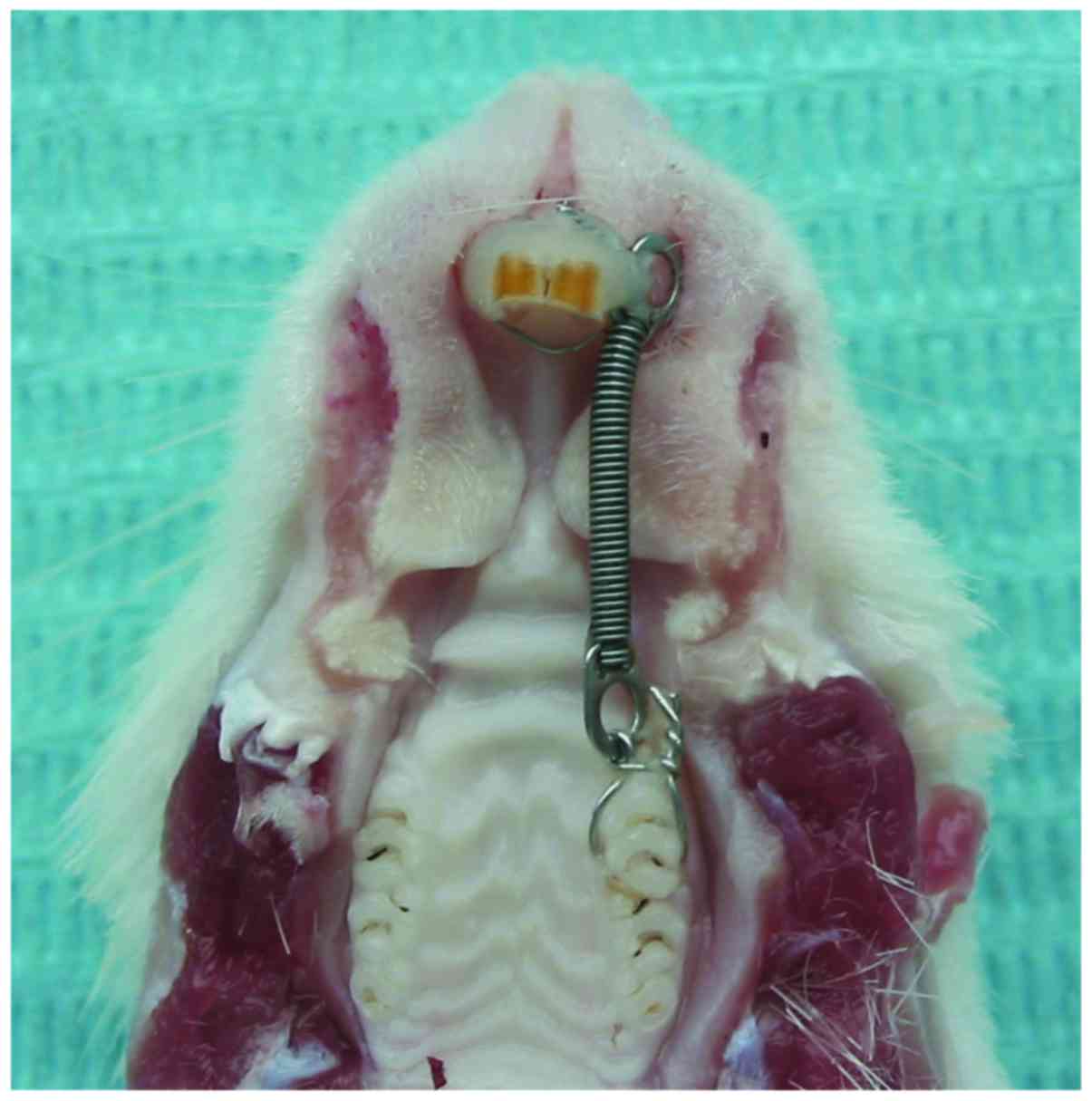
A total of 30 rats of 25-g group and 30 rats of 75-g
group were anesthetized by intraperitoneal injection of 10% chloral
hydrate, a cervical groove was prepared on the incisors using a
round burr with a dental low-speed handpiece (NSK Ltd., Tokyo,
Japan). The maxillary left first molar was connected with both
maxillary central incisors with nickel-titanium coil spring 0.008
inch in diameter (Shinye Odontology Materials Corp. Co., Hangzhou,
China) using a 0.2 mm ligature wire (Changsha Tiantian Dental
Equipment Co., Ltd., Changsha, China) to perform the mesial
movement (Fig. 1). The ligature wire
was then secured with bond adhesive (Transbond; 3M Unitek,
Monrovia, CA, USA) on the incisors. Forces of 25 and 75 g were
applied separately to observe the reactions of the alveolar bone.
Spring retention was checked daily to ensure the stability of the
applied force. The incisors, molar, and spring were cleaned and
irrigated with tap water as needed to prevent potential trauma and
irritation to the gingival and periodontal tissues.
Experimental samples
A total of 10 rats as control without installing
orthodontic appliance were euthanized by perfusion though heart
with 4% paraformaldehyde on day 0, and every 10 rats in each group
were euthanized by the same way on days 3, 7 and 14, respectively.
Then the maxillae were dissected and stored in 4%
paraformaldehyde.
Micro-CT scanning
A total of 70 samples were scanned with a micro-CT
scanner (µCT 35; Scanco Medical AG, Bassersdorf, Switzerland) with
a 7 µm voxel size using the following parameters: 114 mA, 70 kVp,
exposure time of 300 msec, and a horizontal scan angle. The
scanning procedure lasted ~1 h per sample and generated ~1,000
two-dimensional images with a resolution of 1,024×1,024 pixels.
Evaluating the microstructural
parameters
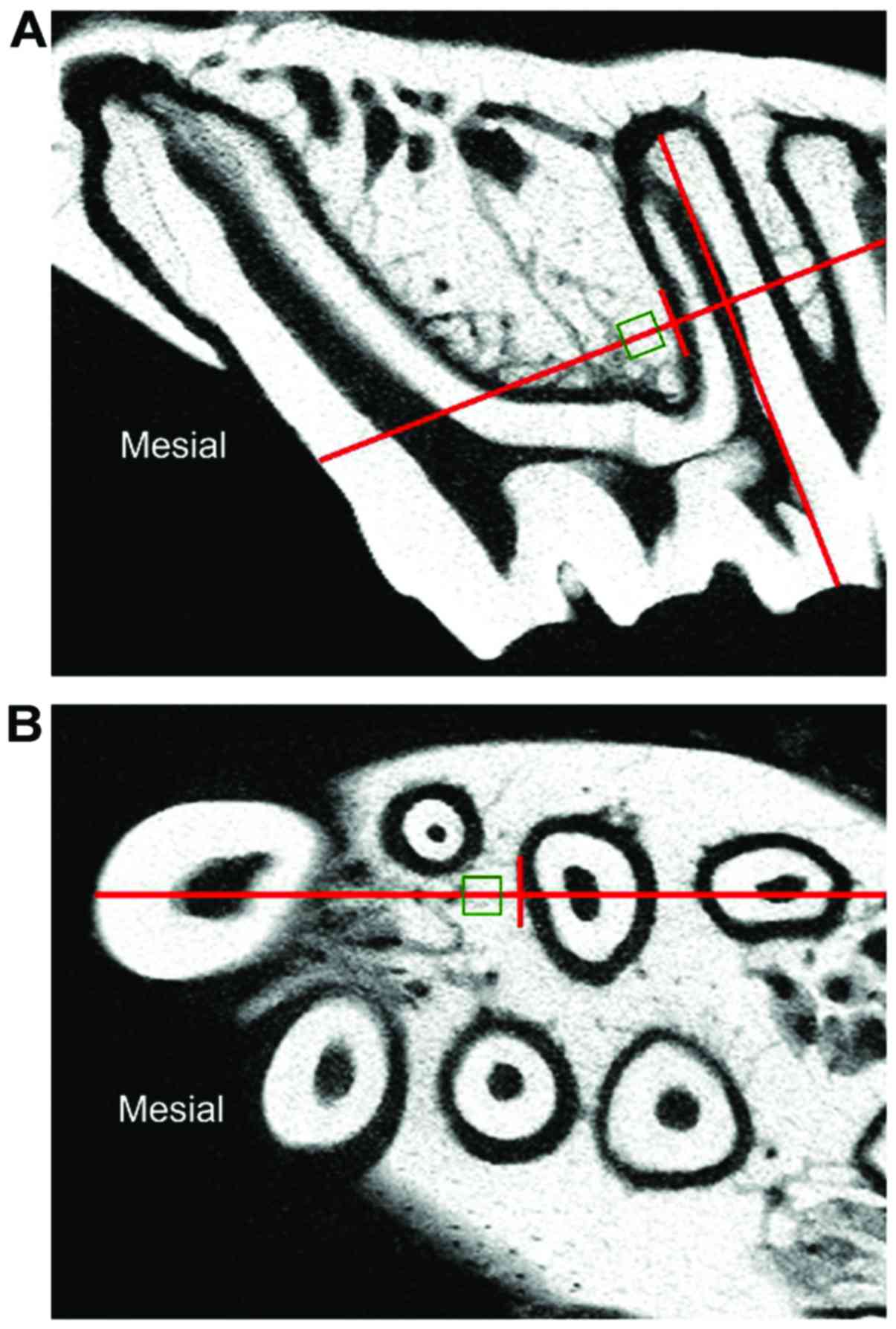
The region of interest (ROI) of the alveolar bone
was selected according to previously described method which showed
the integral microstructure of alveolar bone (6). A 210×210×210 µm cube of trabecular bone
mesial to the cervial third of the distobuccal root of the
maxillary left first molar was selected separately for analysis.
The distance between the cube and the root was 100 µm (Fig. 2).
Three-dimensional microstructure of the alveolar
bone was analyzed using the software affiliated to the micro-CT
(Scanco® software ver. 3.7; Scanco Medical
AG®, Brüttisellen, Switzerland). We evaluated the bone
mineral density (BMD, mg cm−3); bone volume/total volume
(BV/TV, %); trabecular thickness (Tb.Th, mm); trabecular number
(Tb.N, mm−1); and trabecular separation (Tb.Sp, mm),
microstructure model index (SMI), showing whether the trabecular
bone is microstructured as rod-like or plate-like. Theoretical
value for a complete rod-like microstructure is 3, while
theoretical value for an ideal plate-like microstructure is 0. The
stress that the tabular microstructure can bear is larger than that
of clave microstructure (6,7).
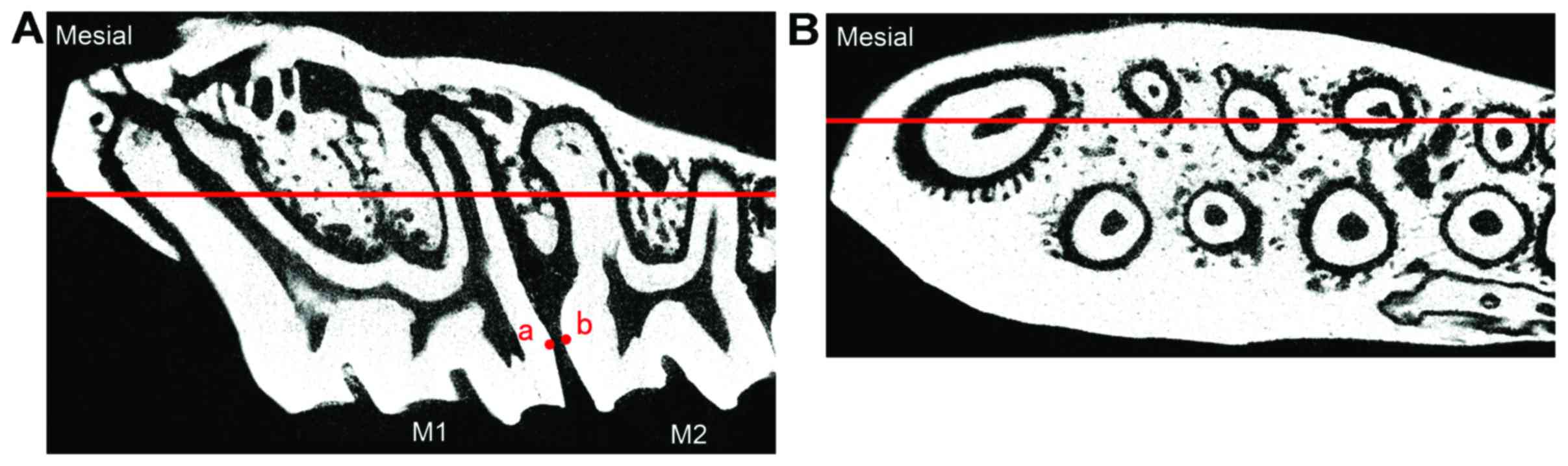
Measuring the distance for mesial
movement
Distance for mesial movement of the molar were
measured between the nearest two landmark points (a,b) in the
crowns of the first molar (M1) and second molar (M2) which were
indentified on the longitudinal section through maximal diameter of
the distobuccal root and mesial root of the first molar (Fig. 3).
Statistical analysis
The data were analyzed by using SPSS, version 21.0
(SPSS Inc., Chicago, IL, USA). Parameters of 25-g and 75-g groups
on days 0, 3, 7 and 14 were compared using Student-Newman-Keuls of
ANOVA. P<0.05 was considered to indicate a statistically
significant difference.
Results
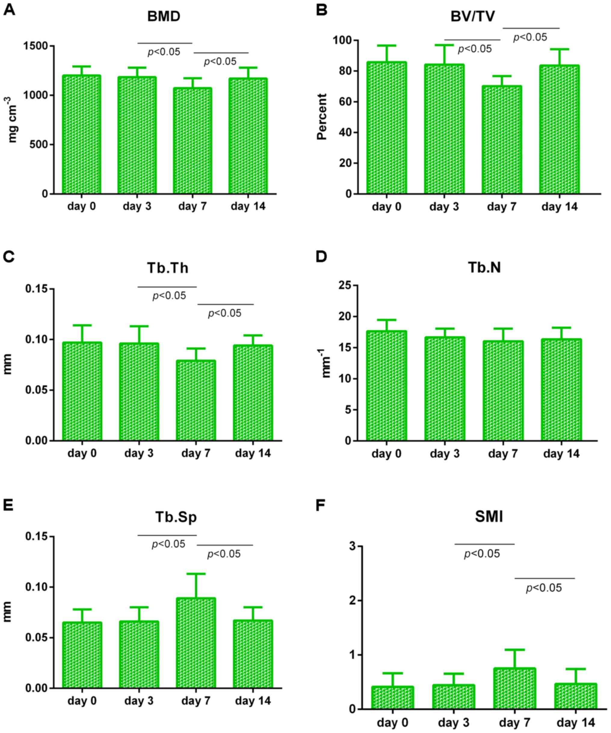
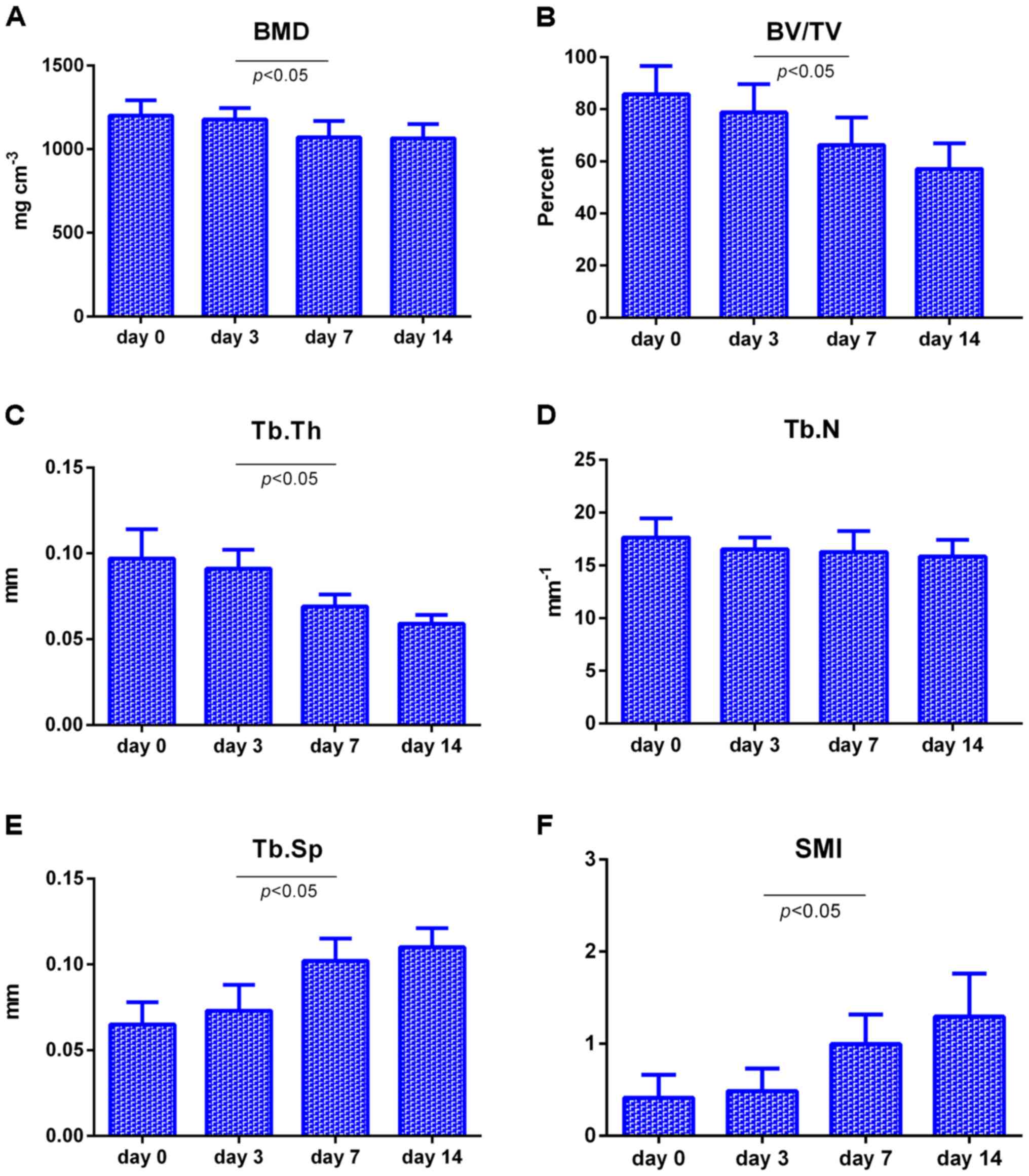
Microstructural parameters for trabecular bone
mesial to the distobuccal root of the maxillary left first molar of
25-g group at different time-points are summarized in Fig. 4. From day 0 to day 3, the parameters
did not display any significant changes (P>0.05); from day 3 to
day 7, BMD, BV/TV and Tb.Th significantly decreased (P<0.05),
whereas Tb.Sp and SMI significantly increased (P<0.05); while
from day 7 to day 14, BMD, BV/TV and Tb.Th increased significantly
(P<0.05), while Tb.Sp and SMI decreased significantly
(P<0.05). Tb.N did not have any clear change during the whole
process of the experiment (P>0.05).
Microstructural parameters for trabecular bone
mesial to the distobuccal root of the maxillary left first molar of
75-g group at different time points are summarized in Fig. 5. Statistical results of parameters on
day 0 to day 7 are consistent with those of 25-g group, while from
day 7 to day 14, changes of parameters did not carry any
statistical significance (P>0.05), Tb.N did not have any
significant change during the whole process of the experiment
(P>0.05).
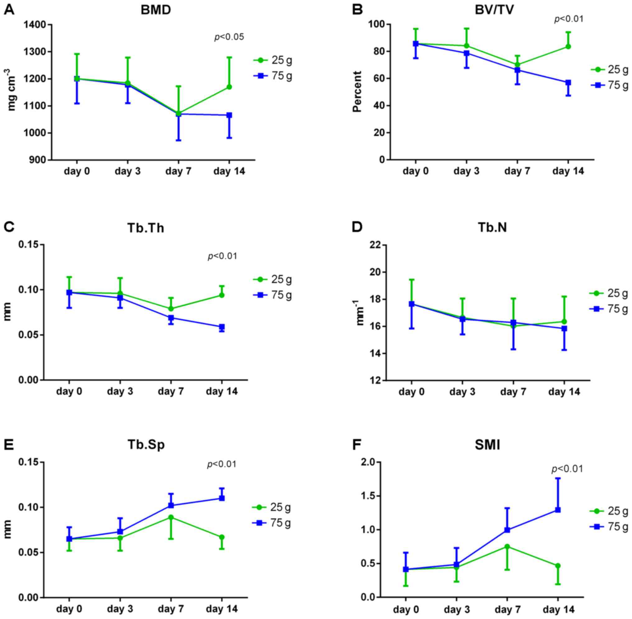
Microstructural parameters for trabecular bone
mesial to the distobuccal root of the maxillary left first molar
between 25-g and 75-g groups at different scanning time points are
summarized in Fig. 6. From day 0 to
day 7, the change trends of microstructural parameters of two
groups were similar, but from day 7 to day 14, the change in trends
between two groups showed significant difference (P<0.05).
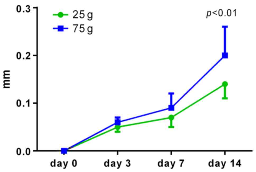
Distance for mesial movement of the molar at
different scanning time points between 25-g and 75-g groups are
summarized in Fig. 7. The 75-g rats
showed larger distance than 25-g only at day 14 (P<0.05).
Discussion
Previous studies frequently employed methods such as
histological analysis to explore microstructure of the alveolar
bone, which was sophisticated in operation and hard to obtain and
describe the internal parameters of the specimens (3–5). The
method of histological sections was also disadvantageous in limited
number of slices and in turn deviation and substantial loss of
information (3). In contrast,
micro-CT is easy to operate with a high resolution of µm that is
capable of accurate qualitative as well as quantitative analysis of
the specimens. It allows improved study of the bone remolding and
later histological or electron microscopic observation of the
specimens, whenever needed, after having completed scanning
(4,5).
The model of mesial movement of the maxillary left
first molar by means of orthodontic extrusion was first proposed by
Waldo and Rothblatt (8). Based on
literature review, Ren et al (9) held the view that one orthodontic force
loading cycle should be 14 days. Repair and reconstruction of
alveolar bone would be dominant after 14 days. Since the molar
volume of human is 50 times than that of the rat, orthodontic
extrusion of 20 g was considered as an ideal value for orthodontic
molar tooth movement mesially, and 75 g belonged to heavy force.
Taking the factor of the basic scale value of 25 g on the
dynamometer used in the experiment, for more accurate measurement,
this study selected 25 and 75 g orthodontic extrusion as the
orthodontic force loading.
Based on the previous study on the three-dimensional
finite elements of the rat molar, under the application of mesial
force loading, the mesial side of the disbuccal root was subject to
the largest compression (10).
During the process of orthodontic tooth movement, alveolar bone on
the compression side would pose the main resistance to the tooth
movement, so if the alveolar bone on the compression side could
always maintain a reasonable and stable rate of resorption and
reconstruction throughout the orthodontic treatment, then the tooth
would be able to move into the ideal position healthily and
effectively. This study chose the alveolar bone mesial to the
cervial third of the distobuccal root of maxillary first molar as
its interest observation region, which is the location of the
largest compression and of typical significance in terms of bone
resorption and remodeling. Some studies using micro-CT system
observed the changes of the microstructure of alveolar bone during
orthodontic movement in vivo (11,12).
However, they did not choose the location of the largest
compression as interest observation region, so the results of the
experiments were affected inevitably. Moreover, our study which
used 70 rats tried to avoid the result error caused by sample
difference as far as possible.
Currently, there exists controversy from
histological experimental studies concerning the time points of
alveolar bone reconstruction during orthodontic tooth movement.
Kohno et al (13) showed that
hyaline change in the alveolar bone appeared on day 7, and bone
resorption and reconstruction took place on day 14 after the force
loading. However, Tomizuka et al (14) indicated that bone resorption happened
on day 10 after force loading through histological observation.
The results showed that on day 0 to day 3 after the
application of the force loading, the parameters of the two groups
did not have any evident change (P>0.05), indicating that
resorption did not occur in the alveolar bone. From day 3 to day 7,
BMD, BV/TV and Tb.Th significantly decreased (P<0.05), while
Tb.Sp and SMI significantly increased (P<0.05), indicating that
resorption occurred in the alveolar bone. The difference was that
from day 7 to day 14, in 25-g group, BMD, BV/TV and Tb.Th increased
significantly (P<0.05), while Tb.Sp and SMI decreased
significantly (P<0.05), indicating that alveolar bone was in the
phase of repair and reconstruction.
Correspondingly, in 75-g group, changes of various
parameters did not carry any statistical significance (P>0.05),
indicating that alveolar bone was still in the phase of resorption.
The comparison of microstructural parameters for trabecular bone
between 25-g and 75-g groups at different scanning time points also
supported above conclusion.
Obviously, on day 14, for the rats of 25-g group
that were already in the repair and reconstruction, it was feasible
to continue applying force loading; however, for the rats of 75-g
group that were still in the phase of resorption, it was disputable
to continue applying any force loading. Thus, further studies are
still needed.
Previous studies showed that orthodontic tooth
movement included three phases: The first phase is initial rapid
tooth movement after the force loading; the second phase is delayed
tooth movement; the third phase is linear tooth movement (2,15–17). In
our study, the tooth movement of both groups conformed with the
description above. Among them, from day 0 to day 3 belonged to
phase 1; from day 3 to day 7 was in phase 2; and from day 7 to day
14 belonged to phase 3. However, 75-g group showed larger distance
than 25-g group only at day 14 (P<0.05). Apparently, this kind
of accelerated tooth movement at the expense of excessive alveolar
bone was not ideal and healthy pattern.
The aforementioned findings may provide reference to
orthodontic practitioners that, in order to maintain the health of
periodontal tissues, low force should be advocated, and it is not
suggestive to frequently apply orthodontic force loading.
Periodontal tissues should be allowed adequate time for repair and
recovery so as to ensure reasonable reconstruction of alveolar bone
and healthy movement of the orthodontic tooth.
In conclusion, these findings indicated that on day
14, the rats of 25-g group that were in repair and reconstruction
phase were feasible to apply continuous force load, however, it was
disputable to continue applying any force loading for the rats of
75-g group.
In addition, the movement distance in 75-g group was
significantly larger only at day 14, compared with 25-g group.
Apparently, the fast tooth movement at the expense of excessive
alveolar bone resorption was not ideal.
Taken together, frequently applying orthodontic
force loading during orthodontic treatment was demonstrated to be
unhelpful. In order to maintain the health of periodontal tissues,
adequate time for repair and recovery is needed to ensure
reasonable remolding of alveolar bone and healthy movement of the
orthodontic tooth.
Acknowledgements
This study was supported by the National Natural
Science Foundation of China (grant no. 81170960) and the Special
Foundation for Sino-Russian Translational Medicine Research Center
of Harbin Medical University (grant no. CR201412).
References
|
1
|
Henneman S, Von den Hoff JW and Maltha JC:
Mechanobiology of tooth movement. Eur J Orthod. 30:299–306. 2008.
View Article : Google Scholar : PubMed/NCBI
|
|
2
|
Wise GE and King GJ: Mechanisms of tooth
eruption and orthodontic tooth movement. J Dent Res. 87:414–434.
2008. View Article : Google Scholar : PubMed/NCBI
|
|
3
|
Salazar M, Hernandes L, Ramos AL,
Micheletti KR, Albino CC and Cuman Nakamura RK: Effect of
teriparatide on induced tooth displacement in ovariectomized rats:
A histomorphometric analysis. Am J Orthod Dentofacial Orthop.
139:e337–e344. 2011. View Article : Google Scholar : PubMed/NCBI
|
|
4
|
Martin-Badosa E, Amblard D, Nuzzo S,
Elmoutaouakkil A, Vico L and Peyrin F: Excised bone structures in
mice: imaging at three-dimensional synchrotron radiation micro CT.
Radiology. 229:921–928. 2003. View Article : Google Scholar : PubMed/NCBI
|
|
5
|
Waarsing JH, Day JS, van der Linden JC,
Ederveen AG, Spanjers C, De Clerck N, Sasov A, Verhaar JA and
Weinans H: Detecting and tracking local changes in the tibiae of
individual rats: A novel method to analyse longitudinal in vivo
micro-CT data. Bone. 34:163–169. 2004. View Article : Google Scholar : PubMed/NCBI
|
|
6
|
Dempster DW, Compston JE, Drezner MK,
Glorieux FH, Kanis JA, Malluche H, Meunier PJ, Ott SM, Recker RR
and Parfitt AM: Standardized nomenclature, symbols, and units for
bone histomorphometry: a 2012 update of the report of the ASBMR
Histomorphometry Nomenclature Committee. J Bone Miner Res. 28:2–17.
2013. View Article : Google Scholar : PubMed/NCBI
|
|
7
|
Parfitt AM, Mathews CH, Villanueva AR,
Kleerekoper M, Frame B and Rao DS: Relationships between surface,
volume, and thickness of iliac trabecular bone in aging and in
osteoporosis. Implications for the microanatomic and cellular
mechanisms of bone loss. J Clin Invest. 72:1396–1409. 1983.
View Article : Google Scholar : PubMed/NCBI
|
|
8
|
Waldo CM and Rothblatt JM: Histologic
response to tooth movement in the laboratory rat; procedure and
preliminary observations. J Dent Res. 33:481–486. 1954. View Article : Google Scholar : PubMed/NCBI
|
|
9
|
Ren Y, Maltha JC and Kuijpers-Jagtman AM:
The rat as a model for orthodontic tooth movement - a critical
review and a proposed solution. Eur J Orthod. 26:483–490. 2004.
View Article : Google Scholar : PubMed/NCBI
|
|
10
|
Gonzales C, Hotokezaka H, Arai Y, Ninomiya
T, Tominaga J, Jang I, Hotokezaka Y, Tanaka M and Yoshida N: An in
vivo 3D micro-CT evaluation of tooth movement after the application
of different force magnitudes in rat molar. Angle Orthod.
79:703–714. 2009. View Article : Google Scholar : PubMed/NCBI
|
|
11
|
Xu Y, Zhao T, Xu W and Ding Y: Periodontal
microstructure change and tooth movement pattern under different
force magnitudes in ovariectomized rats: An in-vivo microcomputed
tomography study. Am J Orthod Dentofacial Orthop. 143:828–836.
2013. View Article : Google Scholar : PubMed/NCBI
|
|
12
|
Xu X, Zhou J, Yang F, Wei S and Dai H:
Using micro-computed tomography to evaluate the dynamics of
orthodontically induced root resorption repair in a rat model. PLoS
One. 11:e01501352016. View Article : Google Scholar : PubMed/NCBI
|
|
13
|
Kohno T, Matsumoto Y, Kanno Z, Warita H
and Soma K: Experimental tooth movement under light orthodontic
forces: rates of tooth movement and changes of the periodontium. J
Orthod. 29:129–135. 2002. View Article : Google Scholar : PubMed/NCBI
|
|
14
|
Tomizuka R, Shimizu Y, Kanetaka H, Suzuki
A, Urayama S, Kikuchi M, Mitani H and Igarashi K: Histological
evaluation of the effects of initially light and gradually
increasing force on orthodontic tooth movement. Angle Orthod.
77:410–416. 2007. View Article : Google Scholar : PubMed/NCBI
|
|
15
|
Reitan K and Kvam E: Comparative behavior
of human and animal tissue during experimental tooth movement.
Angle Orthod. 41:1–14. 1971.PubMed/NCBI
|
|
16
|
Storey E: The nature of tooth movement. Am
J Orthod. 63:292–314. 1973. View Article : Google Scholar : PubMed/NCBI
|
|
17
|
King GJ, Keeling SD, McCoy EA and Ward TH:
Measuring dental drift and orthodontic tooth movement in response
to various initial forces in adult rats. Am J Orthod Dentofacial
Orthop. 99:456–465. 1991. View Article : Google Scholar : PubMed/NCBI
|