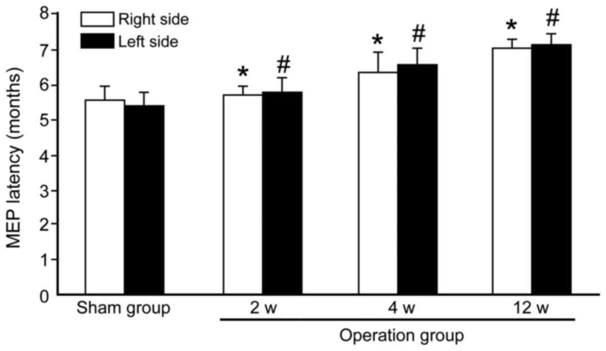
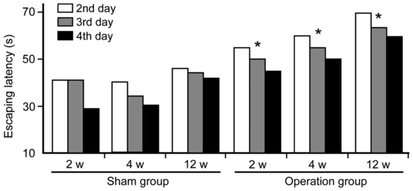
|
1
|
Akinyemi RO, Mukaetova-Ladinska EB, Attems
J, Ihara M and Kalaria RN: Vascular risk factors and
neurodegeneration in ageing related dementias: Alzheimer's disease
and vascular dementia. Curr Alzheimer Res. 10:642–653. 2013.
View Article : Google Scholar : PubMed/NCBI
|
|
2
|
Hainsworth AH, Brittain JF and Khatun H:
Pre-clinical models of human cerebral small vessel disease: utility
for clinical application. J Neurol Sci. 322:237–240. 2012.
View Article : Google Scholar : PubMed/NCBI
|
|
3
|
Miki K, Ishibashi S, Sun L, Xu H, Ohashi
W, Kuroiwa T and Mizusawa H: Intensity of chronic cerebral
hypoperfusion determines white/gray matter injury and
cognitive/motor dysfunction in mice. J Neurosci Res. 87:1270–1281.
2009. View Article : Google Scholar : PubMed/NCBI
|
|
4
|
Brown WR, Moody DM, Thore CR, Anstrom JA
and Challa VR: Microvascular changes in the white mater in
dementia. J Neurol Sci. 283:28–31. 2009. View Article : Google Scholar : PubMed/NCBI
|
|
5
|
Fernando MS, Simpson JE, Matthews F,
Brayne C, Lewis CE, Barber R, Kalaria RN, Forster G, Esteves F,
Wharton SB, et al: MRC Cognitive Function and Ageing Neuropathology
Study Group: White matter lesions in an unselected cohort of the
elderly: molecular pathology suggests origin from chronic
hypoperfusion injury. Stroke. 37:1391–1398. 2006. View Article : Google Scholar : PubMed/NCBI
|
|
6
|
Nave K-A: Myelination and support of
axonal integrity by glia. Nature. 468:244–252. 2010. View Article : Google Scholar : PubMed/NCBI
|
|
7
|
Salzer JL: Clustering sodium channels at
the node of Ranvier: close encounters of the axon-glia kind.
Neuron. 18:843–846. 1997. View Article : Google Scholar : PubMed/NCBI
|
|
8
|
Arroyo EJ and Scherer SS: On the molecular
architecture of myelinated fibers. Histochem Cell Biol. 113:1–18.
2000. View Article : Google Scholar : PubMed/NCBI
|
|
9
|
Rios JC, Rubin M, St Martin M, Downey RT,
Einheber S, Rosenbluth J, Levinson SR, Bhat M and Salzer JL:
Paranodal interactions regulate expression of sodium channel
subtypes and provide a diffusion barrier for the node of Ranvier. J
Neurosci. 23:7001–7011. 2003.PubMed/NCBI
|
|
10
|
Susuki K and Rasband MN: Molecular
mechanisms of node of Ranvier formation. Curr Opin Cell Biol.
20:616–623. 2008. View Article : Google Scholar : PubMed/NCBI
|
|
11
|
Peles E and Salzer JL: Molecular domains
of myelinated axons. Curr Opin Neurobiol. 10:558–565. 2000.
View Article : Google Scholar : PubMed/NCBI
|
|
12
|
Girault JA, Oguievetskaia K, Carnaud M,
Denisenko-Nehrbass N and Goutebroze L: Transmembrane scaffolding
proteins in the formation and stability of nodes of Ranvier. Biol
Cell. 95:447–452. 2003. View Article : Google Scholar : PubMed/NCBI
|
|
13
|
Gasser A, Ho TS-Y, Cheng X, Chang K-J,
Waxman SG, Rasband MN and Dib-Hajj SD: An ankyrinG-binding motif is
necessary and sufficient for targeting Nav1.6 sodium channels to
axon initial segments and nodes of Ranvier. J Neurosci.
32:7232–7243. 2012. View Article : Google Scholar : PubMed/NCBI
|
|
14
|
Leterrier C, Brachet A, Dargent B and
Vacher H: Determinants of voltage-gated sodium channel clustering
in neurons. Semin Cell Dev Biol. 22:171–177. 2011. View Article : Google Scholar : PubMed/NCBI
|
|
15
|
Coman I, Aigrot MS, Seilhean D, Reynolds
R, Girault JA, Zalc B and Lubetzki C: Nodal, paranodal and
juxtaparanodal axonal proteins during demyelination and
remyelination in multiple sclerosis. Brain. 129:3186–3195. 2006.
View Article : Google Scholar : PubMed/NCBI
|
|
16
|
Howell OW, Palser A, Polito A, Melrose S,
Zonta B, Scheiermann C, Vora AJ, Brophy PJ and Reynolds R:
Disruption of neurofascin localization reveals early changes
preceding demyelination and remyelination in multiple sclerosis.
Brain. 129:3173–3185. 2006. View Article : Google Scholar : PubMed/NCBI
|
|
17
|
Howell OW, Rundle JL, Garg A, Komada M,
Brophy PJ and Reynolds R: Activated microglia mediate axoglial
disruption that contributes to axonal injury in multiple sclerosis.
J Neuropathol Exp Neurol. 69:1017–1033. 2010. View Article : Google Scholar : PubMed/NCBI
|
|
18
|
Wolswijk G and Balesar R: Changes in the
expression and localization of the paranodal protein Caspr on axons
in chronic multiple sclerosis. Brain. 126:1638–1649. 2003.
View Article : Google Scholar : PubMed/NCBI
|
|
19
|
Zoupi L, Markoullis K, Kleopa KA and
Karagogeos D: Alterations of juxtaparanodal domains in two rodent
models of CNS demyelination. Glia. 61:1236–1249. 2013. View Article : Google Scholar : PubMed/NCBI
|
|
20
|
Mao Q, Jia F, Zhang XH, Qiu YM, Ge JW, Bao
WJ, Luo QZ and Jiang JY: The up-regulation of voltage-gated sodium
channel Nav1.6 expression following fluid percussion traumatic
brain injury in rats. Neurosurgery. 66:1134–1139; discussion 1139.
2010. View Article : Google Scholar : PubMed/NCBI
|
|
21
|
Wang JALW, Lin W, Morris T, Banderali U,
Juranka PF and Morris CE: Membrane trauma and Na+ leak
from Nav1.6 channels. Am J Physiol Cell Physiol. 297:C823–C834.
2009. View Article : Google Scholar : PubMed/NCBI
|
|
22
|
Hunanyan AS, Alessi V, Patel S, Pearse DD,
Matthews G and Arvanian VL: Alterations of action potentials and
the localization of Nav1.6 sodium channels in spared axons after
hemisection injury of the spinal cord in adult rats. J
Neurophysiol. 105:1033–1044. 2011. View Article : Google Scholar : PubMed/NCBI
|
|
23
|
Wienecke J, Westerdahl A-C, Hultborn H,
Kiehn O and Ryge J: Global gene expression analysis of rodent motor
neurons following spinal cord injury associates molecular
mechanisms with development of postinjury spasticity. J
Neurophysiol. 103:761–778. 2010. View Article : Google Scholar : PubMed/NCBI
|
|
24
|
Hassen GWFJ, Feliberti J, Kesner L,
Stracher A and Mokhtarian F: Prevention of axonal injury using
calpain inhibitor in chronic progressive experimental autoimmune
encephalomyelitis. Brain Res. 1236:206–215. 2008. View Article : Google Scholar : PubMed/NCBI
|
|
25
|
Black JA, Newcombe J, Trapp BD and Waxman
SG: Sodium channel expression within chronic multiple sclerosis
plaques. J Neuropathol Exp Neurol. 66:828–837. 2007. View Article : Google Scholar : PubMed/NCBI
|
|
26
|
Liao Y, Deprez L, Maljevic S, Pitsch J,
Claes L, Hristova D, Jordanova A, Ala-Mello S, Bellan-Koch A,
Blazevic D, et al: Molecular correlates of age-dependent seizures
in an inherited neonatal-infantile epilepsy. Brain. 133:1403–1414.
2010. View Article : Google Scholar : PubMed/NCBI
|
|
27
|
Yao C, Williams AJ, Cui P, Berti R, Hunter
JC, Tortella FC and Dave JR: Differential pattern of expression of
voltage-gated sodium channel genes following ischemic brain injury
in rats. Neurotox Res. 4:67–75. 2002. View Article : Google Scholar : PubMed/NCBI
|
|
28
|
Blumenfeld H, Lampert A, Klein JP, Mission
J, Chen MC, Rivera M, Dib-Hajj S, Brennan AR, Hains BC and Waxman
SG: Role of hippocampal sodium channel Nav1.6 in kindling
epileptogenesis. Epilepsia. 50:44–55. 2009. View Article : Google Scholar : PubMed/NCBI
|
|
29
|
Yao C, Williams AJ, Hartings JA, Lu XC,
Tortella FC and Dave JR: Down-regulation of the sodium channel
Na(v)1.1 α-subunit following focal ischemic brain injury in rats:
in situ hybridization and immunohistochemical analysis. Life Sci.
77:1116–1129. 2005. View Article : Google Scholar : PubMed/NCBI
|
|
30
|
Reimer MM, McQueen J, Searcy L, Scullion
G, Zonta B, Desmazieres A, Holland PR, Smith J, Gliddon C, Wood ER,
et al: Rapid disruption of axon-glial integrity in response to mild
cerebral hypoperfusion. J Neurosci. 31:18185–18194. 2011.
View Article : Google Scholar : PubMed/NCBI
|