Introduction
Ankylosing spondylitis (AS) is a chronic,
progressive inflammatory disease, primarily affecting the
sacroiliac joints and the axial skeleton (spine) and less
frequently, the peripheral joints and other extra-articular organs
including the eyes, skin and cardiovascular system (1). At present, the pathogenesis of AS
remains unclear. It has been determined that the number of cluster
of differentiation (CD) 27+ B cells is decreased while
the number of CD86+ and CD27−CD95+
B cell subsets is increased in patients with AS (2,3).
Furthermore, the percentage of circulating
CD4+CD28− and CD8+
CD28− T cells are increased in patients with AS compared
with healthy people (4,5). A biased balance between Th17 and
Regulatory T cells (Treg) has also been observed in patients with
AS, with increased Th17 and reduced Treg levels in patients with AS
(6,7). Previous studies have demonstrated that
the concentration of serum and intra-articular proinflammatory
cytokines, such as tumor necrosis factor (TNF)-α, may reflect
disease activity, indicating that inhibiting TNF-α may alleviate
the symptoms of AS (8–10).
As well as traditional therapeutics including
non-steroidal anti-inflammatory drugs (NSAID) and
salazosulfadimidine (SSZ), a number of novel strategies have been
developed to target different pathways implicated in the
pathogenesis of AS (11).
Cyclooxygenase-2 inhibitors, including celecoxib, rofecoxib and
pamidronate and anti-TNF-α therapy have been applied as first
line-treatments of AS (12–14). Hematopoietic stem cell
transplantation has also been clinically used to treat severe
autoimmune diseases, such as AS (15).
Mesenchymal stem cells (MSCs) are adult stem cells
that exhibit potent immune modulated activity. MSCs are able to
inhibit B cell differentiation, T cell activation and
proliferation, and induce the generation of regulatory T (Treg)
cells (16,17). Additionally, MSCs suppress T helper
(Th)17 cell generation and interleukin (IL)-17 secretion (18). Previous studies have determined the
treatment efficiency of MSCs for various types of autoimmune
diseases and diabetes (19–21). Considering the effects of MSCs on the
regulation of T and B cell activity, it has been suggested that
administration of MSCs may provide therapeutic effects in patients
with AS. In the current study (Clinical Trials.gov
Identifier: NCT01420432), the therapeutic and adverse effects of
umbilical cord MSC (uMSC) transplantation were evaluated in
patients with AS.
Patients and methods
Patients
From July 2009 to October 2012, 5 AS patients (4
males and 1 female) aged 17–44 years were enrolled from the
Department of Hematology and Cellular Therapy of the Second
Hospital of Shandong University (Jinan, China). All patients met
the diagnostic criteria according to the modified New York criteria
for AS (22). All patients were
treated with non-steroidal anti-inflammatory drugs (NSAIDs),
methotrexate (MTX), salazosulfadimidine (SSZ), hydroxychloroquine
or low dose steroids, and the doses of these drugs remained
unchanged for 4 weeks prior to beginning the study. Patients with
one or more of the following conditions were excluded from the
current study: i) Organ failure, ii) psychosis, iii) severe
infectious diseases including tuberculosis (TB), acquired immune
deficiency syndrome (AIDS), iv) pregnancy, and v) allergies to
medium components for MSC culture and human albumin. The present
study was approved by the Ethics Committee of the Second Hospital
of Shandong University and written informed consent was obtained
from each patient.
uMSC culture, expansion and
treatment
Umbilical cords were obtained from healthy puerperae
(aged 25–30) in the obstetrical department of the Second Hospital
of Shandong University. The donors had no family history of cancer
or genetic diseases. Sera were assessed to exclude hepatitis B,
hepatitis C, human immunodeficiency virus, Epstein Barr virus,
cytomegalovirus, and syphilis infection. The Ethics Committee of
the Second Hospital of Shandong University approved the study and
informed consents were obtained from these donors. MSCs were
isolated from umbilical cord tissue and expanded under the standard
of the Good Manufacturing Practice (GMP) laboratory of the Second
Hospital of Shandong University using a previously described method
(23). Briefly, the umbilical cord
was cut into small sections (1 mm3) following two washes
with phosphate-buffered saline (PBS). The pieces were digested with
collagenase for 1 h and trypsin-EDTA (Gibco; Thermo Fisher
Scientific, Inc., Waltham, MA, USA) for 30 min. The tissue was then
filtered and cells were cultured in Dulbecco's modified Eagle's
medium/nutrient mixture F-12 (DMEM/F12; Gibco; Thermo Fisher
Scientific, Inc.), fixed with 10% defined fetal bovine serum (FBS;
Gibco; Thermo Fisher Scientific Inc.; cat no. 10100147),
L-glutamine (Gibco; Thermo Fisher Scientific Inc.; cat no.
25030081), epidermal growth factor (Invitrogen, Thermo Fisher
Scientific, Inc.; cat no. PHG0311), basic fibroblast growth factor
(Invitrogen, Thermo Fisher Scientific, Inc.; cat no. 13256029) and
penicillin-streptomycin solution (HyClone; GE Healthcare Life
Sciences, Logan, UT, USA; cat no. SV30010) in a humidified
atmosphere with 5% CO2 at 37°C for 72 h. After 3–5 days,
non-adherent cells were removed and the medium was replenished.
When the density of the cells reached 80%, they were digested with
trypsin-EDTA at room temperature and passaged into three new cell
culture dishes. The whole process was performed in a GMP
laboratory. The total number of uMSCs used for each transfusion was
1.2–3.5×10⁶/kg according to the number of cells and each
patient was administered with uMSCs1-3 times via intravenous
infusion.
uMSCs differentiation assay
To assess the differentiation pluripotency of uMSCs,
uMSCs were differentiated into adipocytes, osteocytes and
chondrocytes using a StemPro Adipogenesis or Osteogenesis
differentiation kit according to the manufacturer's protocol
(Gibco; Thermo Fisher Scientific, Inc.). Briefly, the cells
(1×10⁴ cells/cm2) were seeded and cultured at
37°C in an atmosphere containing 5% CO2 for 21 days,
separately, to induce differentiation into adipocytes and
osteoblasts. Following culture, cells were fixed with 4%
formaldehyde at room temperature for 30 min and stained with oil
red O (Sigma-Aldrich; Merck KGaA, Darmstadt, Germany; cat no.
O1391) and Alizarin red (Sigma-Aldrich, Merck KGaA; cat no. A5533)
for 2 h at room temperature, respectively, and assessed under an
inverted microscope (Nikon Corp., Tokyo, Japan). uMSCs were
incubated in DMEM complete culture media at 1.6×10⁷
cells/ml to induce differentiation into chondrocytes. They were
subsequently seeded (8 ×10⁴ cells/well) in a 6-well
plate within the StemPro Chondrogenesis Differentiation kit (Gibco;
cat no. A1007101; Thermo Fisher Scientific, Inc.) for 14 days at
37°C in an atmosphere containing 5% CO2, fixed with 4%
formaldehyde at room temperature for 30 min and stained with Alcian
blue (Sigma-Aldrich; Merck KGaA; cat no. B8438) for 2 h at room
temperature. Cells were subsequently assessed under an inverted
microscope (Nikon Corp.). The whole process was performed in the
GMP lab. The viability of MSCs was assessed using 0.4% trypan blue
staining. A total of 5 µl cells were mixed with 5 µl 0.4% trypan
blue solution and added to the cell count plate for light
microscopy (Nikon Corp.). Suspension cells in the cultured
supernatant and shaded cells under trypan blue were sterilized. To
determine the immunophenotypes of MSCs, 1×10⁶ cells/tube
were washed twice with PBS containing 0.5% FBS and resuspended in
100 µl PBS. Cell suspensions were incubated for 20 min in the dark
at room temperature with 20 µl of the following antibodies:
Fluorescein isothiocyanate (FITC) fluorescence-labeled mouse
anti-human CD34, human-leukocyte antigen-antigen D related
(HLA-DR), FITC-anti-CD105, FITC-anti-CD90, PerCP-anti-CD45,
PerCP-anti-CD14, phycoerythrin (PE) fluorescence-labeled mouse
anti-human CD166, PE-anti-CD11b and PE-anti-CD73 antibody (all
ready to use; BD Biosciences, Franklin Lakes, NJ, USA; cat nos.
560942, 555560, 561443, 561969, 561865, 562692, 559263, 557321 and
561254, respectively). Cells were subsequently washed with PBS and
resuspended in 500 µl PBS. Finally, the single-cell suspension was
assessed using a BD FACScalibur™ analyzer (BD Biosciences) for
direct detection. The results were assessed using FlowJo 7.6 (BD
Biosciences).
Routine microbiological tests
A total of 100 µl of cell culture supernatant was
removed to test for endotoxin (Tachypleus Amebocyte Lysate For
Endotoxin Detection; Chinese Horseshoe Crab Reagent Manufactory
Co., Ltd., Xiamen, China), according to the manufacturer's
protocol. Briefly, the supernatant was used to coat a blood agarose
plate and incubated at 37°C for 48 h. Plates were then assessed
with the naked eye for colony formation. A total of 1 ml of cell
culture supernatant was used to test for mycoplasma (Mycoplasma
Detection kit; ExCell Bio, Shanghai, China; cat no. MB000-1392),
according to the manufacturer's protocol.
Endpoints
Patients were examined prior to MSC infusion to
evaluate the severity of AS using the erythrocyte sedimentation
rate (ESR), C-reactive protein (CRP) levels and the Bath Ankylosing
Spondylitis Disease Activity Index (BASDAI) (24). Physical function was assessed using
the Bath Ankylosing Spondylitis Functional Index (BASFI) (25) and mobility was assessed using the
Bath Ankylosing Spondylitis Metrology Index (BASMI) (26), occiput to wall distance (to evaluate
the reduction level of the range of the cervical spine motion),
Schober's test (27) (to evaluate
the degree of the lumber spine motion) and Patrick's test (28) (Gaenslen test, to check the
abnormality of articulatio coxae and articulatio genus). The
primary efficacy endpoint was to achieve 20% improvement according
to the assessment of Spondyloarthritis International Society
response criteria (ASAS20). ASAS20 is defined as at least three
domains with improvements of >20% from the following four
domains: Patient's global assessment of disease activity (PaGA),
total back pain [on a Visual Analogue Scale (VAS)], functionality
(assessed via BASFI, on a VAS scale) and inflammation (mean of
BASDAI scores for questions 5 and 6), with no deterioration
(defined as a worsening of ≥20% and a net worsening of ≥10 U) in
the remaining domain (29). The
secondary efficacy endpoints were ESR, CRP levels, Schober's test,
and Patrick's test. ESR and CRP levels reduced to the normal range;
Schober's and Patrick's test results changing from positive to
negative were taken as confirmation of efficacy in the present
study. Thalidomide, SSZ and/or NASID had been administered to all
the patients for at least 4 weeks prior to uMSC transfusion. The
dose of SSZ was 0.5–1 g, administered three times a day (tid).
Thalidomide, an anti-TNF-α drug, was administered at a dose of 100
mg once per day. NSAIDs, such as 75 mg/day diclofenac sodium
(Beijing Novartis Pharma, Ltd., Beijing, China) and 60 mg/day
Loxoprofen Sodium (Daiichi-Sankyo, Tokyo, Japan), were
administered. The safety assessment included all the patients who
received uMSC infusions during the study. Any adverse events
associated with uMSC transfusions were recorded and their severity
was assessed by the investigator. The adverse events caused by
underlying diseases were excluded from the safety evaluation.
Statistical analysis
Data are expressed as the mean ± standard deviation.
The unpaired Student's t test was used to compare differences
between groups. SPSS 20.0 software (IBM Corp., Armonk, NY, USA) was
used for statistical analysis. P<0.05 was considered to indicate
a statistically significant difference.
Results
Bio-characteristics of uMSCs
uMSCs were observed to be spindle shaped and adhered
to the plastic flask (Fig. 1A). The
immunophenotype of the uMSCs was detected using flow cytometry
prior to infusion, with fluorescence-labeled monoclonal antibodies
CD34, HLA-DR, CD105, CD90, CD45, CD14, CD116, CD11b and CD73. The
results indicated that uMSCs were positive for CD73, CD90, CD105
and CD166, and negative for CD34, CD14, CD11b, CD45 and HLA-DR
(Fig. 1B). Trypan blue staining
revealed that the viability of expanded MSCs was >95%. The
supernatant of the culture media was determined to be free from
pathogenic microorganisms as it was cultured in both aerobic and
anaerobic conditions. Routine microbiological tests were also
performed prior to cell transplantation, including tests for
endotoxin, aerobic and anaerobic bacteria, fungus and mycoplasma.
Any contaminated cell preparation was eliminated upon
identification. In addition, concentrations of alanine
aminotransferase and endotoxins in the supernatants of each cell
preparation were strictly controlled to remain below 40 IU/l and 5
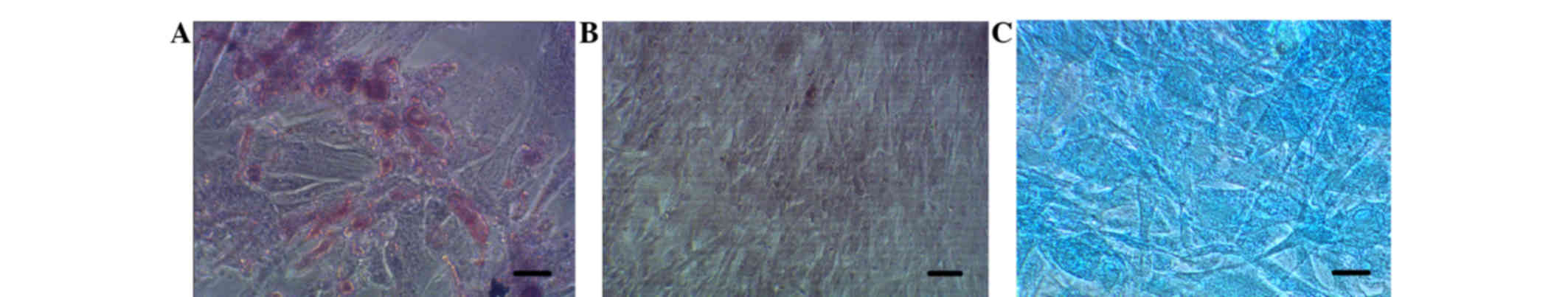
endotoxin units, respectively. To assess the differentiation
pluripotency of uMSCs, differentiation of these cells into
adipocytes, osteocytes and chondrocytes was induced. These
differentiations were confirmed by staining with Oil red O,
Alizarin red and Alcian blue, respectively (Fig. 2A-C, respectively). Taken together,
these results indicate that uMSCs were separated and cultured
successfully.
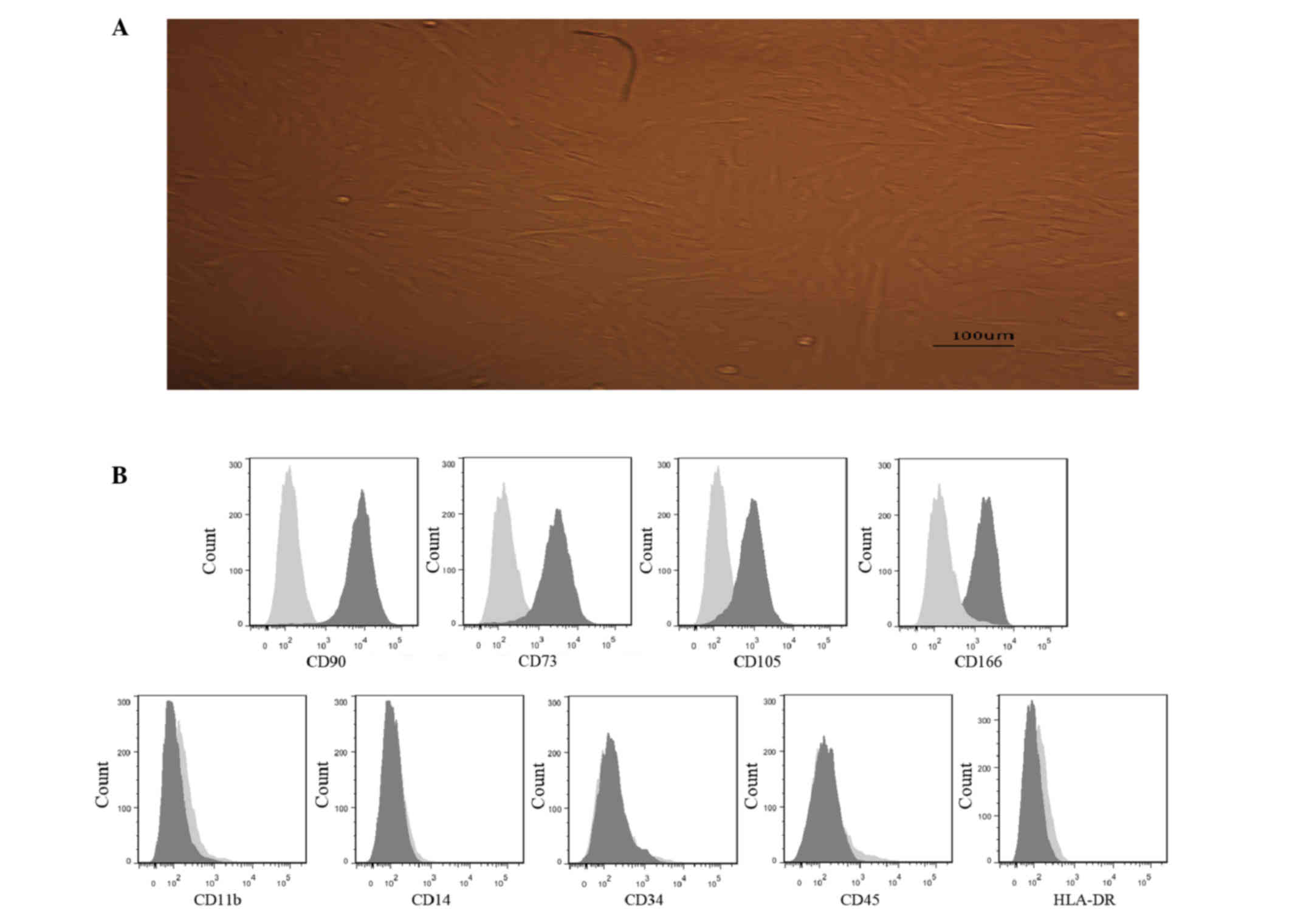 | Figure 1.Characteristics of the uMSCs. (A)
Image of uMSCs at a magnification of ×200. Scale bar=100 µm. (B)
Identification of uMSC immunophenotypes. The surface molecules of
uMSCs were detected using flow cytometry. The cultured umbilical
cord-derived cells consisted of a homogenous mesenchymal population
that were positive for CD73, CD90, CD105 and CD166, and were
negative for CD11b, CD14, CD34, CD45 and HLA-DR. uMSC, umbilical
mesenchymal stem cells; CD, cluster of differentiation; HLA-DR,
human leukocyte antigen-antigen D related; FITC, fluorescein
isothiocyanate; PE, phycoerythrin. |
Clinical outcomes
Case 1
The patient was male, 34 years old and had suffered
with lower back pain for 20 years. The bilateral articulatio coxae
were alternately painful with morning stiffness for ~20 min each
day. Physical examinations determined that the Schober's test was
positive, Patrick's test was positive, occiput to wall distance was
5 cm and thoracic mobility was 3 cm. As a result, BASDAI, BASMI and
BASFI scores were 2.95, 4.3 and 7, respectively. Computed
tomography indicated that the bilateral sacroiliac articular
surface was jagged and revealed sacroiliitis of AS. Laboratory
examinations included assessments of ESR, CRP levels and HLA-B27,
in which ESR was 10 mm/h, CRP was 0.6 mg/l and HLA-B27 was
positive. The patient was therefore diagnosed with AS, according to
the modified New York criteria for AS. The patient was infused with
8 IU uMSCs (1 IU=1×10⁷ uMSCs) initially and administered
SSZ (orally; 1,500 mg/day), thalidomide (orally, 100 mg daily) and
diclofenac sodium (orally, 75 mg daily) continuously. The patient
was then discharged the next day. Following 3 months, the patient
received a second infusion of uMSCs at a dose of 7 IU. Lower back
pain was relieved. The physical examination demonstrated that
Schober's test was negative, Patrick's test of the right side was
positive, but the left side was negative, occiput to wall distance
was 2.5 cm and thoracic mobility was 3 cm. BASDAI, BASMI and BASFI
scores were 0.6, 6 and 6, respectively. The laboratory examination
determined that CRP was 1 mg/l and ESR was 22 mm/h. Following 6
months, the patient felt that back pain had almost disappeared and
returned for examination. The ESR was 7 mm/h, CRP levels were 0.5
ng/l and BASDAI, BASMI and BASFI scores were 0.5, 6 and 6,
respectively. Schober's test and Patrick's test were both negative,
and the occipital to wall distance was 2.5 cm. After 1 year, the
patient had stopped all medication and remained in a stable
physical condition.
Case 2
The patient was male, 20 years old, with a 2-year
history of bilateral knee joint pain and lower back pain. The
physical examination demonstrated that Schober's test was positive,
the bilateral Patrick sign was positive; occiput to wall distance
was 3 cm and thoracic mobility was 3 cm. BASDAI, BASMI and BASFI
scores were 4.9, 5 and 57, respectively. CT indicated a change of
the bilateral hip joint in accordance with AS. Laboratory
examinations determined that HLA-B27 was positive, ESR was 53 mm/h
and CRP levels were 7.8 mg/l. The patient was diagnosed with AS,
according to the modified New York criteria for AS. The patient
received an infusion of 6 IU uMSCs. Treatment was continued, using
1 g SSZ three times per day, 100 mg thalidomide nightly and 60 mg
Loxoprofen tid. After 3 months, the patient felt that knee joint
and lower back pain were relieved and returned for examination. The
physical examination indicated that Schober's test was positive,
bilateral Patrick's test was negative, occiput to wall distance was
2 cm and Thoracic mobility was 3 cm. BASDAI, BASMI and BASFI scores
were 1.35, 5 and 9, respectively. The laboratory examination
determined that CRP levels were 0.3 mg/l and ESR was 3 mm/h. After
6 months, lower back pain had almost disappeared and laboratory
examinations were completed, with an ESR of 5 mm/h, CRP levels of
0.4 ng/l and BASDAI, BASMI and BASFI scores of 1.3, 8 and 8,
respectively. Bilateral Patrick's test was negative, but Schober's
test was positive. The occipital to wall distance was 2 cm. One
year later, the patient's physical condition remained stable and
treatment was ceased.
Case 3
The patient was female, 41 years old, with a 20-year
history of lumbosacral joint and left knee joint pain. Physical
examinations revealed that Schober's test was negative, the
bilateral Patrick's test was positive, occiput to wall distance was
0 cm and thoracic mobility was 3 cm. As a result, BASDAI, BASMI and
BASFI scores were 5.2, 10 and 37, respectively. CT indicated left
sacroilac articular chondritis and bilateral femoral head necrosis
II degree. Laboratory examinations demonstrated that HLA-B27 was
positive, ESR was 20 mm/h and CRP levels were 5 mg/l. The patient
was diagnosed with AS, according to the modified New York criteria
for AS. The patient received an infusion of 6.1 IU uMSCs. Treatment
was continued, with 1 g SSZ tid, 100 mg thalidomide quaque nocte
(qn) and 75 mg diclofenac sodium qn. Following 3 months, lower back
pain was relieved and the patient returned for a second uMSC
infusion at a dose of 6 IU. At this point, the physical examination
revealed that Schober's test was negative, left Patrick's test was
positive, right Patrick's test was negative, occiput to wall
distance was 0 cm and thoracic mobility was 3 cm. BASDAI, BASMI and
BASFI scores were 2.85, 5 and 34.5, respectively. The laboratory
examination demonstrated that CRP levels were 0.3 mg/l and ESR was
7 mm/h. After 6 months, the patient felt occasional lower back pain
and returned for examination. The results indicated that Schober's
test and Patrick's test were negative, occiput to wall distance was
0 cm and thoracic mobility was 3 cm. BASDAI, BASMI and BASFI scores
were 2.55, 6 and 30.2, respectively. The laboratory examination
demonstrated that CRP was 0.3 ng/l and ESR was 4 mm/h. One year
later, the patient stopped taking all medications, and the physical
condition of the patient remained stable.
Case 4
The patient was male, 44 years old, with a history
of lumbosacral joint pain and sternum pain for ~5 years. Physical
examination demonstrated that Schober's test and the bilateral
Patrick's test was positive. As a result, BASDAI, BASMI and BASFI
scores were 5.48, 2 and 52, respectively. CT indicated that the
bilateral sacroiliac articular surface was jagged and the articular
space was narrower, indicating sacroiliitis of AS. Laboratory
examination demonstrated that HLA-B27 was positive, ESR was 26 mm/h
and CRP levels were 3.7 mg/l. The patient was diagnosed with AS,
according to the modified New York criteria for AS and received an
infusion of 9.2 IU uMSCs. Treatment was continued with 0.5 g SSZ
tid and 100 mg thalidomide qn. Following 3 months, the patient
returned for examination. The physical examination demonstrated
that Schober's test and bilateral Patrick's test were positive.
BASDAI, BASMI and BASFI scores were 0.6, 4 and 5, respectively. The
laboratory examinations indicated that CRP levels were 0.9 mg/l and
ESR was 12 mm/h. This patient did now participate in the 6-month
follow up due to personal reasons. One year later, the patient's
physical condition remained stable and treatment was ceased.
Case 5
The patient was male, 17 years old, with a history
of lower back pain and alternate bilateral knee and hip joints
pain. Pain was more serious in the morning, with a short period of
morning stiffness, meaning that the patient was unable to attend
physical education classes. Physical examination demonstrated that
Schober's test was positive and bilateral Patrick's test was
negative. BASDAI, BASMI and BASFI scores were 4.9, 5 and 57,
respectively and CT indicated right sacroiliitis of AS. Laboratory
examinations indicated that HLA-B27 was positive, ESR was 22 mm/h
and CRP levels were 18.7 mg/l. The patient was diagnosed with AS,
according to the modified New York criteria for AS. The patient
received an infusion of 5.4 IU uMSCs. Treatment was continued with
0.25 g SSZ tid and 75 mg diclofenac sodium qd. After 3 months, the
patient felt that low back pain and knee joint pain was relieved
and returned for a second infusion of uMSCs at a dose of 6 IU. The
physical examination demonstrated that Schober's test and left
Patrick's test were negative and that right Patrick's test was
positive. Occiput to wall distance was 0 cm and thoracic mobility
was 3 cm. BASDAI, BASMI and BASFI scores were 4, 10 and 0,
respectively. The laboratory examination indicated that CRP levels
were 2.1 mg/l and ESR was 12 mm/h. Following 6 months, the patient
received a third infusion of uMSCs at a dose of 6 IU. The physical
examination demonstrated that Schober's test and bilateral
Patrick's test were negative. Occiput to wall distance was 0 cm and
thoracic mobility was 3 cm. BASDAI, BASMI and BASFI scores were 13,
10 and 0, respectively. The laboratory examination indicated that
CRP levels were 1.2 mg/l and ESR was 10 mm/h. The patient was able
to partake in physical education classes and undergo exercise. One
year later, the patient stopped taking all medications and physical
condition remained stable.
Analysis of results
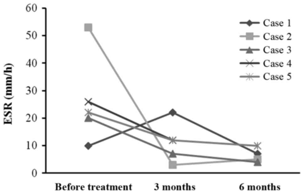
The ESR in 3 of the patients was markedly reduced
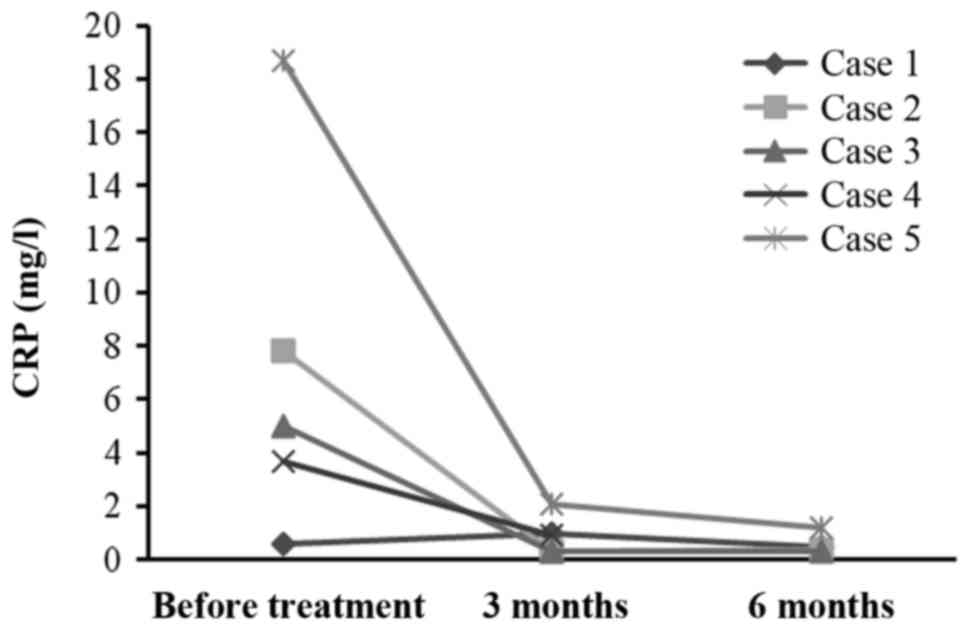
(Fig. 3). CRP levels were reduced in
1 patient and remained stable and were within normal limits (≤10
mg/l) in the other 4 patients (Fig.
4). The marked decline of BASDAI and BASFI scores indicated a
marked improvement in patients' spinal functions and symptoms. The
increase in BASMI scores indicated an improvement in spinal
movement of patients following treatment. The specific results were
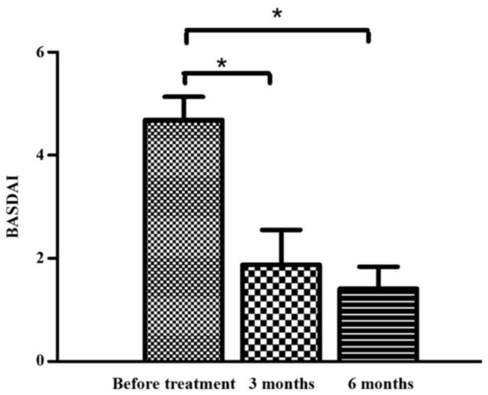
as follows: i) Changes in BASDAI prior to and following treatment:
There was a significant reduction in BASDAI in patients following 3
months treatment, compared with the results prior to treatment
(P<0.05; Table I; Fig. 5). There was also a significant
decrease in BASDAI in patients after 6 months treatment, compared
with the results prior to treatment (P<0.05; Table II; Fig.
5). There was no significant difference observed in BASDAI in
patients between 3 and 6 months following treatment. ii) Changes in
BASFI prior to and following treatment: There were no significant
differences in BASFI in patients observed prior to and following 3
or 6 months of treatment (both P>0.05; Tables I and II). iii) Changes in BASMI prior to and
following treatment: There were no significant differences in BASMI
observed in patients prior to and following 3 and 6 months of
treatment (P>0.05; Tables I and
II). No significant differences
were observed in the BASMI in patients following 6 months
treatment.
 | Table I.Comparison of BASDAI, BASFI and BASMI
prior to and following 3 months of treatment. |
Table I.
Comparison of BASDAI, BASFI and BASMI
prior to and following 3 months of treatment.
| Index | Before treatment
(n=5) | Following treatment
(n=5) | t | P-values |
|---|
| BASDAI | 4.686±0.999 | 1.880±1.499 | 4.205 | 0.014a |
| BASFI | 42.000±21.213 | 10.900±13.585 | 2.568 | 0.062 |
| BASMI | 5.260±2.922 | 6.000±2.345 | −0.45 | 0.676 |
| Table II.Comparison of BASDAI, BASFI and BASMI
prior to and following 6 months of treatment. |
Table II.
Comparison of BASDAI, BASFI and BASMI
prior to and following 6 months of treatment.
| Index | Prior to treatment
(n=4) | Following treatment
(n=4) | t | P-value |
|---|
| BASDAI | 4.488±1.035 | 1.413±0.847 | 10.054 | 0.002 |
| BASFI | 39.5002±23.269 | 11.050±13.211 | 1.987 | 0.141 |
| BASMI | 6.075±2.637 | 7.500±1.914 | −0.735 | 0.514 |
Safety evaluation
Following infusion of uMSCs, 60% of patients (3/5)
presented with a fever (body temperature, 38.5–39°C). One patient
had a temperature of 39°C and was administered 5 mg dexamethasone
(Qilu Pharmaceutical Co., Ltd., Shandong, China) via intravenous
injection and 25 mg promethazine hydrochloride (Qilu Pharmaceutical
Co., Ltd.) via intramuscular injection. The body temperature of
this patient then returned to normal. The temperature of the other
two patients remained normal and they did not require any
treatment. There were no other adverse reactions associated with
uMSC infusion during and following the transplantation. These
results indicate that uMSC infusion is safe for patients with
AS.
Discussion
AS is an autoimmune disease that leads to
progressive ankylosis of the vertebrae and ossification of
paravertebral ligaments (30). The
exact pathogenesis of AS remains unclear, however a number of
molecules are involved in the pathogenic process. TNF-α serves an
important role in pathogenesis, activating inflammation and
destroying the target tissue. Furthermore, IL-17 secretion from
induced by IL-23 serves an important role (31). It has been demonstrated that levels
of IL-17 and IL-23 in the serum of such patients is increased
compared with the healthy population (32). In addition,
CD4+CD25+CD127low Treg cells serve
a role in AS. It has been determined that the ratio of Treg in
patients with active AS is lower than in healthy controls (33). B cell subsets are also abnormal in
patients with AS (34). A previous
study indicated that there are various types of autoantibodies in
patients with AS, suggesting that autoantibodies are potentially
associated with the pathogenesis of AS (35).
As MSCs serve an important role in immunomodulation,
they are used to treat autoimmune diseases including systemic lupus
erythematous, multiple sclerosis and acute graft-verse-host disease
following allogeneic hematopoietic stem cell transplantation
(36). It has been demonstrated that
MSCs may favor the emergence of Th2 phenotype T cells with a potent
reduction in the Th1/Th17 cell-related cytokines TNF-a and IL-17
(37–39). MSCs are also able to suppress the
activity of human Th17 cells, inhibit the differentiation of native
CD4+ T cells into Th17 cells and significantly suppress
cytokine production, leading to the upregulation of Tregs (40,41). One
proposed mechanism is that MSCs induce the production of IL-10 by
plasmacytoid dendritic cells, which in turn may induce Tregs
(42,43). Due to their multi-differentiation
capacity, MSCs may induce the synthesis of proteoglycans and
restore disc structure and may also suppress B cell function
(44). In vitro studies have
demonstrated that MSCs may prevent B cells from activating,
proliferating and differentiating into immunoglobulin
(Ig)-producing cells (45,46). Furthermore, MSCs may be a potential
treatment for cardiovascular diseases, including ischemic heart
disease and peripheral artery disease (47). MSCs may differentiate into
insulin-producing cells and thus may be beneficial in the treatment
of type 1 diabetes mellitus, which leads to pancreas β cell
destruction and attenuates insulin production (48). MSC therapy may also be considered as
an effective treatment of hepatic diseases such as hepatic fibrosis
(49). Transplantation of MSCs is a
novel method of treatment for osteoarthritis, osteonecrosis and
bone regeneration (50–52). MSCs are able to differentiate into
neurons and therefore may be used for to treat neurodegenerative
diseases including Alzheimer's disease, amylotrophic lateral
sclerosis, Parkinson's disease and retinal diseases (53,54).
MSCs may be derived from both bone marrow and
umbilical cord. However, the collection of MSCs from the bone
marrow (BM) is invasive and the amount of MSCs in the BM is low
(55). The yield of BM-MSCs also
significantly decreases with donor age and the rate of BM-MSC
regeneration is markedly low (56).
uMSCs may be harvested without invasive procedures and the
percentage of MSCs in the umbilical cord blood is higher than in
the bone marrow (57,58). Furthermore, compared with BM-MSC,
there are fewer ethical issues associated with uMSCs. Therefore, in
the present study, patients with AS underwent transfusion with
uMSCs.
In the current study, patients were treated with
thalidomide, SSZ and NSAIDs for at least 4 weeks prior to MSC
transfusion. However, there were no marked improvements in the
symptoms of patients during this period. ESR, CRP levels and BASFI
scores were determined 1 day prior to transfusion to ensure that
any changes in the aforementioned indexes were caused by uMSC
transfusion. All patients experienced pain relief and the scores
assessing AS activity and severity were decreased following uMSC
infusion. A number of patients were able to perform certain
exercises following treatment, whereas these activities had been
limited prior to uMSC transfusion. Physical examinations
demonstrated that the majority of patients exhibited less marked
symptoms of AS. ESR and CRP levels returned to and remained within
normal ranges, suggesting that AS activity was reduced,
inflammation was inhibited and the disease was not progressing.
BASDAI and BASFI scores markedly decreased, indicating improved
function of articulation and alleviated symptoms. However, the
improvement in BASMI was not significant at any point, indicating
that spine mobility only improved slightly. The results of the
present study indicate that uMSC transplantation is both feasible
and safe and induces limited side effects.
There were a number of limitations of the present
study. The number of patients with AS included was very low, due to
the low morbidity of AS (~0.18% in Asia). One patient failed to
complete examinations due to personal reasons thus incomplete
information was collected, limiting statistical analysis. Levels of
IL-17, TNF-α, Ig and Treg was not measured, therefore no
comparisons were completed regarding the changes that occurred
following transfusion of uMSCs. The current study also did not
include a control group that did not receive an infusion, which
would have allowed a comparison. All the aforementioned limitations
should be noted and future studies should aim to ameliorate these
limitations.
Acknowledgements
The present study was supported by the following
grants: The key laboratory of kidney regenerative medicine of
Shandong University (2015QY002-07), the Project of Science and
Technology of Shenzhen City (JCYJ20150402105524048) and Natural
Science Foundation of Shandong Provence (ZR2011HQ042). The authors
would like to thank all the staff at the Department of Hematology
and Cellular therapy and Laboratory for Molecular Medicine of the
Second Hospital and Institute of Biotherapy for Hematological
Malignancies, Shandong University for their support and all the
patients included in the current study.
References
|
1
|
Singh A and Karrar S: The role of
intracellular organisms in the pathogenesis of inflammatory
arthritis. Int J Inflam. 2014:1587932014.PubMed/NCBI
|
|
2
|
Niu XY, Zhang HY, Liu YJ, Zhao D, Shan YX
and Jiang YF: Peripheral B-cell activation and exhaustion markers
in patients with ankylosing spondylitis. Life Sci. 93:687–692.
2013. View Article : Google Scholar : PubMed/NCBI
|
|
3
|
Lin Q, Gu JR, Li TW, Zhang FC, Lin ZM,
Liao ZT, Wei QJ, Cao SY and Li L: Value of the peripheral blood
B-cells subsets in patients with ankylosing spondylitis. Chin Med J
(Engl). 122:1784–1789. 2009.PubMed/NCBI
|
|
4
|
Schirmer M, Goldberger C, Würzner R,
Duftner C, Pfeiffer KP, Clausen J, Neumayr G and Falkenbach A:
Circulating cytotoxic CD8(+) CD28(−) T cells in ankylosing
spondylitis. Arthritis Res. 4:71–76. 2002. View Article : Google Scholar : PubMed/NCBI
|
|
5
|
Duftner C, Goldberger C, Falkenbach A,
Würzner R, Falkensammer B, Pfeiffer KP, Maerker-Hermann E and
Schirmer M: Prevalence, clinical relevance and characterization of
circulating cytotoxic CD4+CD28-T cells in ankylosing spondylitis.
Arthritis Res Ther. 5:R292–R300. 2003. View
Article : Google Scholar : PubMed/NCBI
|
|
6
|
Smith JA and Colbert RA: Review: The
interleukin-23/interleukin-17 axis in spondyloarthritis
pathogenesis: Th17 and beyond. Arthritis Rheumatol. 66:231–241.
2014. View Article : Google Scholar : PubMed/NCBI
|
|
7
|
Zambrano-Zaragoza JF, Agraz-Cibrian JM,
González-Reyes C, Durán-Avelar Mde J and Vibanco-Pérez N:
Ankylosing spondylitis: From cells to genes. Int J Inflam.
2013:5016532013.PubMed/NCBI
|
|
8
|
Hreggvidsdottir HS, Noordenbos T and
Baeten DL: Inflammatory pathways in spondyloarthritis. Mol Immunol.
57:28–37. 2014. View Article : Google Scholar : PubMed/NCBI
|
|
9
|
Scarpa R, Costa L, Atteno M, Del Puente A,
Caso F and Moll JM: Psoriatic arthritis: Advances in
pharmacotherapy based on molecular target. Expert Opin
Pharmacother. 14:2311–2313. 2013. View Article : Google Scholar : PubMed/NCBI
|
|
10
|
Fiocco U, Sfriso P, Oliviero F, Lunardi F,
Calabrese F, Scagliori E, Cozzi L, Di Maggio A, Nardacchione R,
Molena B, et al: Blockade of intra-articular TNF in peripheral
spondyloarthritis: Its relevance to clinical scores, quantitative
imaging and synovial fluid and synovial tissue biomarkers. Joint
Bone Spine. 80:165–170. 2013. View Article : Google Scholar : PubMed/NCBI
|
|
11
|
Toussirot E and Wendling D: Current
guidelines for the drug treatment of ankylosing spondylitis. Drugs.
56:225–240. 1998. View Article : Google Scholar : PubMed/NCBI
|
|
12
|
Sampaio-Barros PD, Costallat LT, Bertolo
MB, Neto JF and Samara AM: Methotrexate in the treatment of
ankylosing spondylitis. Scand J Rheumatol. 29:160–162. 2000.
View Article : Google Scholar : PubMed/NCBI
|
|
13
|
Dougados M, Béhier JM, Jolchine I, Calin
A, van der Heijde D, Olivieri I, Zeidler H and Herman H: Efficacy
of celecoxib, a cyclooxygenase 2-specific inhibitor, in the
treatment of ankylosing spondylitis: A six-week controlled study
with comparison against placebo and against a conventional
nonsteroidal antiinflammatory drug. Arthritis Rheum. 44:180–185.
2001. View Article : Google Scholar : PubMed/NCBI
|
|
14
|
Slobodin G, Rosner I, Rimar D, Boulman N,
Rozenbaum M and Odeh M: Ankylosing spondylitis: Field in progress.
Isr Med Assoc J. 14:763–767. 2012.PubMed/NCBI
|
|
15
|
Farge D, Terriou L, Badoglio M, Cras A,
Desreumaux P, Hadj-Khelifa S, Marjanovic Z, Moisan A, Dulery R,
Faucher C, et al: Autologous stem cell transplantation for
autoimmune diseases: Recommendations from the SFGM-TC. Pathol Biol
(Paris). 62:204–208. 2014.(In French). View Article : Google Scholar : PubMed/NCBI
|
|
16
|
Prevosto C, Zancolli M, Canevali P, Zocchi
MR and Poggi A: Generation of CD4+ or CD8+ regulatory T cells upon
mesenchymal stem cell-lymphocyte interaction. Haematologica.
92:881–888. 2007. View Article : Google Scholar : PubMed/NCBI
|
|
17
|
Di Ianni M, Del Papa B, De Ioanni M,
Moretti L, Bonifacio E, Cecchini D, Sportoletti P, Falzetti F and
Tabilio A: Mesenchymal cells recruit and regulate T regulatory
cells. Exp Hematol. 36:309–318. 2008. View Article : Google Scholar : PubMed/NCBI
|
|
18
|
Gu J, Lin CM, Gu W, Cai XZ, Li Z, Ren MM,
Sun X, Ni J, Shen LJ, Wu W, et al: Immunomodulatory effect of
UC-MSC on function of immunocytes of rats with collagen type II
induced arthritis. Zhongguo Shi Yan Xue Ye Xue Za Zhi. 22:166–170.
2014.(In Chinese). PubMed/NCBI
|
|
19
|
Wang D, Li J, Zhang Y, Zhang M, Chen J, Li
X, Hu X, Jiang S, Shi S and Sun L: Umbilical cord mesenchymal stem
cell transplantation in active and refractory systemic lupus
erythematosus: A multicenter clinical study. Arthritis Res Ther.
16:R792014. View
Article : Google Scholar : PubMed/NCBI
|
|
20
|
El-Jawhari JJ, El-Sherbiny YM, Jones EA
and McGonagle D: Mesenchymal stem cells, autoimmunity and
rheumatoid arthritis. QJM. 107:505–514. 2014. View Article : Google Scholar : PubMed/NCBI
|
|
21
|
Kong D, Zhuang X, Wang D, Qu H, Jiang Y,
Li X, Wu W, Xiao J, Liu X, Liu J, et al: Umbilical cord mesenchymal
stem cell transfusion ameliorated hyperglycemia in patients with
type 2 diabetes mellitus. Clin Lab. 60:1969–1976. 2014. View Article : Google Scholar : PubMed/NCBI
|
|
22
|
van der Linden S, Valkenburg HA and Cats
A: Evaluation of diagnostic criteria for ankylosing spondylitis. A
proposal for modification of the New York criteria. Arthritis
Rheum. 27:361–368. 1984. View Article : Google Scholar : PubMed/NCBI
|
|
23
|
Qu H, Liu X, Ni Y, Jiang Y, Feng X, Xiao
J, Guo Y, Kong D, Li A, Li X, et al: Laminin 411 acts as a potent
inducer of umbilical cord mesenchymal stem cell differentiation
into insulin-producing cells. J Transl Med. 12:1352014. View Article : Google Scholar : PubMed/NCBI
|
|
24
|
Garrett S, Jenkinson T, Kennedy LG,
Whitelock H, Gaisford P and Calin A: A new approach to defining
disease status in ankylosing spondylitis: The bath ankylosing
spondylitis disease activity index. J Rheumatol. 21:2286–2291.
1994.PubMed/NCBI
|
|
25
|
Calin A, Garrett S, Whitelock H, Kennedy
LG, O'Hea J, Mallorie P and Jenkinson T: A new approach to defining
functional ability in ankylosing spondylitis: The development of
the bath ankylosing spondylitis functional index. J Rheumatol.
21:2281–2285. 1994.PubMed/NCBI
|
|
26
|
Jenkinson TR, Mallorie PA, Whitelock HC,
Kennedy LG, Garrett SL and Calin A: Defining spinal mobility in
ankylosing spondylitis (AS). The bath AS metrology index. J
Rheumatol. 21:1694–1698. 1994.PubMed/NCBI
|
|
27
|
Thomas E, Silman AJ, Papageorgiou AC,
Macfarlane GJ and Croft PR: Association between measures of spinal
mobility and low back pain. An analysis of new attenders in primary
care. Spine (Phila Pa 1976). 23:343–347. 1998. View Article : Google Scholar : PubMed/NCBI
|
|
28
|
White FA: Physical Signs in Medicine and
Surgery: An Atlas of Rare, Lost and Forgotten Physical Signs.
Museum Press; Ocala, FL: pp. 1772009
|
|
29
|
Anderson JJ, Baron G, van der Heijde D,
Felson DT and Dougados M: Ankylosing spondylitis assessment group
preliminary definition of short-term improvement in ankylosing
spondylitis. Arthritis Rheum. 44:1876–1886. 2001. View Article : Google Scholar : PubMed/NCBI
|
|
30
|
Daikh DI and Chen PP: Advances in managing
ankylosing spondylitis. F1000Prime Rep. 6:782014. View Article : Google Scholar : PubMed/NCBI
|
|
31
|
Chen MH, Chen HA, Chen WS, Chen MH, Tsai
CY and Chou CT: Upregulation of BMP-2 expression in
peripheral-blood mononuclear cells by proinflammatory cytokines and
radiographic progression in ankylosing spondylitis. Mod Rheumatol.
25:913–918. 2015. View Article : Google Scholar : PubMed/NCBI
|
|
32
|
Wang X, Lin Z, Wei Q, Jiang Y and Gu J:
Expression of IL-23 and IL-17 and effect of IL-23 on IL-17
production in ankylosing spondylitis. Rheumatol Int. 29:1343–1347.
2009. View Article : Google Scholar : PubMed/NCBI
|
|
33
|
Ji W, Li H, Gao F, Chen Y, Zhong L and
Wang D: Effects of Tripterygium glycosides on interleukin-17 and
CD4+CD25+CD127low regulatory T-cell expression in the peripheral
blood of patients with ankylosing spondylitis. Biomed Rep.
2:517–520. 2014.PubMed/NCBI
|
|
34
|
Lin Q, Gu JR, Li TW, Zhang FC, Lin ZM,
Liao ZT, Wei QJ, Cao SY and Li L: Value of the peripheral blood
B-cells subsets in patients with ankylosing spondylitis. Chin Med J
(Engl). 122:1784–1789. 2009.PubMed/NCBI
|
|
35
|
Wright C, Sibani S, Trudgian D, Fischer R,
Kessler B, LaBaer J and Bowness P: Detection of multiple
autoantibodies in patients with ankylosing spondylitis using
nucleic acid programmable protein arrays. Mol Cell Proteomics.
11:M9.003842012. View Article : Google Scholar : PubMed/NCBI
|
|
36
|
Doğan SM, Kılınç S, Kebapçı E, Tuğmen C,
Gürkan A, Baran M, Kurtulmuş Y, Olmez M and Karaca C: Mesenchymal
stem cell therapy in patients with small bowel transplantation:
Single center experience. World J Gastroenterol. 20:8215–8220.
2014. View Article : Google Scholar : PubMed/NCBI
|
|
37
|
Ren G, Zhang L, Zhao X, Xu G, Zhang Y,
Roberts AI, Zhao RC and Shi Y: Mesenchymal stem cell-mediated
immunosuppression occurs via concerted action of chemokines and
nitric oxide. Cell Stem Cell. 2:141–150. 2008. View Article : Google Scholar : PubMed/NCBI
|
|
38
|
De Miguel MP, Fuentes-Julián S,
Blázquez-Martínez A, Pascual CY, Aller MA, Arias J and
Arnalich-Montiel F: Immunosuppressive properties of mesenchymal
stem cells: Advances and applications. Curr Mol Med. 12:574–591.
2012. View Article : Google Scholar : PubMed/NCBI
|
|
39
|
Bai L, Lennon DP, Eaton V, Maier K, Caplan
AI, Miller SD and Miller RH: Human bone marrow-derived mesenchymal
stem cells induce Th2-polarized immune response and promote
endogenous repair in animal models of multiple sclerosis. Glia.
57:1192–1203. 2009. View Article : Google Scholar : PubMed/NCBI
|
|
40
|
Gao W, Thompson L, Zhou Q, Putheti P,
Fahmy TM, Strom TB and Metcalfe SM: Treg versus Th17 lymphocyte
lineages are cross-regulated by LIF versus IL-6. Cell Cycle.
8:1444–1450. 2009. View Article : Google Scholar : PubMed/NCBI
|
|
41
|
Awasthi A and Kuchroo VK: Th17 cells: From
precursors to players in inflammation and infection. Int Immunol.
21:489–498. 2009. View Article : Google Scholar : PubMed/NCBI
|
|
42
|
Aggarwal S and Pittenger MF: Human
mesenchymal stem cells modulate allogeneic immune cell responses.
Blood. 105:1815–1822. 2005. View Article : Google Scholar : PubMed/NCBI
|
|
43
|
Maccario R, Podestà M, Moretta A, Cometa
A, Comoli P, Montagna D, Daudt L, Ibatici A, Piaggio G, Pozzi S, et
al: Interaction of human mesenchymal stem cells with cells involved
in alloantigen-specific immune response favors the differentiation
of CD4+ T-cell subsets expressing a regulatory/suppressive
phenotype. Haematologica. 90:516–525. 2005.PubMed/NCBI
|
|
44
|
Mwale F, Wang HT, Roughley P, Antoniou J
and Haglund L: Link N and mesenchymal stem cells can induce
regeneration of the early degenerate intervertebral disc. Tissue
Eng Part A. 20:2942–2949. 2014. View Article : Google Scholar : PubMed/NCBI
|
|
45
|
Rafei M, Hsieh J, Fortier S, Li M, Yuan S,
Birman E, Forner K, Boivin MN, Doody K, Tremblay M, et al:
Mesenchymal stromal cell-derived CCL2 suppresses plasma cell
immunoglobulin production via STAT3 inactivation and PAX5
induction. Blood. 112:4991–4998. 2008. View Article : Google Scholar : PubMed/NCBI
|
|
46
|
Healy ME, Bergin R, Mahon BP and English
K: Mesenchymal stromal cells protect against caspase 3-mediated
apoptosis of CD19(+) peripheral B cells through contact-dependent
upregulation of VEGF. Stem Cells Dev. 24:2391–2402. 2015.
View Article : Google Scholar : PubMed/NCBI
|
|
47
|
Tao H, Han Z, Han ZC and Li Z:
Proangiogenic features of mesenchymal stem cells and their
therapeutic applications. Stem Cells Int. 2016:13147092016.
View Article : Google Scholar : PubMed/NCBI
|
|
48
|
Hashemian SJ, Kouhnavard M and
Nasli-Esfahani E: Mesenchymal stem cells: Rising concerns over
their application in treatment of type one diabetes mellitus. J
Diabetes Res. 2015:6751032015. View Article : Google Scholar : PubMed/NCBI
|
|
49
|
Eom YW, Shim KY and Baik SK: Mesenchymal
stem cell therapy for liver fibrosis. Korean J Intern Med.
30:580–589. 2015. View Article : Google Scholar : PubMed/NCBI
|
|
50
|
Ham O, Lee CY, Kim R, Lee J, Oh S, Lee MY,
Kim J, Hwang KC, Maeng LS and Chang W: Therapeutic potential of
differentiated mesenchymal stem cells for treatment of
osteoarthritis. Int J Mol Sci. 16:14961–14978. 2015. View Article : Google Scholar : PubMed/NCBI
|
|
51
|
Wang C, Wang Y, Meng HY, Yuan XL, Xu XL,
Wang AY, Guo QY, Peng J and Lu SB: Application of bone marrow
mesenchymal stem cells to the treatment of osteonecrosis of the
femoral head. Int J Clin Exp Med. 8:3127–3135. 2015.PubMed/NCBI
|
|
52
|
Asatrian G, Pham D, Hardy WR, James AW and
Peault B: Stem cell technology for bone regeneration: Current
status and potential applications. Stem Cells Cloning. 10:39–48.
2015.
|
|
53
|
Ullah I, Subbarao RB and Rho GJ: Human
mesenchymal stem cells-current trends and future prospective.
Biosci Rep. 35(pii): e001912015.PubMed/NCBI
|
|
54
|
Ng TK, Fortino VR, Pelaez D and Cheung HS:
Progress of mesenchymal stem cell therapy for neural and retinal
diseases. World J Stem Cells. 6:111–119. 2014. View Article : Google Scholar : PubMed/NCBI
|
|
55
|
Secunda R, Vennila R, Mohanashankar AM,
Rajasundari M, Jeswanth S and Surendran R: Isolation, expansion and
characterisation of mesenchymal stem cells from human bone marrow,
adipose tissue, umbilical cord blood and matrix: A comparative
study. Cytotechnology. 67:793–807. 2015. View Article : Google Scholar : PubMed/NCBI
|
|
56
|
Bieback K, Kern S, Klüter H and Eichler H:
Critical parameters for the isolation of mesenchymal stem cells
from umbilical cord blood. Stem Cells. 22:625–634. 2004. View Article : Google Scholar : PubMed/NCBI
|
|
57
|
Lu LL, Liu YJ, Yang SG, Zhao QJ, Wang X,
Gong W, Han ZB, Xu ZS, Lu YX, Liu D, et al: Isolation and
characterization of human umbilical cord mesenchymal stem cells
with hematopoiesis-supportive function and other potentials.
Haematologica. 91:1017–1026. 2006.PubMed/NCBI
|
|
58
|
Wang Y, Li Y, Song L, Li Y, Jiang S and
Zhang S: The transplantation of Akt-overexpressing amniotic
fluid-derived mesenchymal stem cells protects the heart against
ischemia-reperfusion injury in rabbits. Mol Med Rep. 14:234–242.
2016.PubMed/NCBI
|