Introduction
Non-alcoholic fatty liver disease (NAFLD) consists
of a spectrum of illnesses ranging from simple hepatic steatosis to
non-alcoholic steatohepatitis (NASH) and irreversible cirrhosis
(1). This syndrome has become the
most common cause of liver disease (2). Indeed, NAFLD is currently recognized as
an aetiological factor in numerous conditions previously labelled
as cryptogenic cirrhosis (3). To
date, the pathogenesis and progression of NAFLD are not fully
understood, while current therapeutic treatment methods require
improvement (1,4). Initial theories of the pathogenesis of
NAFLD/NASH were based on the ‘2-hit’ hypothesis. Briefly, the
‘first hit’ involves hepatic triglyceride (TG) accumulation or
steatosis, while the ‘second hit’ is usually linked to mediators,
including inflammatory cytokines and oxidative stress (1). However, there is an increasing
recognition of the function that free fatty acids (FFA) have in
directly promoting liver injury, leading to the modification of
this theory (1). Additionally, a
‘third-hit’ has been included to reflect the inadequate hepatocyte
proliferation (1).
In the progression of chronic liver injury, the
development of fibrosis/cirrhosis depends on the efficacy of
hepatocyte regeneration (5). Hence,
cell death with impaired proliferation of hepatocyte progenitors
represents the proposed ‘third hit’ in NAFLD pathogenesis (5). Furthermore, hepatic TG accumulation may
occur as a result of increased fat synthesis and delivery, as well
as decreased export and/or oxidation of fat (6). Additionally, inflammatory cytokines,
including tumour necrosis factor (TNF)-α, interleukin (IL)-6 and
IL-8, mediate steatohepatitis in patients with NAFLD (7) and may be regulated by the peroxisome
proliferator-activated receptor (PPAR)-γ. PPAR-γ may be activated
by the PPARγ coactivator (PGC)-1α following sirtuin (Sirt)-1
activation (8).
Chinese herbal medicine (CHM), which has been
traditionally used in China and other parts of Asian countries for
thousands of years, is now spreading worldwide (9). A specific and basic feature of Chinese
medicine is the use of formulas containing several herbs (herbal
cocktail) to ameliorate a series of abnormal symptoms associated
with a particular disease. Gegen Qinlian decoction (GGQLD), a
classical formula from the Treatise on Febrile Diseases, is one of
the well-known traditional Chinese medicines and consists of Kudzu
root, Rhizoma coptidis, Scutellaria baicalensis
Georgi and honey-fried liquorice root (10). Furthermore, it is widely used to
treat diarrhoea (11). In previous
studies, it was revealed that GGQLD improved diabetes (12,13) and
had an anti-inflammatory effect based on its constituents (14–16).
Furthermore, glucose and lipid metabolism disorder and inflammation
are important in NAFLD. A previous study demonstrated that GGQLD
therapeutically managed NAFLD by improving the PPARγ-mediated
regulation of lipids and suppression of inflammation (17). However, the mechanisms by which GGQLD
triggers PPARγ are unknown, including whether it has a potential
Sirt1 agonist activity. Therefore, following the guidelines of the
traditional Chinese medicine theory of ‘same treatment for
different diseases’ and based on the ameliorating effects of PPARγ
in NAFLD management (17), it was
hypothesized that GGQLD could improve NAFLD as a Sirt1 agonist. In
the present study, this hypothesis was investigated.
Materials and methods
Preparation of GGQLD and resveratrol
(Resl)
GGQLD granules were provided by the Department of
Pharmacy of Dongfang Hospital, Beijing University of Chinese
Medicine (Beijing, China). The granules consisted of the following
ingredients of the Gegen Qinlian formula: Kudzu root, Rhizoma
coptidis, Scutellaria baicalensis Georgi and main
liquorice at 24, 9, 9 and 6 g, respectively. Resl was purchased
from Zelangyiy Co., Ltd., (Nanjing, China).
Animals and treatment
A total of 30 male Sprague-Dawley rats (7 weeks old;
weight, 200±20 g) were supplied by Vital River Laboratory Animal
Technology Co., Ltd. (Beijing, China). All animal experimental
procedures were approved by the Animal Ethics Committee of Beijing
University of Chinese Medicine under the guidelines issued by
Regulations of Beijing Laboratory Animal Management (18). The rats were maintained on a 12-h
light/dark cycle under a constant temperature (22±2°C) and humidity
of 50±10% with ad libitum access to standard chow diet
(control group) or a high-fat diet (HFD; 34% fat, 19% protein and
47% carbohydrate by energy composition; n=6 per group) for 8 weeks
to induce NAFLD. Additionally, the GGQLD granules and Resl were
dissolved in 100 ml of distilled water, and stored at 2–8°C until
further use. The five groups of animals (n=6) were treated orally
for 8 weeks as follows: GGQLD low- and high-dose (GGQLD-L and
GGQLD-H) groups treated with 5.04 and 10.08 g/kg/day GGQLD,
respectively; Resl group, treated with 400 mg/kg/day Resl; and the
HFD model and control groups were treated with 10 ml/kg/day saline
for 8 weeks from the beginning. The GGQLD and Resl groups were
administered the respective treatments at the beginning of the HFD
feeding, and the control received the standard chow diet.
Determination of metabolic parameters
and liver enzymes
At the end of the treatment, the animals were
anaesthetized using 4% chloral hydrate (Sinopharm Chemical Reagent
Co. Ltd, Shanghai, China) after a 12-h overnight fast and blood
samples were collected from the abdominal aorta of rats. The
fasting serum TGs (Jing 20142401132), total cholesterol (TC, Jing
20162400910), high-density lipoprotein cholesterol (HDL-C, Jing
20152400950) and low-density lipoprotein cholesterol (LDL-C, Jing
20142400518) as well as fasting serum alanine aminotransferase
(ALT, Jing 20142401158) and aspartate aminotransferase (AST, Jing
20142401158) were analysed using ELISA kits (BioSino Biotechnology
and Science, Inc., Beijing, China).
Histological analysis
Paraffin sections of the liver tissues were fixed
with 10% formaldehyde solution for 48 h at room temperature, and
the tissue slices (thickness, 4 µm) were prepared for hematoxylin
staining for 10 min and eosin staining for 2 min at room
temperature. Hematoxylin and eosin (H&E) were purchased from
Beijing Zeping Bioscience & Technologies Co., Ltd. (Beijing,
China). The Oil Red O (ORO; Sigma-Aldrich; Merck KGaA, Darmstadt,
Germany) staining was performed with freshly frozen liver tissues
(−196°C, liquid nitrogen) for 3 min at room temperature. The
H&E- and ORO-stained slides were visualized using a light
microscope in order to investigate architecture of the liver and
hepatic lipid droplets. The liver tissue samples from 3–5 rats from
each group were prepared, stained and the images were subsequently
captured using an Olympus digital camera (BX40; Olympus
Corporation, Japan) and the NIS Element SF 4.00.06 software (Nikon
Corporation, Tokyo, Japan).
Western blotting of Sirt1, PGC-1α and
nuclear factor κ-light-chain-enhancer of activated B cells (NF-κB)
p65
Proteins in 100 mg liver tissue homogenate were
extracted using ice-cold tissue lysis buffer (WB0006; TDY Biotech
Co., Ltd., Beijing, China) at 4°C in an electric tissue homogenizer
at 250 × g for 5 sec, 5 times at 5 sec intervals, then incubated on
ice for for 20 min. The supernatant was extracted by centrifugation
at 12,000 × g for 20 min at 4°C. Protein concentrations were
determined using a bicinchoninic acid protein assay kit (Promega
Corporation, Madison, WI, USA). Samples (30 µg) were separated by
10% SDS-PAGE and transferred onto polyvinylidene difluoride
membranes. The membranes were then incubated in blocking buffer (5%
skimmed milk powder) for 2 h prior to the addition of primary
antibodies, which were then incubated at 4°C overnight. The
membranes were immunoblotted with primary antibodies against Sirt1
(1:2,000; ab104833; Abcam, Cambridge, UK), PGC-1α (1:1,000;
sc-13067; Santa Cruz Biotechnology, Inc., Dallas, TX, USA), NF-κB
(p65; 1:2,000; TDY076) and β-actin (1:10,000; TDY041; both TDY
Biotech Co., Ltd.) at 4°C overnight. Peroxidase-conjugated goat
anti-rabbit secondary antibodies (1:20,000; cat. no. S001; TDY
Biotech, Co., Ltd.) were incubated at room temperature for 2 h and
an ECL detection system (EMD Millipore, Billerica, MA, USA) was
used according to routine methods (19). The intensities of the protein bands
were analysed using Gel-Pro 3.2 software (Media Cybernetics, Inc.,
Rockville, MD, USA). β-actin protein was used as the internal
control to normalize the protein loading. The experiment was
performed in triplicate.
Reverse transcription quantitative
polymerase chain reaction (RT-qPCR) for Sirt1, PGC-1α and NF-κB
mRNA expression
Total RNA was isolated from the liver samples using
TRIzol reagent (Invitrogen; Thermo Fisher Scientific, Inc.,
Waltham, MA, USA) and subsequently reverse-transcribed at 42°C for
60 min using the oligodT primers and SuperScript Reverse
Transcriptase (Invitrogen; Thermo Fisher Scientific, Inc.). The RT
reaction was performed with 1 µg of total RNA in a 12-µl reaction
using a standard cDNA synthesis kit (CWBIO, Beijing, China),
according to the manufacturer's instructions. The qPCR primer
sequences were as follows: Sirt1, forward
5′-CCAGATTTCAAGGCTGTTGGTTCC-3′ and reverse
5′-CCACAGGAACTAGAGGATAAGGCGT-3′; PGC-1α, forward
5′-TTCAGTGTCACCACCGAAATCCTTAT-3′ and reverse
5′-AGAGGATCTACTGCCTGGGGACC-3′; NF-κB, forward
5′-ACGATCTGTTTCCCCTCATC-3′ and reverse 5′-TGCTTCTCTCCCCAGGAATA-3′;
and GAPDH, forward 5′-TGGAGTCTACTGGCGTCTT-3′ and reverse
5′-TGTCATATTTCTCGTGGTTCA-3′. For each qPCR, a TaqMan®
Real-Time PCR assay (Thermo Fisher Scientific, Inc.) was used and
the typical thermal cycling conditions were an initial activation
step at 95°C for 5 min, followed by 45 cycles of amplification:
94°C for 15 sec, 60°C for 30 sec, 72°C for 30 sec and a final
extension of 72°C for 5 min. To compare the Sirt1, PGC-1α and NF-κB
mRNA levels, the cDNA concentrations were normalized to GAPDH PCR
products and the data were analysed using the 2−ΔΔCq
method (20).
Statistical analysis
Data were expressed as the mean ± standard deviation
unless otherwise indicated. The data were analysed using a one-way
analysis of variance and a post hoc test. Statistical analysis was
performed using SPSS v17.0 (IBM Corp, Armonk, NY, USA). P<0.05
was considered to indicate a statistically significant
difference.
Results
Effect of GGQLD on lipid metabolism
and liver enzymes in HFD rats
All rats tolerated the experimental procedures well
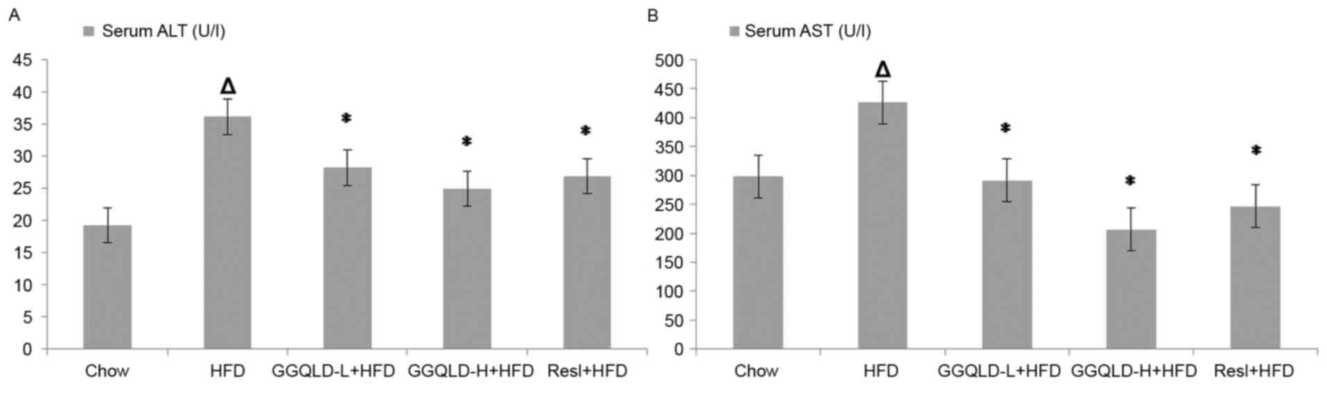
and no mortalities occurred during the study. The serum ALT and AST
(36.10±3.92 and 425.85±13.84 U/l, respectively) concentrations in
the HFD-fed rats were significantly higher than those in the
chow-fed rats (19.27±3.36 and 298.10±5.13 U/l, respectively;
P<0.01). Treatment with GGQLD-L and GGQLD-H significantly
attenuated ALT levels (28.20±3.01 and 24.98±1.55 U/l, respectively;
P<0.01) and AST (291.24±22.36 and 206.68±20.48 U/l,
respectively; P<0.01) compared with the HFD-fed rats. Similarly,
Resl also significantly reduced the ALT and AST levels (26.88±1.45
and 246.66±20.47 U/l, respectively) compared with rats fed with HFD
(P<0.01; Fig. 1).
The HDL-C levels rose significantly in the GGQLD-H
and Resl groups (1.90±0.10 and 1.30±0.11 mmol/l, respectively)
compared with those in the HFD model group (0.95±0.04 mmol/l;
P<0.01). Furthermore, GGQLD-H also decreased the LDL-C
concentration (0.98±0.15 mmol/l) compared with the HFD group, but
not significantly (P>0.05), whereas the decrease in the GGQLD-L
and Resl groups (0.79±0.09 and 0.78±0.09 mmol/l, respectively) was
significant compared to that of the HFD model group (1.07±0.20
mmol/l; P<0.01). Treatment with GGQLD-L, GGQLD-H and Resl
significantly attenuated the elevated TC (1.94±0.39, 1.53±0.11 and
1.63±0.13 mmol/l, respectively) compared with that of the HFD group
(2.81±0.79 mmol/l; P<0.05; Fig.
2). Treatment with GGQLD-L, GGQLD-H and Resl had no significant
effect on serum TG levels compared with those in the HFD group.
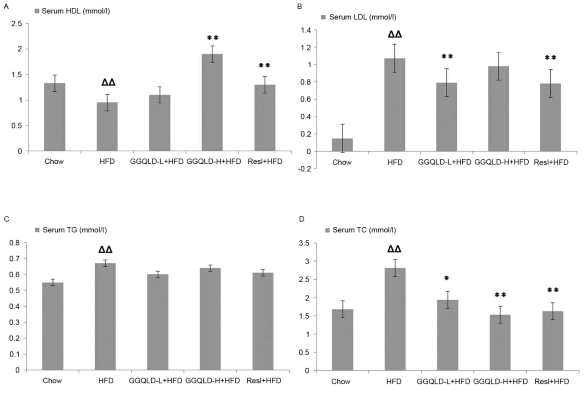 | Figure 2.Effect of GGQLD on the concentration
of HDL, LDL, TG and TC. (A) HDL, (B) LDL, (C) TG and (D) TC levels
in serum. ∆∆P<0.01 vs. chow; *P<0.05 and
**P<0.01 vs. HFD. GGQLD, Gegen Qinlian decoction; TG,
triglyceride; TC, total cholesterol; HDL, high-density lipoprotein;
LDL, low-density lipoprotein; HFD, high-fat diet; GGQLD-L, Gegen
Qinlian decoction low dose; GGQLD-H, Gegen Qinlian decoction high
dose; Resl, resveratrol. |
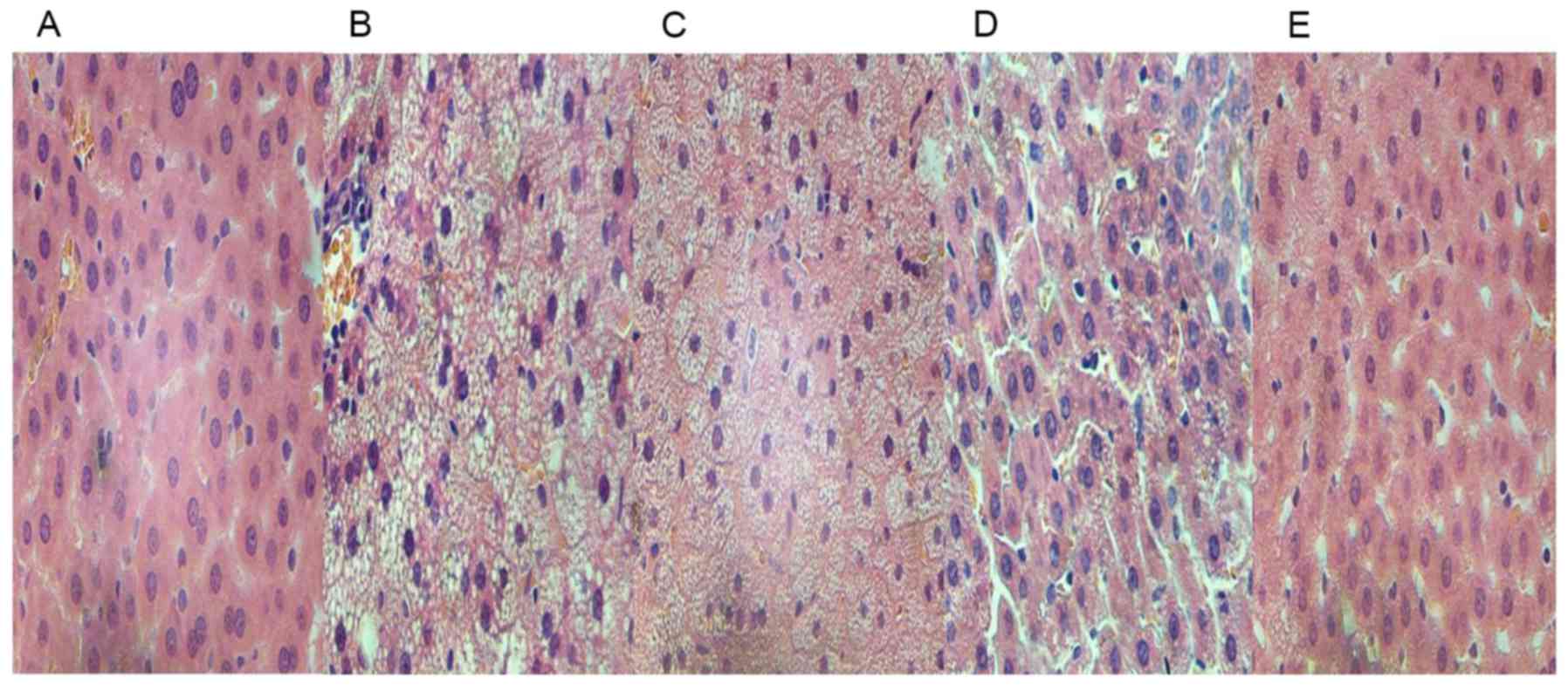
Histopathological changes in rats
An increase in the number of H&E-positive
hepatocytes was evident in the HFD rats compared to those in the
chow group (Fig. 3A and B). The
H&E-stained images also exhibited steatosis and infiltration
with inflammatory cells in the intercellular substance (Fig. 3B). The treatment of HFD rats with
GGQLD and Resl reduced the fat deposits in the liver (Fig. 3C-E) and the GGQLD-treated rats
revealed histological features similar to the chow group with
little steatosis (Fig. 3C and
D).
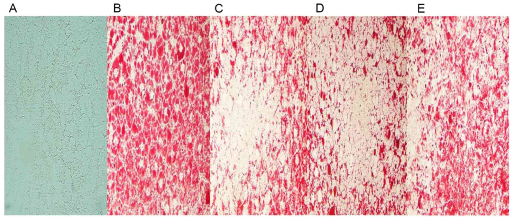
Furthermore, few lipid droplets were detected in the
liver sections from the chow group in ORO staining analysis
(Fig. 4A). Compared to that of
HFD-fed model rats (Fig. 4B), GGQLD
and Res1 markedly restrained the deposition of lipid droplets in
hepatocytes (Fig. 4C-E).
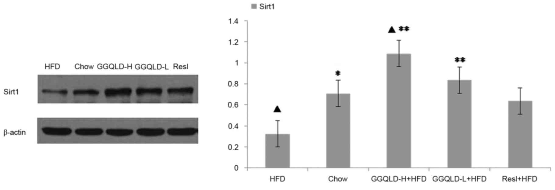
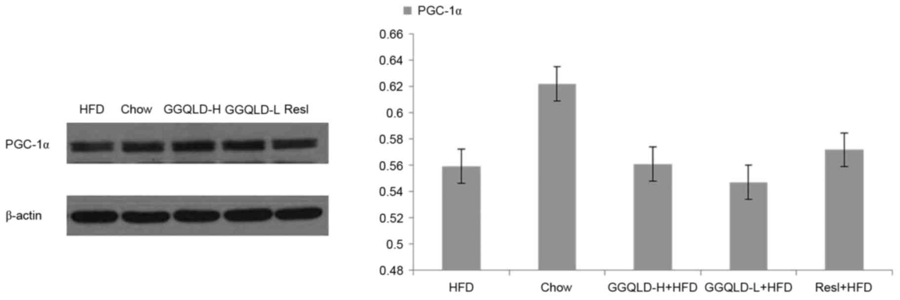
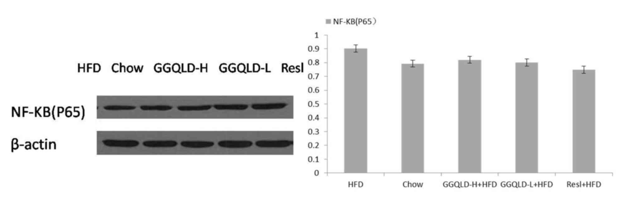
GGQLD regulates Sirt1, PGC-1α and
NF-κB (p65) expression in rats with HFD-induced NAFLD
The possibility of GGQLD having a regulatory effect
on Sirt1, PGC-1α and NF-κB (p65) expression was investigated. As
demonstrated in Fig. 5, the Sirt1
protein level was significantly decreased in the HFD group
(P<0.05) compared with the chow group. Notably, both GGQLD
groups significantly improved the Sirt1 level compared with the HFD
group (P<0.01; Fig. 5). Resl also
improved the Sirt1 level compared with the HFD group, however this
difference was not significant. As demonstrated in Figs. 6 and 7, although the PGC-1α and NF-κB (p65)
levels were improved by GGQLD-L, GGQLD-H and Resl, the effect was
not significant (P>0.05 vs. HFD group).
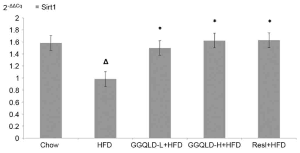
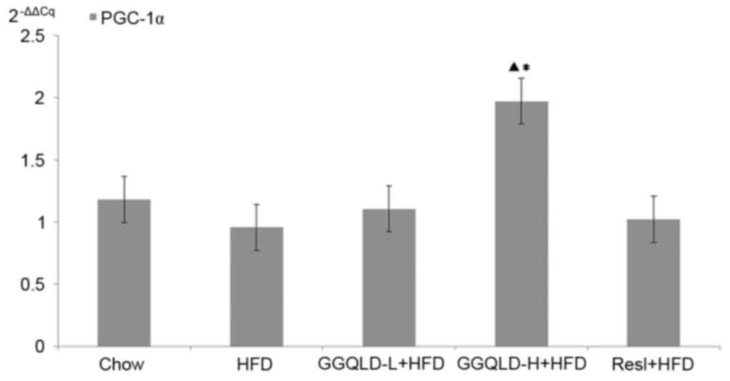
Validation of GGQLD effects on Sirt1,
PGC-1α and NF-κB gene expression using RT-qPCR
To confirm the action of GGQLD on liver Sirt1,
PGC-1α and NF-κB protein expression levels, their gene expression
levels were also measured. Sirt1 gene expression in the HFD group
significantly decreased compared with that of the chow group
(P<0.05; Fig. 8). Notably,
GGQLD-L, GGQLD-H and Resl significantly increased Sirt1 gene
expression compared with the HFD group (P<0.05; Fig. 8). As demonstrated in Fig. 9, PGC-1α gene expression in the HFD
group decreased slightly compared with that of the chow group, but
not significantly (P>0.05). However, a significant increase in
PGC-1α gene expression was evident in the GGQLD-H group (P<0.01)
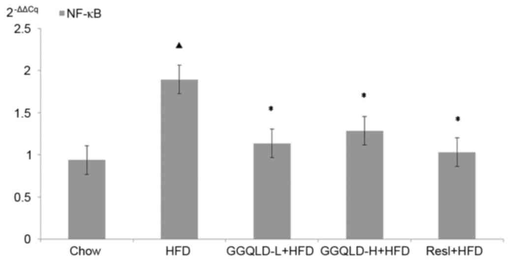
compared with the HFD group. Furthermore, NF-κB gene expression in
the HFD rat group increased significantly compared with that of the
chow group (P<0.01; Fig. 10).
Notably, GGQLD and Resl significantly decreased the NF-κB gene
expression compared with the HFD group (P<0.01; Fig. 10).
Discussion
NAFLD, which is a multi-factorial disorder
associated with a variety of genetic and environmental contributory
factors, is considered to be the most common cause of liver disease
(21). Whether the initial ‘2-hit
hypothesis’ or ‘3-hit hypothesis’ is applied in determining the
etiology, insulin resistance, oxidative stress and inflammatory
cascades are believed to serve integral roles in the pathogenesis
and progression (22). The presence
of steatosis induced by the overaccumulation of FFA/TGs and
cholesterol is closely associated with chronic hepatic inflammation
(23), which is partly mediated by
the activation of the inhibitor of the NF-κB kinase subunit β/NF-κB
signalling pathway. In murine models of HFD-induced steatosis,
increased NF-κB activity is associated with elevated hepatic
expression of inflammatory cytokines, including TNF-α and
activation of Kupffer cells (23).
PPARγ is one of three subtypes of the PPARs. Its agonists stimulate
adipose tissue to absorb and store FFAs, and thereby, inhibits
liver fatty acid synthesis (24) by
activating adenosine monophosphate (AMP)-activated protein kinase
(AMPK). PPARγ not only has anti-inflammatory effects but also
effectively improves insulin resistance (25). The activation of PPARγ in the immune
system modulates the inflammatory response (26). Furthermore, PGC-1α is a key
transcriptional regulator of energy homeostasis, and is important
in modulating liver fatty acid oxidation (27). The induction of this coactivator has
been documented in experimental models of obesity and diabetes
(28). The activation of PGC-1α
increased the expression of genes that are critical to
gluconeogenesis, fatty acid oxidation, lipid transport and
mitochondrial biogenesis (29–31).
PGC-1α is not only an important coactivator in liver
biology, but may be activated by Sirt1. Sirt1, which is the
receptor for Resl, targets numerous proteins, including PPARγ,
PGC-1α, uncoupling protein 2 and NF-κB, which serve key roles in
various metabolic disorders (32).
Furthermore, they lead to the activation of mitochondrial fatty
acid oxidation genes. At present, accumulating evidence has
indicated an important role for Sirts (33). In total, seven Sirts (Sirt1-7) have
been identified in mammals, of which the nuclear-located Sirt1 is
the closest homologue of the Sir2 protein, based on a
highly-conserved family of nicotinamide adenine
dinucleotide+-dependent enzymes that are easily
activated by Resl (34). Sirt1 has
been demonstrated to increase genomic stability and cellular stress
resistance, and it regulates energy metabolism and cellular
senescence via deacetylation of target proteins, including p53,
forkhead box transcription factors and PGC-1α (35–38). It
maintains the oxidation of fatty acids at low glucose
concentrations and is a regulator of PGC-1α, which activates PPARγ
and induces the transcription of metabolically relevant genes for
the oxidation of mitochondrial fatty acid (8). In recent years, numerous studies
(39,40) have concluded that Sirt1 activation
has a negative regulatory effect on the inflammatory processes. One
of the key proteins in these processes is NF-κB, which critically
modulates DNA transcription in inflammatory, infectious and
apoptotic processes (41). The
dysregulation of NF-κB may lead to inflammatory and autoimmune
diseases. Increasing evidence has revealed that NF-κB activation
contributes to the pathogenesis of NAFLD/NASH and the development
of hepatocellular carcinoma (23).
The activation of Sirt1 deacetylates the RelA/p65 subunit and
thereby inhibits NF-κB signalling (42,43).
An animal model of HFD-induced NAFLD has been widely
used to identify the pathogenesis and evaluate novel treatments for
NAFLD (44,45). In the present study, 8 weeks of an
HFD-induced fatty liver disease in Sprague-Dawley rats, in which
key biochemical features and histological abnormalities were
revealed by H&E- and ORO-stained liver samples, were consistent
with existing reports (44,46). Although studies (47) have focused on therapies for NAFLD, no
pharmacological agents have been approved for its treatment.
Therefore, the majority of the clinical efforts have currently
shifted their focus to strategies associated with metabolic
syndromes, namely obesity, diabetes, dyslipidaemia and hypertension
(48). Other interventions are
directed at specific pathways that are potentially involved in the
pathogenesis of NAFLD, including oxidative stress, insulin
resistance and pro-inflammatory cytokines (49).
Numerous studies have used adipocytes to demonstrate
that Resl has an anti-obesity potential by decreasing adipocyte
proliferation, inducing adipocyte apoptosis, inhibiting
preadipocyte differentiation, decreasing lipogenesis and promoting
lipolysis and fatty acid β-oxidation (50–54).
These effects may be mediated by the central regulators of
adipogenesis, lipogenesis and fatty acid β-oxidation, including the
aforementioned AMPK, Sirt1 and PGC-1α (55). Resl, a deacetylase, is an indirect
activator of Sirt1, which increases the intracellular cyclic AMP
(cAMP) concentration by inhibiting cAMP phosphodiesterases that
downregulate cAMP (56).
Additionally, increased cAMP concentrations activate AMPK, which
finally binds to the promoter of PGC-1α, demonstrating its
important enzymatic role in regulating cellular energy homeostasis
(57).
CHM has been demonstrated to exhibit an extremely
beneficial effect on numerous diseases (58–60), and
the formulations used have a relatively wide safety. Our previous
study (14) demonstrated that GGQLD
therapeutically managed NAFLD by improving PPARγ dysregulation,
thereby regulating lipids and suppressing inflammation. In the
present study, treatment with GGQLD for 8 weeks significantly
lowered the liver hepatic aminotransferases (ALT and AST) to a
level that was comparable to that of the normal chow-fed control
group. Additionally, it was effective in impeding fat infiltration,
which was evidenced by the decreased hepatic TC, LDL and lipid
droplets. Although the pathogenic mechanisms of NAFLD are still
under investigation, fat accumulation, particularly TG filtration
into hepatocytes, is considered the first step in the development
of NAFLD (1). Hence, lipid
accumulation serves a confirmed vital role in NAFLD. The results of
the biochemical and histological assays demonstrated that GGQLD-L
and Resl reduced the serum LDL-C and TC, respectively.
Additionally, the HDL-C levels were raised in the GGQLD-H group
while the histological staining revealed that GGQLD-L, GGQLD-H and
Resl decreased the lipid droplets in the hepatocytes and normalized
the steatosis in HFD rats.
Sirt1, a regulator of PGC-1α that activates PPARγ,
induces the transcription of metabolically relevant genes for the
oxidation of mitochondrial fatty acids (8). This cascade has a negative regulatory
effect on inflammatory processes. The present study demonstrated
that GGQLD and Resl evidently improved the Sirt1 protein and gene
expression level. Although GGQLD and Resl considerably and
significantly decreased NF-κB gene expression, the protein
expression demonstrated a declining trend. PGC-1α gene and protein
expression in the HFD group was slightly decreased compared to that
in the chow group but not significantly. However, an evident
increase in the PGC-1α gene expression was observed in the GGQLD-H
group compared with the chow and HFD groups. Notably, GGQLD
increased the Sirt1 protein and gene expression, with effects that
were comparable to those of Resl.
Thus, based on the data of the present study, GGQLD
had a positive effect on NAFLD by improving the regulation of
Sirt1, which has a critical role in lipid and inflammation
regulation, thereby additional experimental evidence was provided
to support its clinical use. Since the herbal content of GGQLD has
been used for thousands of years in traditional medicine, it is
considered relatively safe, reliable and tolerable. In summary, the
present study explored GGQLD as a potential optional approach to
treat NAFLD by managing lipid metabolism, inflammation and
histological abnormalities via the Sirt1 pathway. Further
experiments would focus on the intestinal immune response in NAFLD
based on the involvement of the gut liver axis using systems
biology and omics methods.
Acknowledgements
The present study was supported by the Youth Fund of
National Natural Science Foundation of China (grant no. 81503407)
and the Self-selected subject of the Beijing University of Chinese
Medicine Grants (grant nos. 2015-JYB-JSMS125 and
2013-JYBZZ-XS-153).
References
|
1
|
Dowman JK, Tomlinson JW and Newsome PN:
Pathogenesis of non-alcoholic fatty liver disease. QJM. 103:71–83.
2010. View Article : Google Scholar : PubMed/NCBI
|
|
2
|
de Alwis NM and Day CP: Non-alcoholic
fatty liver disease: The mist gradually clears. J Hepatol.
48:S104–S112. 2008. View Article : Google Scholar : PubMed/NCBI
|
|
3
|
Clark JM and Diehl AM: Nonalcoholic fatty
liver disease: An underrecognized cause of cryptogenic cirrhosis.
JAMA. 289:3000–3004. 2003. View Article : Google Scholar : PubMed/NCBI
|
|
4
|
Enjoji M, Yasutake K, Kohjima M and
Nakamuta M: Nutrition and nonalcoholic Fatty liver disease: The
significance of cholesterol. Int J Hepatol. 2012:9258072012.
View Article : Google Scholar : PubMed/NCBI
|
|
5
|
Jou J, Choi SS and Diehl AM: Mechanisms of
disease progression in nonalcoholic fatty liver disease. Semin
Liver Dis. 28:370–379. 2008. View Article : Google Scholar : PubMed/NCBI
|
|
6
|
Postic C and Girard J: Contribution of de
novo fatty acid synthesis to hepatic steatosis and insulin
resistance: Lessons from genetically engineered mice. J Clin
Invest. 118:829–838. 2008. View
Article : Google Scholar : PubMed/NCBI
|
|
7
|
Kugelmas M, Hill DB, Vivian B, Marsano L
and McClain CJ: Cytokines and NASH: A pilot study of the effects of
lifestyle modification and vitamin E. Hepatology. 38:413–419. 2003.
View Article : Google Scholar : PubMed/NCBI
|
|
8
|
Zhao Y, Ling F, Griffin TM, He T, Towner
R, Ruan H and Sun XH: Up-regulation of the Sirtuin 1 (Sirt1) and
peroxisome proliferator-activated receptor γ coactivator-1α
(PGC-1α) genes in white adipose tissue of Id1 protein-deficient
mice: Implications in the protection against diet and age-induced
glucose intolerance. J Biol Chem. 289:29112–29122. 2014. View Article : Google Scholar : PubMed/NCBI
|
|
9
|
Xue CC, Zhang AL, Greenwood KM, Lin V and
Story DF: Traditional chinese medicine: An update on clinical
evidence. J Altern Complement Med. 16:301–312. 2010. View Article : Google Scholar : PubMed/NCBI
|
|
10
|
Shi Z, Li Z, Zhang S, Fu H and Zhang H:
Subzero-Temperature Liquid-Liquid Extraction Coupled with
UPLC-MS-MS for the simultaneous determination of 12 bioactive
components in traditional chinese medicine gegen-qinlian decoction.
J Chromatogr Sci. 53:1407–1413. 2015. View Article : Google Scholar : PubMed/NCBI
|
|
11
|
Ling X, Xiang Y, Tang Q, Chen F and Tan X:
Comparative pharmacokinetics of eight major bioactive components in
normal and bacterial diarrhea mini-pigs after oral administration
of Gegen Qinlian Decoction. J Chromatogr B Analyt Technol Biomed
Life Sci. 1044–1045:132–141. 2017. View Article : Google Scholar
|
|
12
|
Zhang CH, Xu GL, Liu YH, Rao Y, Yu RY,
Zhang ZW, Wang YS and Tao L: Anti-diabetic activities of Gegen
Qinlian Decoction in high-fat diet combined with
streptozotocin-induced diabetic rats and in 3T3-L1 adipocytes.
Phytomedicine. 20:221–229. 2013. View Article : Google Scholar : PubMed/NCBI
|
|
13
|
Tong XL, Zhao LH, Lian FM, Zhou Q, Xia L,
Zhang JC, Chen XY and Ji HY: Clinical observations on the
dose-effect relationship of gegen qin lian decoction on 54
out-patients with type 2 diabetes. J Tradit Chin Med. 31:56–59.
2011. View Article : Google Scholar : PubMed/NCBI
|
|
14
|
Song S, Qiu M, Chu Y, Chen D, Wang X, Su A
and Wu Z: Berberine down-regulates cellular JNK and NF-kappaB
activation and this may result in an inhibition of HSV replication.
Antimicrob Agents Chemother. 58:5068–5078. 2014. View Article : Google Scholar : PubMed/NCBI
|
|
15
|
Wu Q, Ye H, Zhu YZ, Guo M, He XX and Zheng
XB: Protective effect of baicalin against LPS-induced intestinal
injury. Zhongguo Zhong Yao Za Zhi. 38:2854–2858. 2013.(In Chinese).
PubMed/NCBI
|
|
16
|
Yang X, Yang J and Zou H: Baicalin
inhibits IL-17-mediated joint inflammation in murine
adjuvant-induced arthritis. Clin Dev Immunol. 2013:2680652013.
View Article : Google Scholar : PubMed/NCBI
|
|
17
|
Wang YL, Liu LJ, Zhao WH and Li JX:
Intervening TNF-α via PPARγ with Gegenqinlian decoction in
experimental nonalcoholic fatty liver disease. Evid Based
Complement Alternat Med. 2015:7156382015.PubMed/NCBI
|
|
18
|
Regulations of Beijing Laboratory Animal
Management. Laboratory Animal Science. 5:1–3. 2005.(In
Chinese).
|
|
19
|
Vink EI, Yondola MA, Wu K and Hearing P:
Adenovirus E4-ORF3-dependent relocalization of TIF1α and TIF1γ
relies on access to the Coiled-Coil motif. Virology. 422:317–325.
2012. View Article : Google Scholar : PubMed/NCBI
|
|
20
|
Livak KJ and Schmittgen TD: Analysis of
relative gene expression data using real-time quantitative PCR and
the 2(-Delta Delta C(T)) method. Methods. 25:402–408. 2001.
View Article : Google Scholar : PubMed/NCBI
|
|
21
|
Petta S, Gastaldelli A, Rebelos E,
Bugianesi E, Messa P, Miele L, Svegliati-Baroni G, Valenti L and
Bonino F: Pathophysiology of non alcoholic fatty liver disease. Int
J Mol Sci. 17:pii: E2082. 2016. View Article : Google Scholar : PubMed/NCBI
|
|
22
|
Lewis JR and Mohanty SR: Nonalcoholic
fatty liver disease: A review and update. Dig Dis Sci. 55:560–578.
2010. View Article : Google Scholar : PubMed/NCBI
|
|
23
|
Cai D, Yuan M, Frantz DF, Melendez PA,
Hansen L, Lee J and Shoelson SE: Local and systemic insulin
resistance resulting from hepatic activation of IKK-beta and
NF-kappaB. Nat Med. 11:183–190. 2005. View
Article : Google Scholar : PubMed/NCBI
|
|
24
|
Browning JD and Horton JD: Molecular
mediators of hepatic steatosis and liver injury. J Clin Invest.
114:147–152. 2004. View Article : Google Scholar : PubMed/NCBI
|
|
25
|
Gastaldelli A, Miyazaki Y, Mahankali A,
Berria R, Pettiti M, Buzzigoli E, Ferrannini E and DeFronzo RA: The
effect of pioglitazone on the liver: Role of adiponectin. Diabetes
Care. 29:2275–2281. 2006. View Article : Google Scholar : PubMed/NCBI
|
|
26
|
Liu Y, Shi J, Lu J, Meng G, Zhu H, Hou Y,
Yin Y, Zhao S and Ding B: Activation of peroxisome
proliferator-activated receptor-gamma potentiates pro-inflammatory
cytokine production, and adrenal and somatotropic changes of weaned
pigs after Escherichia coli lipopolysaccharide challenge. Innate
Immun. 15:169–178. 2009. View Article : Google Scholar : PubMed/NCBI
|
|
27
|
Nassir F and Ibdah JA: Role of
mitochondria in nonalcoholic fatty liver disease. Int J Mol Sci.
15:8713–8742. 2014. View Article : Google Scholar : PubMed/NCBI
|
|
28
|
Estall JL, Kahn M, Cooper MP, Fisher FM,
Wu MK, Laznik D, Qu L, Cohen DE, Shulman GI and Spiegelman BM:
Sensitivity of lipid metabolism and insulin signaling to genetic
alterations in hepatic peroxisome proliferator-activated
receptor-gamma coactivator-1 alpha expression. Diabetes.
58:1499–1508. 2009. View Article : Google Scholar : PubMed/NCBI
|
|
29
|
Finck BN and Kelly DP: PGC-1 coactivators:
Inducible regulators of energy metabolism in health and disease. J
Clin Invest. 116:615–622. 2006. View
Article : Google Scholar : PubMed/NCBI
|
|
30
|
St-Pierre J, Drori S, Uldry M, Silvaggi
JM, Rhee J, Jäger S, Handschin C, Zheng K, Lin J, Yang W, et al:
Suppression of reactive oxygen species and neurodegeneration by the
PGC-1 transcriptional coactivators. Cell. 127:397–408. 2006.
View Article : Google Scholar : PubMed/NCBI
|
|
31
|
Lee WJ, Kim M, Park HS, Kim HS, Jeon MJ,
Oh KS, Koh EH, Won JC, Kim MS, Oh GT, et al: AMPK activation
increases fatty acid oxidation in skeletal muscle by activating
PPARalpha and PGC-1. Biochem Biophys Res Commun. 340:291–295. 2006.
View Article : Google Scholar : PubMed/NCBI
|
|
32
|
Chaudhary N and Pfluger PT: Metabolic
benefits from Sirt1 and Sirt1 activators. Curr Opin Clin Nutr Metab
Care. 12:431–437. 2009. View Article : Google Scholar : PubMed/NCBI
|
|
33
|
Sacconnay L, Carrupt PA and Nurisso A:
Human sirtuins: Structures and flexibility. J Struct Biol.
196:534–542. 2016. View Article : Google Scholar : PubMed/NCBI
|
|
34
|
Tauriainen E, Luostarinen M, Martonen E,
Finckenberg P, Kovalainen M, Huotari A, Herzig KH, Lecklin A and
Mervaala E: Distinct effects of calorie restriction and resveratrol
on diet-induced obesity and Fatty liver formation. J Nutr Metab.
2011:5250942011. View Article : Google Scholar : PubMed/NCBI
|
|
35
|
Finkel T, Deng CX and Mostoslavsky R:
Recent progress in the biology and physiology of sirtuins. Nature.
460:587–591. 2009. View Article : Google Scholar : PubMed/NCBI
|
|
36
|
Feige JN and Auwerx J: Transcriptional
targets of sirtuins in the coordination of mammalian physiology.
Curr Opin Cell Biol. 20:303–309. 2008. View Article : Google Scholar : PubMed/NCBI
|
|
37
|
Denu JM and Gottesfeld JM: Minireview
series on sirtuins: From biochemistry to health and disease. J Biol
Chem. 287:42417–42418. 2012. View Article : Google Scholar : PubMed/NCBI
|
|
38
|
Liu Y, Dentin R, Chen D, Hedrick S,
Ravnskjaer K, Schenk S, Milne J, Meyers DJ, Cole P, Yates J III, et
al: A fasting inducible switch modulates gluconeogenesis via
activator/coactivator exchange. Nature. 456:269–273. 2008.
View Article : Google Scholar : PubMed/NCBI
|
|
39
|
Cui X, Chen Q, Dong Z, Xu L, Lu T, Li D,
Zhang J, Zhang M and Xia Q: Inactivation of Sirt1 in mouse livers
protects against endotoxemic liver injury by acetylating and
activating NF-κB. Cell Death Dis. 7:e24032016. View Article : Google Scholar : PubMed/NCBI
|
|
40
|
Iskender H, Dokumacioglu E, Sen TM, Ince
I, Kanbay Y and Saral S: The effect of hesperidin and quercetin on
oxidative stress, NF-κB and SIRT1 levels in a STZ-induced
experimental diabetes model. Biomed Pharmacother. 90:500–508. 2017.
View Article : Google Scholar : PubMed/NCBI
|
|
41
|
Valenzuela R, Illesca P, Echeverría F,
Espinosa A, Rincón-Cervera MÁ, Ortiz M, Hernandez-Rodas MC,
Valenzuela A and Videla LA: Molecular adaptations underlying the
beneficial effects of hydroxytyrosol in the pathogenic alterations
induced by a high-fat diet in mouse liver: PPAR-α and Nrf2
activation, and NF-κB down-regulation. Food Funct. 8:1526–1537.
2017. View Article : Google Scholar : PubMed/NCBI
|
|
42
|
Yeung F, Hoberg JE, Ramsey CS, Keller MD,
Jones DR, Frye RA and Mayo MW: Modulation of NF-kappaB-dependent
transcription and cell survival by the SIRT1 deacetylase. EMBO J.
23:2369–2380. 2004. View Article : Google Scholar : PubMed/NCBI
|
|
43
|
Schug TT, Xu Q, Gao H, Peres-da-Silva A,
Draper DW, Fessler MB, Purushotham A and Li X: Myeloid deletion of
SIRT1 induces inflammatory signaling in response to environmental
stress. Mol Cell Biol. 30:4712–4721. 2010. View Article : Google Scholar : PubMed/NCBI
|
|
44
|
Barbuio R, Milanski M, Bertolo MB, Saad MJ
and Velloso LA: Infliximab reverses steatosis and improves insulin
signal transduction in liver of rats fed a high-fat diet. J
Endocrinol. 194:539–550. 2007. View Article : Google Scholar : PubMed/NCBI
|
|
45
|
Samuel VT, Liu ZX, Qu X, Elder BD, Bilz S,
Befroy D, Romanelli AJ and Shulman GI: Mechanism of hepatic insulin
resistance in non-alcoholic fatty liver disease. J Biol Chem.
279:32345–32353. 2004. View Article : Google Scholar : PubMed/NCBI
|
|
46
|
Fraulob JC, Ogg-Diamantino R,
Fernandes-Santos C, Aguila MB and Mandarim-de-Lacerda CA: A mouse
model of metabolic syndrome: Insulin resistance, fatty liver and
non-alcoholic fatty pancreas disease (NAFPD) in C57BL/6 mice fed a
high fat diet. J Clin Biochem Nutr. 46:212–223. 2010. View Article : Google Scholar : PubMed/NCBI
|
|
47
|
Lombardi R, Onali S, Thorburn D, Davidson
BR, Gurusamy KS and Tsochatzis E: Pharmacological interventions for
non-alcohol related fatty liver disease (NAFLD): An attempted
network meta-analysis. Cochrane Database Syst Rev.
3:CD0116402017.PubMed/NCBI
|
|
48
|
Dietrich CG, Rau M, Jahn D and Geier A:
Changes in drug transport and metabolism and their clinical
implications in non-alcoholic fatty liver disease. Expert Opin Drug
Metab Toxicol. 13:625–640. 2017. View Article : Google Scholar : PubMed/NCBI
|
|
49
|
Lam B and Younossi ZM: Treatment options
for nonalcoholic fatty liver disease. Therap Adv Gastroenterol.
3:121–137. 2010. View Article : Google Scholar : PubMed/NCBI
|
|
50
|
Wang S, Moustaid-Moussa N, Chen L, Mo H,
Shastri A, Su R, Bapat P, Kwun I and Shen CL: Novel insights of
dietary polyphenols and obesity. J Nutr Biochem. 25:1–18. 2014.
View Article : Google Scholar : PubMed/NCBI
|
|
51
|
Ahn J, Lee H, Kim S and Ha T: Resveratrol
inhibits TNF-alpha-induced changes of adipokines in 3T3-L1
adipocytes. Biochem Biophys Res Commun. 364:972–977. 2007.
View Article : Google Scholar : PubMed/NCBI
|
|
52
|
Chen S, Li Z, Li W, Shan Z and Zhu W:
Resveratrol inhibits cell differentiation in 3T3-L1 adipocytes via
activation of AMPK. Can J Physiol Pharmacol. 89:793–799.
2011.PubMed/NCBI
|
|
53
|
Chen S, Xiao X, Feng X, Li W, Zhou N,
Zheng L, Sun Y, Zhang Z and Zhu W: Resveratrol induces
Sirt1-dependent apoptosis in 3T3-L1 preadipocytes by activating
AMPK and suppressing AKT activity and survivin expression. J Nutr
Biochem. 23:1100–1112. 2012. View Article : Google Scholar : PubMed/NCBI
|
|
54
|
Costa Cdos S, Rohden F, Hammes TO, Margis
R, Bortolotto JW, Padoin AV, Mottin CC and Guaragna RM: Resveratrol
upregulated SIRT1, FOXO1, and adiponectin and downregulated
PPARγ1-3 mRNA expression in human visceral adipocytes. Obes Surg.
21:356–361. 2011. View Article : Google Scholar : PubMed/NCBI
|
|
55
|
Maruyama H, Kiyono S, Kondo T, Sekimoto T
and Yokosuka O: Palmitate-induced Regulation of PPARγ via PGC1α: A
mechanism for lipid accumulation in the liver in nonalcoholic fatty
liver disease. Int J Med Sci. 13:169–178. 2016. View Article : Google Scholar : PubMed/NCBI
|
|
56
|
Park SJ, Ahmad F, Philp A, Baar K,
Williams T, Luo H, Ke H, Rehmann H, Taussig R, Brown AL, et al:
Resveratrol ameliorates aging-related metabolic phenotypes by
inhibiting cAMP phosphodiesterases. Cell. 148:421–433. 2012.
View Article : Google Scholar : PubMed/NCBI
|
|
57
|
Simmons GE Jr, Pruitt WM and Pruitt K:
Diverse roles of SIRT1 in cancer biology and lipid metabolism. Int
J Mol Sci. 16:950–965. 2015. View Article : Google Scholar : PubMed/NCBI
|
|
58
|
Lee MY, Seo CS, Shin IS, Kim YB, Kim JH
and Shin HK: Evaluation of oral subchronic toxicity of soshiho-tang
water extract: The traditional herbal formula in rats. Evid Based
Complement Alternat Med. 2013:5901812013.PubMed/NCBI
|
|
59
|
Bose S and Kim H: Evaluation of in vitro
anti-inflammatory activities and protective effect of fermented
preparations of Rhizoma Atractylodis Macrocephalae on intestinal
barrier function against lipopolysaccharide insult. Evid Based
Complement Alternat Med. 2013:3630762013. View Article : Google Scholar : PubMed/NCBI
|
|
60
|
Wang L, Qiu XM, Hao Q and Li DJ:
Anti-inflammatory effects of a Chinese herbal medicine in
atherosclerosis via estrogen receptor β mediating nitric oxide
production and NF-κB suppression in endothelial cells. Cell Death
Dis. 4:e5512013. View Article : Google Scholar : PubMed/NCBI
|