Introduction
Missed abortion (MA) is a common obstetrical and
gynecological disease and is a complication of early pregnancy,
which is characterized by the arrest of embryonic or fetal
development (1). The prevalence of
MA is ~2% in singleton pregnancies and ~90% of MA cases occur in
the first trimester (2). In China,
MA is a widespread and serious clinical occurrence that not only
damages the woman's health, but also has a great influence on
population quality (3). Currently,
the etiology of MA is not fully understood. An increasing number of
researchers are focusing on the underlying pathogenesis of this
disorder.
Several causes, such as chromosomal anomalies,
uterine abnormalities, hormonal problems and autoimmune disorders,
have been demonstrated to be associated with the occurrence of MA
(4); however, no one cause is
identified in certain cases. It has been reported that normal
pregnancy has a certain degree of trophoblast apoptosis, which is
conducive to the formation and development of villi and chorionic
villi branch (5). However, excessive
apoptosis of trophoblasts may result in villi dysplasia or
degeneration of cytotrophoblast cells, or even pregnancy failure
and MA (6–8). Additionally, a study by Chen et
al (9) reported that sufficient
angiogenesis in villi has an important role in the maintenance of
early pregnancy. Therefore, apoptosis and angiogenesis may be
associated with the occurrence of MA.
A study by Nelissen et al (10) revealed that epigenetic regulation
governing the control of gene expression is an important factor of
placental development and function. Genome-wide analysis has
demonstrated that there are >600 microRNA (miR) expressed in
human placenta (11). Notably, a
study by Hosseini (12) investigated
the differentially expressed miR in maternal plasma and placenta in
patients that had experienced a miscarriage and observed that the
expression level of miR-575 was upregulated in these patients.
However, the role of miR-575 in MA has not been investigated.
The present study investigated the relative
expression level of miR-575 in embryo villus tissues in patients
with MA. In addition, the effects of miR-575 on the apoptosis and
angiogenesis of villus cells were assessed. The present study aimed
to explore the role of miR-575 in MA and to further analyze the
potential molecular mechanisms.
Materials and methods
Patients
A total of 10 childless women aged 25–30 years with
MA at ~6 gestational weeks at Southern Medical University
(Guangzhou, China) between June 2013 and December 2015 were
included in the present study. An additional 10 fertile women aged
between 25–30 years with ≥1 child and no history of MA were used as
controls. Embryo villus tissue samples of the 20 cases were
extracted, as previously described (13). Samples were snap frozen in liquid
nitrogen and stored at −80°C until use. The present study was
approved by the local Ethics Committee at the hospital of Southern
Medical University and all patients provided written informed
consent.
Cell line and cell transfection
Human choriocarcinoma cell line, JEG-3, was used in
the present study, which was purchased from American Type Culture
Collection (Manassas, VA, USA). The cell line was cultured in
Dulbecco's modified Eagle medium (DMEM)/F12 (Gibco; Thermo Fisher
Scientific, Inc., Waltham, MA, USA) supplemented with 10% fetal
bovine serum (Gibco; Thermo Fisher Scientific, Inc.) and 100 U/ml
penicillin (Sigma-Aldrich; Merck KGaA, Darmstadt, Germany) in an
atmosphere of 5% CO2 at 37°C.
For cell transfection, miR-575 mimic, inhibitor and
scramble, and the pc-SOD2 (pcDNA 3.1 vector with SOD2 coding
sequence) were purchased from Sangon Biotech Co., Ltd., (Shanghai,
China). Vectors were transfected into JEG-3 cells using
Lipofectamine 2000 (Invitrogen; Thermo Fisher Scientific, Inc.),
according to the manufacturer's instructions.
Reverse transcription-quantitative
polymerase chain reaction (RT-qPCR)
Total RNA from tissues (MA tissues and control
tissues) or the cultured cells (JEG-3 cells transfected with or
without vectors) was isolated using TRIzol reagent (Invitrogen;
Thermo Fisher Scientific, Inc.), according to the manufacturer's
protocol. The concentration and purity of the isolated DNA was
determined using an SMA 400 UV-VIS spectrophotometer (Merinton,
Ltd., Shanghai, China). Purified RNA (0.5 µg/µl) was mixed with
nuclease-free water for the cDNA synthesis using Script cDNA
Synthesis kit (Bio-Rad Laboratories, Inc., Hercules, CA, USA).
Targets (mRNA and miR) were synthesized using the SYBR
ExScriptqRT-PCR kit (Takara Biotechnology Co., Ltd., Dalian,
China). Primers used for target amplification are demonstrated in
Table I. U6 and GAPDH were used as
internal controls. The reaction system was in a final volume of 20
µl, which contained the following: 1 µl cDNA, 10 µl SYBR Premix EX
Taq, 1 µl forward primer (10 µM), 1 µl reverse primer (10 µM) and 7
µl ddH2O. The amplification system was as follows:
Denatureation at 95°C for 5 min, followed by 40 cycles at 95°C for
30 sec, annealing at 54°C for 30 sec and extension at 72°C for 1
min and a final extension at 72°C for 10 min. The mRNA/miR
expression levels were detected by SYBR Green-based qPCR (SYBR
Green Master mix; Thermo Fisher Scientific, Inc.). Three repeats
were performed. The 2−ΔΔCq method (14) was used to calculate the relative
expression levels of mRNA and miR.
 | Table I.Primers used for target
amplification. |
Table I.
Primers used for target
amplification.
| Target name | Forward primer | Reverse primer | Amplicon size,
bp | Melting temperature,
°C |
|---|
| Bcl-2 |
5′-GTGGAGGAGCTCTTCAGGGA-3′ |
5′-AGGCACCCAGGGTGATGCAA-3′ | 157 | 54 |
| Bax |
5′-GGCCCACCAGCTCTGAGCAGA-3′ |
5′-GCCACGTGGGCGTCCCAAAGT-3′ | 484 | 58 |
| p-p53 |
5′-CCCCTCCTGGCCCCTGTCATCTTC-3′ |
5′-GCAGCGCCTCACAACCTCCGTCAT-3′ | 308 | 56 |
| VEGF |
5′-CCTGGTGGACATCTTCCAGGAGTACC-3′ |
5′-GAAGCTCATCTCTCCTATGTGCTGGC-3′ | 505 | 55 |
| Ang-2 |
5′-GTCCACCTGAGGAACTGTCT-3′ |
5′-TTGTGACAGCAGCGTCTGTA-3′ | 106 | 75 |
| U6 |
5′-CTCGCTTCGGCAGCACA-3′ |
5′-AACGCTTCACGAATTTGCGT-3′ | 92 | 65 |
| GAPDH |
5′-GGGAGCCAAAAGGGTCAT-3′ |
5′-GAGTCCTTCCACGATACCAA-3′ | 202 | 65 |
Cell apoptosis assay
Cell apoptosis was assayed by flow cytometry using
an annexin V-fluorescein isothiocyanate (FITC) cell apoptosis kit
(Invitrogen; Thermo Fisher Scientific, Inc.), according to the
manufacturer's protocol. Briefly, after transfection for 48 h,
cells were cultured with fresh serum-free DMEM/F12 medium at 37°C
for 12 h. Subsequently, cells were harvested and washed three times
(5 min/wash) with phosphate-buffered saline (pH 7.4), and then
resuspended in staining buffer. Following this, cells were mixed
with 5 µl annexin-V-FITC and 5 µl propidium iodide (PI). After 10
min, the mixtures were analyzed using a FACScan flow cytometer (BD
Biosciences, San Jose, CA, USA) and in-built software. Annexin
V-positive and PI-negative cells were regarded as apoptotic
cells.
Western blot analysis
Cells were lysed with radioimmunoprecipitation assay
buffer (Sangon Biotech Co., Ltd.). Subsequently, 50 µg protein
sample was separated by 10% SDS-PAGE and blotted onto
polyvinylidene difluoride membranes. Following incubation with
primary antibodies specific for angiopoietin 2 (Ang-2; ab99971),
vascular endothelial growth factor (VEGF; ab32152), B-cell lymphoma
2 (Bcl-2; ab32124), Bcl-2-associated X protein (Bax; ab32503),
phosphorylated-p53 (p-p53; ab1101) and superoxide dismutase 2
(SOD2; ab13534) (all 1:1,000 dilution) at 4°C overnight, the
membranes were incubated with the appropriate horseradish
peroxidase-conjugated secondary antibodies (1:1,000 dilution,
catalog no. ab6721) at room temperature for 0.5–1 h. All antibodies
were purchased from Abcam (Cambridge, MA, USA). The immunoreactive
protein bands were visualized by enhanced chemiluminescence
(Amersham; GE Healthcare, Chicago, IL, USA). GAPDH served as the
internal control.
Target gene prediction
Bioinformatic analysis was performed to predict the
target gene of miR-575, using TargetScanHuman v7.1 software
(http://www.targetscan.org/vert_71).
Luciferase reporter analysis
Vectors of SOD2-3′-untranslated region (UTR),
miR-575 inhibitor and scramble were synthesis by Sangon Biotech
Co., Ltd. The dual-luciferase reporter plasmids, SOD2-WT
(containing the wild-type SOD2 putative 3′-UTR-binding site) and
SOD2-Mut (containing the mutant SOD2 3′-UTR) were constructed.
Lipofectamine 2000 (Invitrogen; Thermo Fisher Scientific, Inc.) was
used to transfect plasmids into cells. Luciferase activities were
measured using the dual-luciferase reporter assay system (Promega
Corp., Madison, WI, USA) after 48 h of cell transfection. The
relative reporter activity was normalized to Renilla
luciferase activity.
Statistical analysis
In the present study, all experiments were conducted
three times, independently. The data were expressed as the mean ±
standard deviation. Statistical analysis was performed using SPSS
v. 19.0 software (IBM Corp., Armonk, NY, USA). All collected data
were tested for the normal distribution using one-sample
Kolmogorov-Smirnov test. Differences between groups were evaluated
using one-way analysis of variance with Tukey's post hoc test.
P<0.05 was considered to indicate a statistically significant
difference.
Results
Expression of miR-575 in villus tissue
and cells
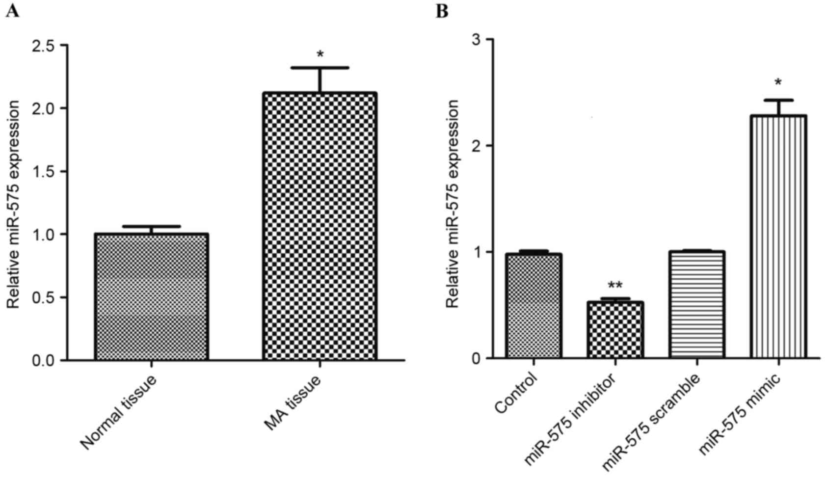
To investigate the miR-575 expression in villus
tissue and cells, RT-qPCR analysis was performed. Results
demonstrated that the expression of miR-575 was significantly
upregulated in MA placental villus tissues compared with normal
tissues (P<0.05; Fig. 1A).
Furthermore, after JEG-3 cells were transfected with miR-575
inhibitor, the expression of miR-575 decreased significantly
compared with the control and miR-575 scramble groups (P<0.05).
No significance difference in miR-575 expression was observed
between the control and miR-575 scramble transfected groups.
Contrastingly, after the JEG-3 cells were transfected with miR-575
mimic, the expression of miR-575 significantly increased compared
with the control group (P<0.01; Fig.
1B).
Effect of miR-575 on cell
apoptosis
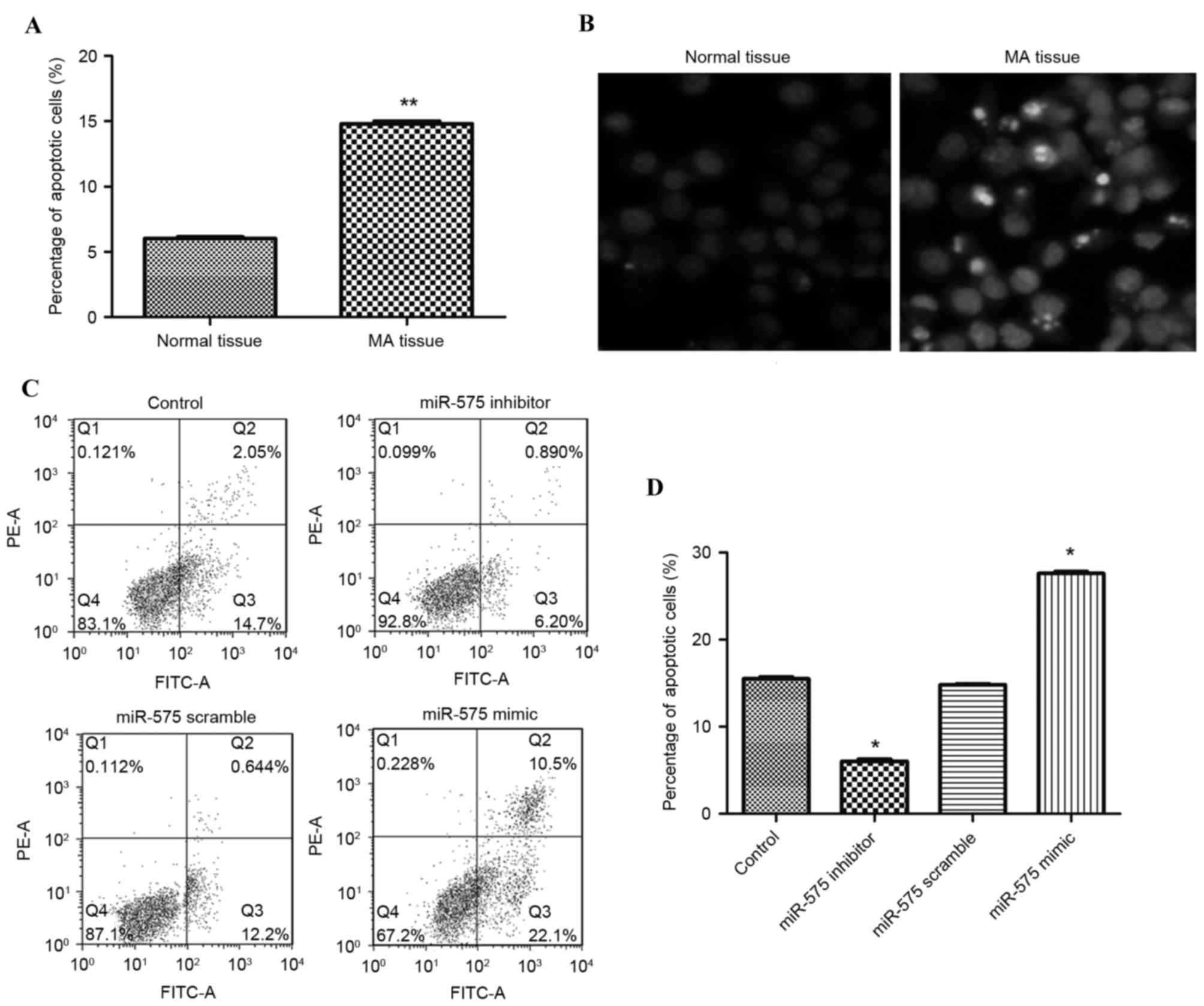
Excessive cell apoptosis may result in pregnancy
failure and MA (8). To characterize
the biological significance of miR-575 in MA development, cell
apoptosis assays were performed using flow cytometry. As
demonstrated in Fig. 2A and B, the
percentage of apoptotic cells in MA villus tissue was significantly
higher than that in normal tissue (P<0.05). Transfection
experiments also indicated that overexpression of miR-575
significantly promoted the apoptosis of JEG-3 cells compared with
the control group (P<0.01; Fig. 2C
and D). No significant difference in cell apoptosis was
observed between the control and miR-575 scramble transfected
groups. When JEG-3 cells were transfected with miR-575 inhibitor,
the percentage of apoptotic cells decreased significantly compared
with the other three groups (P<0.05).
Effect of miR-575 on cell
apoptosis-related protein expression
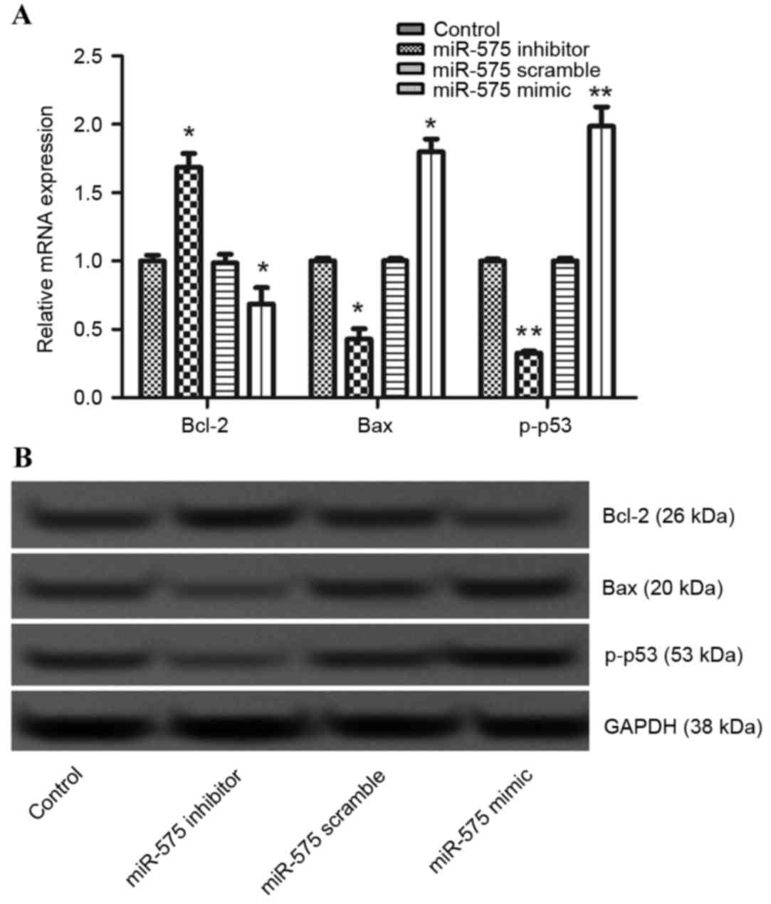
To further explore the underlying molecular
mechanisms of miR-575 on cell apoptosis, the expression levels of
apoptosis-related proteins, Bcl-2, Bax and p-p53, were measured
using RT-qPCR and western blot analyses. Results demonstrated that
overexpression of miR-575 significantly decreased the mRNA
expression level (P<0.05) and the protein expression level of
Bcl-2 compared with that in the control group. Also, overexpression
of miR-575 significantly increased the expression levels of Bax and
p-p53 compared with that in the control group (P<0.05 and
P<0.01, respectively). No significant difference in expression
was observed between the control and miR-575 scramble transfected
groups. However, miR-575 suppression significantly increased the
expression level of Bcl-2 (P<0.05), and decreased the expression
levels of Bax and p-p53 compared with the other three groups
(P<0.05 and P<0.01, respectively; Fig. 3A and B).
Effect of miR-575 on
angiogenesis-related protein expression
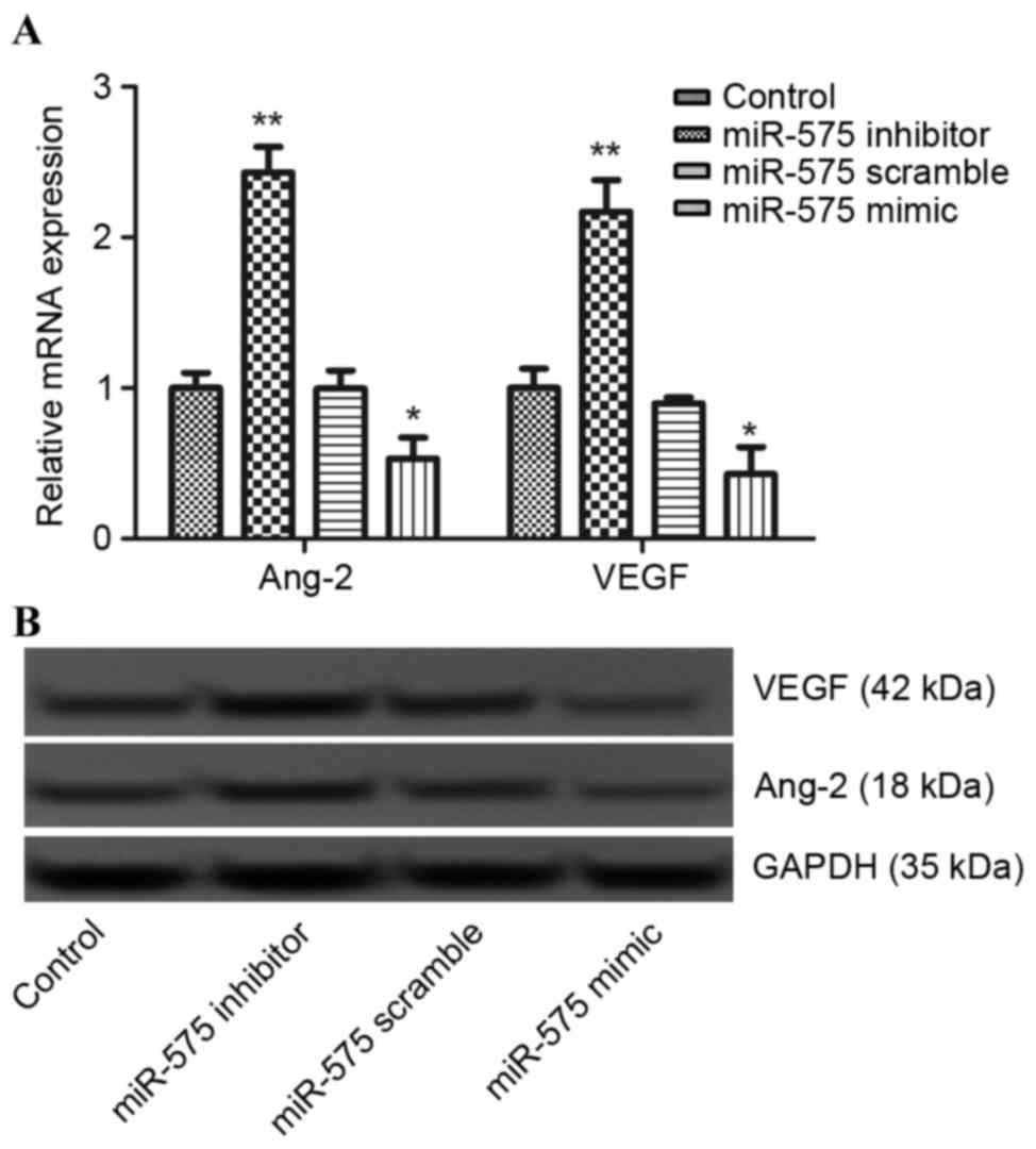
It has been reported that sufficient angiogenesis
has an important role in the maintenance of early pregnancy
(9). Therefore, the role of miR-575
in angiogenesis was investigated by detecting the expression levels
of angiogenesis-related proteins, Ang-2 and VEGF, using RT-qPCR and
western blot analyses. As demonstrated in Fig. 4A and B, miR-575 overexpression
significantly decreased the mRNA expression levels (P<0.05) and
markedly decreased the protein expression levels of both Ang-2 and
VEGF compared with the control group. However, after JEG-3 cells
were transfected with miR-575 inhibitor, the expression levels of
both Ang-2 and VEGF increased significantly compared with the
miR-575 scramble and mimic groups (P<0.01).
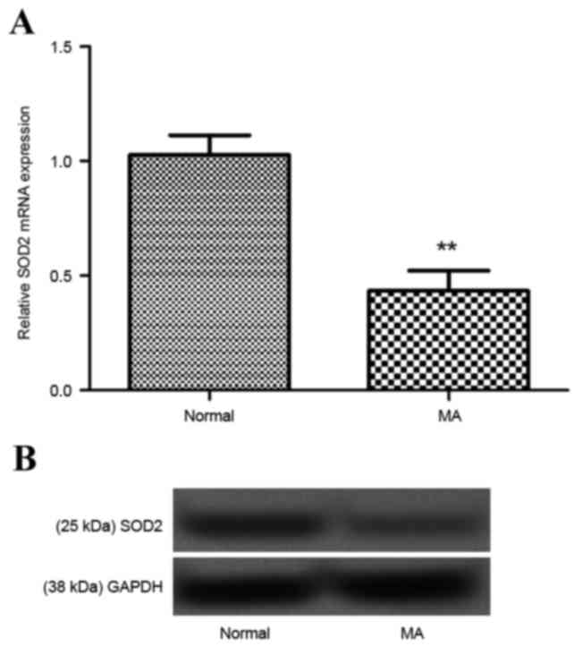
SOD2 is a direct target for miR-575
and miR-575 regulates MA by targeting SOD2
Research has demonstrated that SOD is downregulated
in trophoblasts from women with MA (15). In the present study, the expression
of SOD2 in villus tissue of MA patients was investigated by RT-qPCR
and western blot analyses. As demonstrated in Fig. 5A and B, SOD2 was significantly
downregulated in villus tissue from patients with MA compared with
normal tissues (P<0.01).
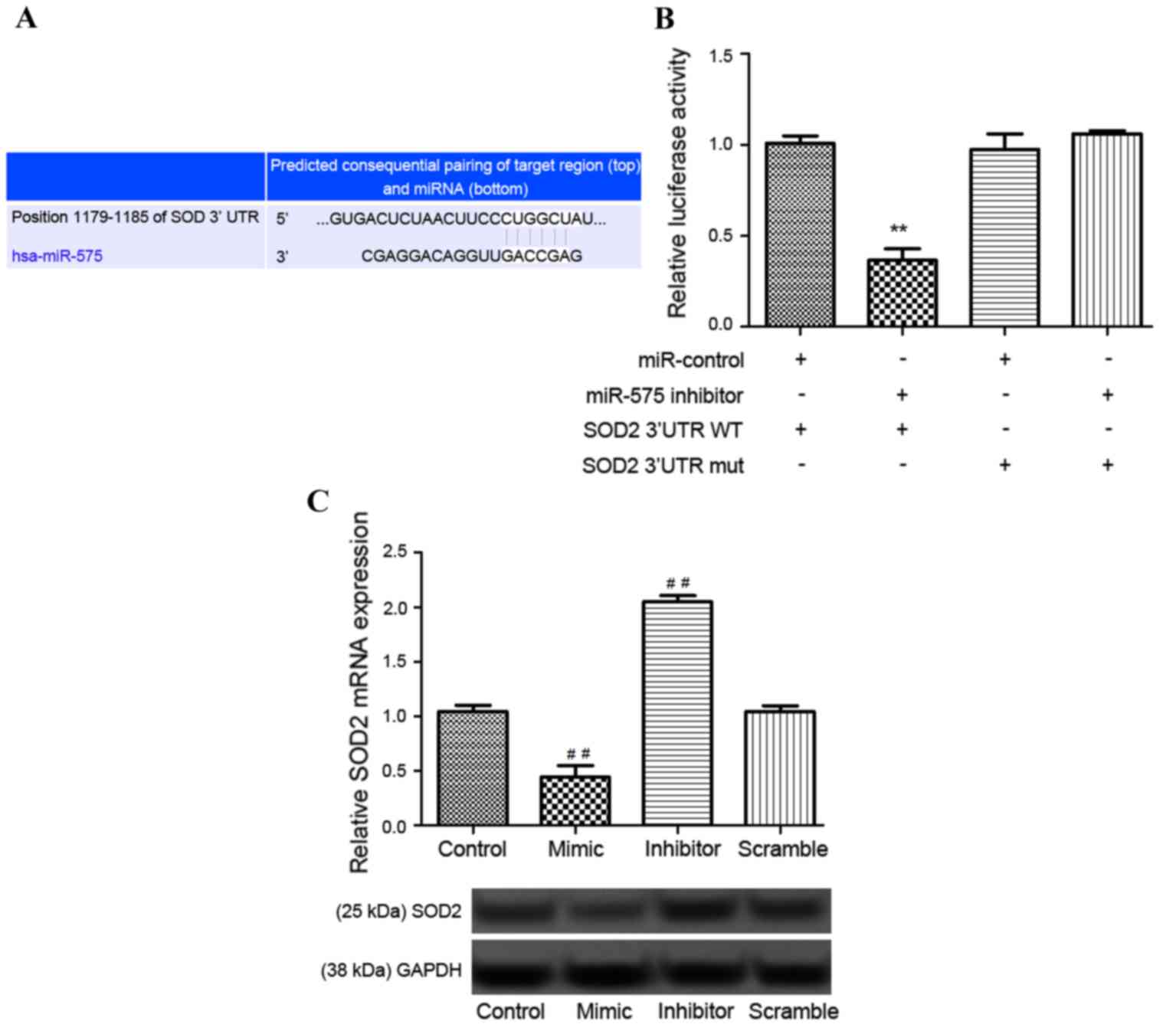
According to TargetScanHuman software (targetscan.org), SOD2 was predicted to be the target
gene of miR-575 (Fig. 6A).
Luciferase reporter analysis demonstrated that the relative
luciferase activity of the reporter that contained SOD2 wild type
3′-UTR reduced significantly in miR-575-inhibitor-transfected cells
compared with the miR-control transfected cells containing the SOD2
wild type 3′UTR (P<0.01; Fig.
6B). Additionally, the relative expression of SOD2 decreased
significantly when miR-575 was overexpressed and increased
significantly when miR-575 was silenced compared with the control
group (P<0.01; Fig. 6C). These
results indicated that miR-575 may directly regulate the SOD2 gene.
Furthermore, after pc-SOD2 was transfected into cells, it was
demonstrated that SOD2 expression was significantly increased
compared with the control group (P<0.01; Fig. 7A). Notably, further investigation
indicated that overexpressed SOD2 was able to significantly reverse
the inhibiting effect of miR-575 inhibitor on apoptosis (P<0.01;
Fig. 7B) and expression of
angiogenesis factors, Ang-2 and VEGF (Fig. 7C). These results suggested that
miR-575 may regulate apoptosis and angiogenesis by targeting the
SOD2 gene.
 | Figure 7.(A) Expression of SOD2 after cells
were transfected with pc-SOD2 assayed by RT-qPCR and western blot
analysis. (B) Percentage of apoptotic JEG-3 cells following
transfection with pc-SOD2, assayed by flow cytometry. (C)
Expression levels of angiogenesis-related proteins, VEGF and Ang-2,
after cells were transfected with pc-SOD2, assayed by RT-qPCR and
western blot analysis. Experiments were repeated three times. Data
are expressed as the mean + standard deviation. *P<0.05 and
**P<0.01 vs. control; #P<0.05 vs. inhibitor group.
SOD2, superoxide dismutase 2; pc-SOD2, pcDNA 3.1 vector with SOD2
coding sequence; RT-qPCR, reverse transcription-quantitative
polymerase chain reaction; VEGF, vascular endothelial growth
factor; Ang-2, angiopoietin 2; FITC, fluorescein isothiocyanate;
PE-A, pseudomonas exotoxin A. |
Discussion
The present study demonstrated that miR-575 was
significantly overexpressed in MA embryo villus tissue compared
with normal tissues. The percentage of apoptotic cells in MA embryo
villus tissues was significantly higher than that in normal
tissues. Transfection experiments revealed that miR-575
overexpression was able to promote the apoptosis of JEG-3 cells and
decreased the expression of angiogenesis-related proteins. After
JEG-3 cells were transfected with miR-575 inhibitor, the percentage
of apoptotic cells decreased significantly, and the expression of
angiogenesis-related proteins, Ang-2 and VEGF, significantly
increased. To the best of our knowledge, the present study is the
first to investigate the role of miR-575 in MA.
Various studies have suggested that the level of
apoptosis is critically important for the successful development of
normal pregnancy (7–17). A low level of apoptosis in placental
villi tissues is a normal physiological phenomenon (8); however, a high level of apoptosis may
result in miscarriage (18,19). In the present study, miR-575 was
overexpressed in MA embryo villus tissue, suggesting the possible
role of miR-575 in promoting the development of MA. Notably,
miR-575 overexpression promoted the apoptosis of JEG-3 cells,
suggesting that miR-575 overexpression may have an important role
in the cell apoptosis in MA.
Previous research has reported that a series of
genes coordinately regulate apoptosis and proliferation during
pregnancy (20). For example, p53, a
key regulator of apoptosis and the cell cycle, has been
demonstrated to be overexpressed in the chorionic villi of females
with spontaneous abortions or MA (21,22). In
the present study, p-p53 was upregulated in the miR-575 mimic
group. However, when JEG-3 cells were transfected with miR-575
inhibitor, the expression of p-p53 decreased significantly,
indicating that miR-575 may have an important role in MA by
regulating the apoptosis of villi cells.
In addition to p53, Bax, one of the pro-apoptotic
members of the Bcl-2 family demonstrated reduced expression in the
miR-575 inhibitor group. Contrastingly, the expression of
anti-apoptotic Bcl-2 increased significantly following miR-575
inhibition. Notably, previous research has revealed that the
abnormal expression of Bcl-2 and Bax in placental villi tissues is
associated with recurrent abortion (23). These results further indicated the
role miR-575 in the apoptosis of villi cells.
A successful pregnancy not only depends on
apoptosis, but also depends on sufficient villous angiogenesis,
which is able to supply adequate oxygen and nutrients (24,25).
Abnormal angiogenesis is considered as an important contributor to
MA (26). VEGF is believed to be a
powerful angiogenesis promoter, which has a critical role in the
development and maintenance of the vasculature (27). A study by Çöl-Madendag et al
(28) revealed that decreased
expression of VEGF in placental villi and decidua may be associated
with early abortion. Additionally, the angiopoietin family is
another important angiogenic protein family, which has been
demonstrated to be critically involved in angiogenesis,
particularly in the female reproductive tract (29–31).
Ang-2, a member of the angiopoietin family, may be expressed in the
early placenta in normal and pathological pregnancy (32,33).
High levels of Ang-2 may confer protection to the placenta
(34). The present study
demonstrated that overexpression of miR-575 significantly decreased
the expression of VEGF and Ang-2. After miR-575 was inhibited, the
expression levels of VEGF and Ang-2 increased again, indicating the
regulatory role of miR-575 in angiogenesis in MA.
It has been reported that oxidative stress is
associated with MA (35). SOD is an
important antioxidant that catalyzes the dismutation of superoxide
into hydrogen peroxide and oxygen molecules, thereby scavenging
free radicals and preventing oxygen toxicity (36). Notably, alterations to SOD
concentration have been closely related to spontaneous abortion
(36). A study by Zhu et al
(15) indicated that SOD was
downregulated in trophoblasts from women with MA. In the present
study, SOD2 was suggested to be a target gene of miR-575, and was
downregulated in MA tissue. Further investigation demonstrated that
overexpression of SOD2 was able to reverse the inhibiting effects
of miR-575 inhibitor on apoptosis and expression of angiogenesis
factors. These results suggested that miR-575 may regulate
apoptosis and angiogenesis by targeting the SOD2 gene.
In conclusion, the results of the present study
suggest distinct roles of miR-575 in apoptosis and angiogenesis in
villi tissue and cells. Inhibition of miR-575 may inhibit apoptosis
and promote angiogenesis to prevent MA. Therefore, miR-575 may
serve as a biomarker of MA, as well as a potential molecular target
for the treatment of MA.
References
|
1
|
Cao W, Wenlin XU, Chen T, Wang X, Wang X,
Qiu J, Chen N and Mao Y: CD4+CD25+FoxP3+ regulatory T cells and
cytokines interact with estradiol in cases of missed abortion. Exp
Ther Med. 7:417–422. 2014. View Article : Google Scholar : PubMed/NCBI
|
|
2
|
Sebire NJ, Thornton S, Hughes K, Snijders
RJ and Nicolaides KH: The prevalence and consequences of missed
abortion in twin pregnancies at 10 to 14 weeks of gestation. Br J
Obstet Gynaecol. 104:847–848. 1997. View Article : Google Scholar : PubMed/NCBI
|
|
3
|
Zhang X, Li J, Gu Y, Zhao Y, Wang Z and
Jia G: A pilot study on environmental and behavioral factors
related to missed abortion. Environ Health Prev Med. 16:273–278.
2011. View Article : Google Scholar : PubMed/NCBI
|
|
4
|
O'Connell RM, Rao DS, Chaudhuri AA and
Baltimore D: Physiological and pathological roles for microRNAs in
the immune system. Nat Rev Immunol. 10:111–122. 2010. View Article : Google Scholar : PubMed/NCBI
|
|
5
|
Longtine MS, Chen B, Odibo AO, Zhong Y and
Nelson DM: Villous trophoblast apoptosis is elevated and restricted
to cytotrophoblasts in pregnancies complicated by preeclampsia,
IUGR, or preeclampsia with IUGR. Placenta. 5:352–359. 2012.
View Article : Google Scholar
|
|
6
|
Chatzaki E, Makrigiannakis A, Margioris
AN, Kouimtzoglou E and Gravanis A: The Fas/FasL apoptotic pathway
is involved in kappa-opioid-induced apoptosis of human endometrial
stromal cells. Mol Hum Reprod. 7:867–874. 2001. View Article : Google Scholar : PubMed/NCBI
|
|
7
|
Jerzak M and Bischof P: Apoptosis in the
first trimester human placenta: The role in maintaining immune
privilege at the maternal-foetal interface and in the trophoblast
remodelling. Eur J Obstet Gynecol Reprod Biol. 100:138–142. 2002.
View Article : Google Scholar : PubMed/NCBI
|
|
8
|
Halperin R, Peller S, Rotschild M,
Bukovsky I and Schneider D: Placental apoptosis in normal and
abnormal pregnancies. Gynecol Obstet Invest. 50:84–87. 2000.
View Article : Google Scholar : PubMed/NCBI
|
|
9
|
Chen H, Deng X, Yang Y, Shen Y, Chao L,
Wen Y and Sun Y: Expression of GRIM-19 in missed abortion and
possible pathogenesis. Fertil Steril. 103:138–146. 2015. View Article : Google Scholar : PubMed/NCBI
|
|
10
|
Nelissen EC, van Montfoort AP, Dumoulin JC
and Evers JL: Epigenetics and the placenta. Hum Reprod Update.
17:397–417. 2011. View Article : Google Scholar : PubMed/NCBI
|
|
11
|
Miura K, Miura S, Yamasaki K, Higashijima
A, Kinoshita A, Yoshiura K and Masuzaki H: Identification of
pregnancy-associated microRNAs in maternal plasma. Clin Chem.
56:1767–1771. 2010. View Article : Google Scholar : PubMed/NCBI
|
|
12
|
Hosseini MK, Gunel T and Gumusoglu E:
MicroRNA profiling in miscarriage patients. IJMM. 34:S94. 2014.
|
|
13
|
Ward RH, Modell B, Petrou M, Karagözlu F
and Douratsos E: Method of sampling chorionic villi in first
trimester of pregnancy under guidance of real time ultrasound. Br
Med J (Clin Res Ed). 286:1542–1544. 1983. View Article : Google Scholar : PubMed/NCBI
|
|
14
|
Livak K and Schmittgen TD: Analysis of
relative gene expression data using real-tie quantitative PCR and
the 2(-Delta Delta C(T)) method. Methods. 25:402–408. 2001.
View Article : Google Scholar : PubMed/NCBI
|
|
15
|
Zhu LJ, Chen YP, Chen BJ and Mei XH:
Changes in reactive oxygen species, superoxide dismutase and
hypoxia-inducible factor-1α levels in missed abortion. Int J Clin
Exp Med. 7:2179–2184. 2014.PubMed/NCBI
|
|
16
|
Savion S, Lepsky E, Orenstein H, Carp H,
Shepshelovich J, Torchinsky A, Fein A and Toder V: Apoptosis in the
uterus of mice with pregnancy loss. Am J Reprod Immunol.
47:118–127. 2002. View Article : Google Scholar : PubMed/NCBI
|
|
17
|
Choi HK, Choi BC, Lee SH, Kim JW, Cha KY
and Baek KH: Expression of angiogenesis- and apoptosis-related
genes in chorionic villi derived from recurrent pregnancy loss
patients. Mol Reprod Dev. 66:24–31. 2003. View Article : Google Scholar : PubMed/NCBI
|
|
18
|
Cinar O, Kara F and Can A: Potential role
of decidual apoptosis in the pathogenesis of miscarriages. Gynecol
Endocrinol. 28:382–385. 2012. View Article : Google Scholar : PubMed/NCBI
|
|
19
|
Nair RR, Khanna A and Singh K: Association
of FAS-1377 G>A and FAS-670 A>G functional polymorphisms of
FAS gene of cell death pathway with recurrent early pregnancy loss
risk. J Reprod Immunol. 93:114–118. 2012. View Article : Google Scholar : PubMed/NCBI
|
|
20
|
Levy R and Nelson D: CURRENT TOPIC: To be,
or not to be, that is the question. Apoptosis In human trophoblast.
Placenta. 21:1–13. 2000. View Article : Google Scholar : PubMed/NCBI
|
|
21
|
Kaare M, Bützow R, Ulander VM, Kaaja R,
Aittomäki K and Painter JN: Study of p53 gene mutations and
placental expression in recurrent miscarriage cases. Reprod Biomed
Online. 18:430–435. 2009. View Article : Google Scholar : PubMed/NCBI
|
|
22
|
Chen Y, Shen D, Gu Y, Zhong P, Xie J and
Song Q: The diagnostic value of Ki-67, P53 and P63 in
distinguishing partial Hydatidiform mole from hydropic abortion.
Wien Klin Wochenschr. 124:184–187. 2012. View Article : Google Scholar : PubMed/NCBI
|
|
23
|
Taylor DD and Gercel-Taylor C: Alterations
in T-cell signal transduction molecules associated with recurrent
spontaneous pregnancy loss. J Reprod Immunol. 63:137–154. 2004.
View Article : Google Scholar : PubMed/NCBI
|
|
24
|
Rajakumar A and Conrad KP: Expression,
ontogeny, and regulation of hypoxia-inducible transcription factors
in the human placenta. Biol Reprod. 63:559–569. 2000. View Article : Google Scholar : PubMed/NCBI
|
|
25
|
Lim KH, Zhou Y, Janatpour M, McMaster M,
Bass K, Chun SH and Fisher SJ: Human cytotrophoblast
differentiation/invasion is abnormal in pre-eclampsia. Am J Pathol.
151:1809–1818. 1997.PubMed/NCBI
|
|
26
|
Fang Y, Yu S, Ma Y, Sun P, Ma D, Ji C and
Kong B: Association of Dll4/notch and HIF-1a-VEGF signaling in the
angiogenesis of missed abortion. PLoS One. 8:e706672013. View Article : Google Scholar : PubMed/NCBI
|
|
27
|
Arjamaa O, Nikinmaa M, Salminen A and
Kaarniranta K: Regulatory role of HIF-1alpha in the pathogenesis of
age-related macular degeneration (AMD). Ageing Res Rev. 8:349–358.
2009. View Article : Google Scholar : PubMed/NCBI
|
|
28
|
Çöl-Madendag I, Madendag Y, Altinkaya SÖ,
Bayramoglu H and Danisman N: The role of VEGF and its receptors in
the etiology of early pregnancy loss. Gynecol Endocrinol.
30:153–156. 2014. View Article : Google Scholar : PubMed/NCBI
|
|
29
|
Folkman J and Klagsbrun M: Angiogenic
factors. Science. 235:442–447. 1987. View Article : Google Scholar : PubMed/NCBI
|
|
30
|
Maisonpierre PC, Suri C, Jones PF,
Bartunkova S, Wiegand SJ, Radziejewski C, Compton D, McClain J,
Aldrich TH, Papadopoulos N, et al: Angiopoietin-2, a natural
antagonist for Tie2 that disrupts in vivo angiogenesis. Science.
277:55–60. 1997. View Article : Google Scholar : PubMed/NCBI
|
|
31
|
Valenzuela DM, Griffiths JA, Rojas J,
Aldrich TH, Jones PF, Zhou H, McClain J, Copeland NG, Gilbert DJ,
Jenkins NA, et al: Angiopoietins 3 and 4: Diverging gene
counterparts in mice and humans. Proc Natl Acad Sci USA.
96:1904–1909. 1999. View Article : Google Scholar : PubMed/NCBI
|
|
32
|
Plaisier M, Dennert I, Rost E, Koolwijk P,
van Hinsbergh VW and Helmerhorst FM: Decidual vascularization and
the expression of angiogenic growth factors and proteases in first
trimester spontaneous abortions. Hum Reprod. 24:185–197. 2009.
View Article : Google Scholar : PubMed/NCBI
|
|
33
|
Seval Y, Sati L, Celik-Ozenci C, Taskin O
and Demir R: The distribution of angiopoietin-1, angiopoietin-2 and
their receptors tie-1 and tie-2 in the very early human placenta.
Placenta. 29:809–815. 2008. View Article : Google Scholar : PubMed/NCBI
|
|
34
|
Daponte A, Deligeoroglou E, Pournaras S,
Tsezou A, Garas A, Anastasiadou F, Hadjichristodoulou C and
Messinis IE: Angiopoietin-1 and angiopoietin-2 as serum biomarkers
for ectopic pregnancy and missed abortion: A case-control study.
Clin Chim Acta. 415:145–151. 2013. View Article : Google Scholar : PubMed/NCBI
|
|
35
|
Xiu-Ju MA and Xiao WX: Relationship
between oxidative stress and missed abortion. J Shanxi Med Univ.
2006.
|
|
36
|
Valdivia A, Pérez-Álvarez S, Aroca-Aguilar
JD, Ikuta I and Jordán J: Superoxide dismutases: A
physiopharmacological update. J Physiol Biochem. 65:195–208. 2009.
View Article : Google Scholar : PubMed/NCBI
|