Introduction
Tuberculosis (TB) is caused by infection of
Mycobacterium tuberculosis (M. tuberculosis), which
remains the leading cause of mortality from infectious disease
worldwide (1). According to a World
Health Organization study in 2012, TB remains a global emergency,
particularly in less developed countries (2). Tuberculosis typically affects the
pulmonary system but may also affect any other organ system within
the body; this type of TB is referred to as extra-pulmonary TB
(3). Extra-pulmonary involvement
accounts for approximately 14% of TB patients and skeletal system
involvement occurs in 1–3% of patients (4,5). Spinal
tuberculosis is the most frequent bone localization, accounting for
40–50% of all skeletal tuberculosis cases, followed by major weight
bearing joints of the lower extremity, including the hip and knee
(6–8). Extra-pulmonary TB often occurs in
patients with reduced immune function. It is well known that the
human immunodeficiency virus (HIV) is the etiological agent
responsible for acquired immune deficiency syndrome. It severely
weakens the immune system and increases the susceptibility of
individuals to opportunistic infections, including TB (9). TB may cause macrophage dysfunction,
anemia and hypoproteinemia, thus making patients highly vulnerable
to other infections. Brucellosis (BS) is a zoonotic bacterial
disease, caused by the gram-negative bacteria Brucella
(10). The involvement of the
musculoskeletal is associated with 10–85% of complications
associated with brucellosis (11).
Similarly to TB, spinal involvement is one of the most commonly
encountered forms of human brucellosis (12). BS may be misdiagnosed as TB as its
clinical features and basic laboratory parameters are comparable.
It is especially difficult to make definitive diagnoses in cases of
co-infection with TB and BS. The present study reports a case of
spinal TB in association with BS. To the best of our knowledge,
this co-infection has not been described in previous
literature.
Case report
History and physical examination
A 45-year-old male was admitted as an outpatient to
the Department of Orthopedics, Yantaishan Hospital (Yantai, China)
with a history of lower back pain and non-tender swelling in the
right flank for 3 months, intermittent hyperpyrexia, sweating and
body aches for 3 weeks and numbness and weakness of right lower
limb for 2 days. The patient had been in excellent health prior to
this presentation with no other remarkable medical or surgical
history. There was no medical history of personal or family
tuberculosis. The patient also had no history of tobacco use,
intravenous drug use or other factors for HIV infection. The
patient had traveled to Inner Mongolia 1-month prior and had
experience weight loss of 6 kilograms during the previous 3
months.
On the day of admission to our hospital, the
patient's temperature rose to 38.8°C from 37.5°C at home, with
sweating and arthralgia, but other vital signs were normal.
Physical examination revealed a 7×16 cm non-tender, fluctuant right
flank swelling but no visible cutaneous changes. Lumbar flexion
movement was limited, hepatosplenomegaly wasn't palpated, and
inguinal and axillary lymph nodes were undetectable. Myodynamia of
right lower limb weakened from Grade 5 to 3 according to the
Frankel grade (13). Pick-up test
and right Lasegue sign was positive. Babinski sign was negative on
both sides. Nerve damage was Frankel Grade D. Systemic examination
was normal.
Laboratory examinations and imaging
findings
On admission a specimen of blood was collected and
sent for culture. Laboratory examinations demonstrated a white
blood cell count of 8,900 cells/mm3 (normal, 3,500–8,500
cells/mm3) with 65% neutrophils. Inflammatory markers
were elevated significantly, with an erythrocyte sedimentation rate
(ESR) of 102 mm/h (normal, 0.0–20 mm/h), C-Reactive Protein (CRP)
level of 46 mg/dl (normal, 0.0–10 mg/dl) and procalcitonin of 0.43
ng/ml (normal, 0–0.05 ng/ml). TB-Interferon Gamma Release assay was
positive. A Tuberculin Purified Protein Derivative (PPD) skin test
was positive, with a 15 mm diameter (normal, 0.0–5 mm) (14). Hepatic and renal functions were
normal. The anti-HIV antibody test was negative. Abdominal
ultrasonography didn't indicate any marked changes, with the
exception of retroperitoneal multiple liquid dark areas. Chest and
hip X-rays were normal, but a lateral radiograph of the lumbar
spinal indicated a narrowed disc space and spondylitis at the L4-5
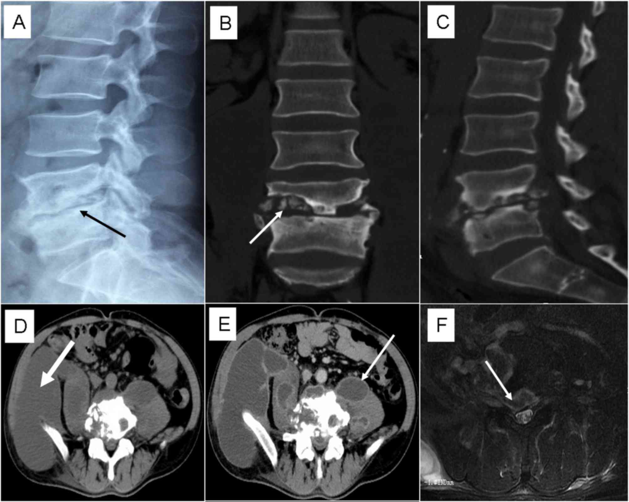
level (Fig. 1A). Computed tomograph
(CT) scan indicated vertebral destruction, formation of sequestra
and bilateral psoas abscesses and right iliac fossa abscess
(Fig. 1B-D). Contrast indicated that
the abscesses were clearly peripherally enhanced (Fig. 1E). The magnetic resonance imaging
(MRI) of the T2-weighted fat suppression axial demonstrated that
the abscess had spread into the spinal canal causing thecal sac
compression (Fig. 1F).
Diagnosis and management
According to the aforementioned results, the
preliminary diagnosis was lumbar TB combined with psoas abscess.
Quadruple anti-tuberculosis drug therapy was initiated empirically
[isoniazid 300 mg, pyrazinamide 25 mg/kg (both Shanghai Shangyin
Xinyi Pharmaceutical Co., Ltd., Shanghai, China) rifampin 600 mg
and ethambutol 20 mg/kg (both Hutchison Whampoa Guangzhou
Baiyunshan Chinese Medicine Co. Ltd., Guangzhou, China) daily]. Due
to the intermittent hyperpyrexia and sweating, pyogenic spondylitis
was also considered as differential diagnosis, although the
patient's white blood cell and neutrophil level was normal. Taken
into consideration the patient's travel history to Inner Mongolia,
which is one of the major pastoral areas of China (15), prior to a fever, a number of
laboratory tests were completed to determine a positive diagnosis
of brucellosis. Rose-Bengal Plate Test (RBPT) was positive
(16) and as a result, a standard
serum agglutination test (SAT) was performed as previously
described (17), using the SAT
antigen (cat. no. SY0070; Kemin Bio-Technology Ltd., Shanghai,
China) which revealed a titer of antibodies to Brucella of 1/80.
Following laboratory confirmation of BS, antibiotic treatment was
initiated with doxycycline 100 mg twice daily. The blood samples
obtained on admission yielded B. melitensis, and a diagnosis
of BS was confirmed. During this period the patient's fever and
sweating gradually disappeared and lower back pain improved. In
order to determine further treatment, a CT-guided percutaneous
drainage of the abscess was performed and four drainage catheters
were inserted. The pus was sent for culture. The abscesses were
aspirated and irrigated with saline solution (5–10 ml) and
isoniazid (200 mg) twice a day. The culture of the pus was positive
for M. tuberculosis. Therefore, the definitive diagnosis was
spinal TB combined with BS. Drainage catheters were removed when
abscesses were minimal (<30 ml), as indicated by a follow-up CT.
Single posterior approach surgery, including decompression,
debridement, posterior instrumentation and fusion, was subsequently
performed under general anesthesia. Inititaly, a CT-guided
percutaneous drainage of the abscess was performed and when the
abscess was minimal (<30 ml) the drainage catheters were
removed. The posterior approach surgery was subsequently performed
to remove the abscess located in the intervertebral space and
intraspinal canal. During surgery, internal fixation (M8;
Medtronic, Minneapolis, MN, USA) was used following decompression
and debridement to maintain the stability of the spine. Any
residual abscess was treated by postoperative anti-tuberculosis
drug therapy. On patient admission anti-tuberculosis drug therapy
was initiated for 7 days and the CT-guided percutaneous drainage
was performed for 10 days. When the catheters were removed the
patient continued quadruple antituberculosis and doxycycline
treatment for a further 7 days, then the operation was performed.
There was a total of 24 days from hospital admission to surgery.
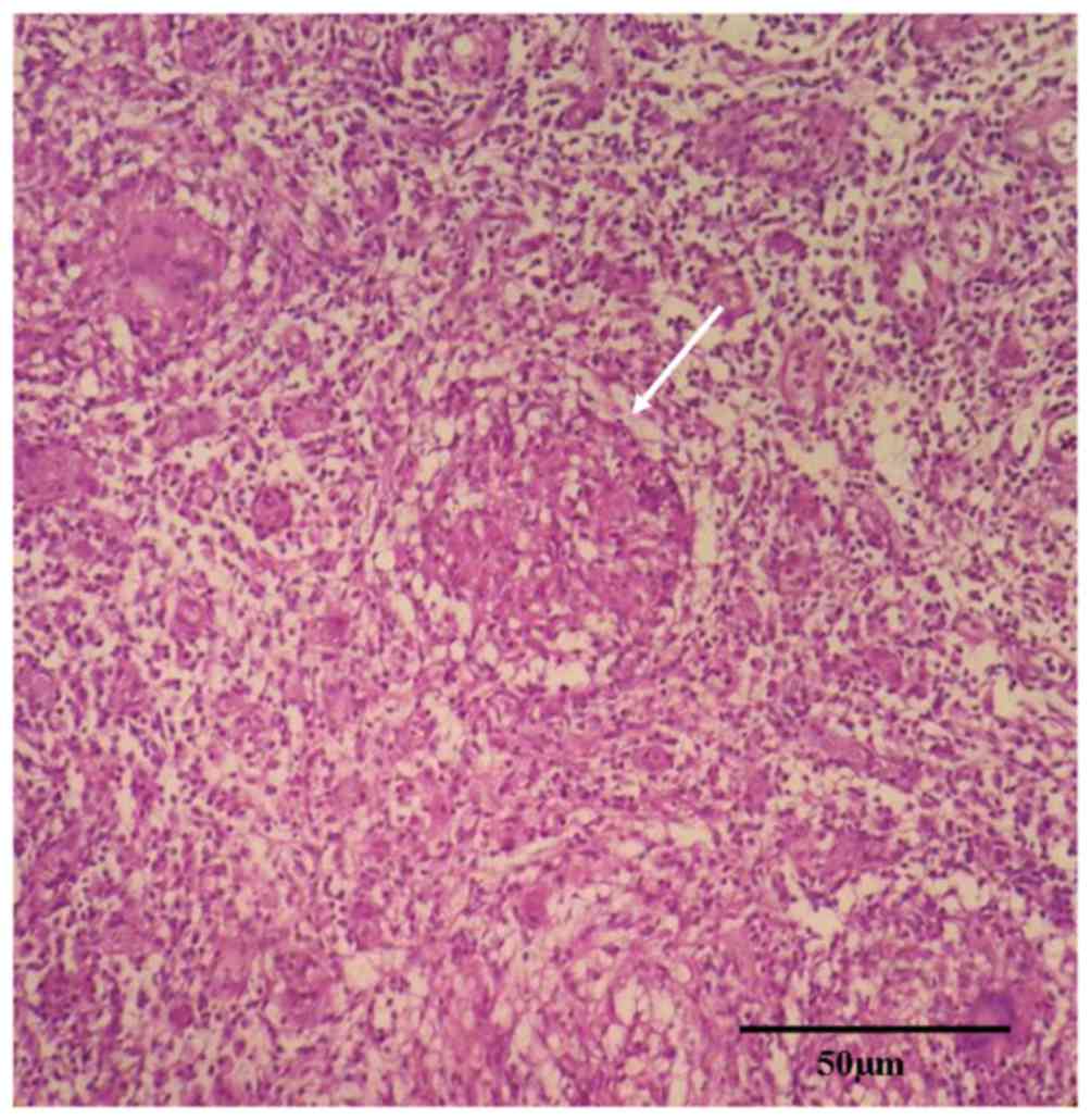
Histopathological examination of the lumbar lesion post surgery
revealed granulomatous inflammation and caseous necrosis (Fig. 2), which is consistent with the
diagnosis of spinal TB. Tissue sections were fixed in 4%
paraformaldehyde at 4°C for 4 h, placed in processing cassettes,
dehydrated through a serial alcohol gradient and embedded in
paraffin wax blocks. Prior to immunostaining, 5-µm-thick tissue
sections were dewaxed in xylene, rehydrated through decreasing
concentrations of ethanol and washed in PBS. The sections were
stained with hematoxylin and eosin at 20°C (hematoxylin for 10 min
and eosin for 30 sec). Following staining the sections were
dehydrated through increasing concentrations of ethanol and xylene.
A light microscope was used to examine the tissue sections at
magnification, ×200. The patient was at rest for 2 weeks and then
began to complete daily activities with a lumbar brace. The patient
no longer experienced fever, sweating or arthralgia. Numbness and
weakness of the right lower limb disappeared and myodynamia
recovered to Grade 5. Treatment with oral doxycycline was completed
and the patient was discharged to continued quadruple
antituberculosis treatment for 6 months (isoniazid 300 mg, rifampin
600 mg, ethambutol 20 mg/kg and pyrazinamide 25 mg/kg) and double
antituberculosis treatment for a further 6 months (isoniazid 300 mg
and rifampin 600 mg). A follow-up radiograph after 3 months
indicated that the internal fixation was in a good position and
preliminary vertebral fusion at L4/5 level, indicating the
formation of new bone at the L4/5 disk space.
Discussion
TB and BS are infectious diseases endemic in certain
areas of the world, generally in developing countries (2,7,18) and are relatively common causes of
vertebral osteomyelitis (19). Due
to similarities in clinical presentation and basic laboratory
parameters, a proportion of patients may be misdiagnosed with
either TB or BS (18). Despite
extensive efforts to control TB, it remains an urgent global health
problem, particularly due to the spread of HIV and emergence of
multi-drug resistant TB. Human BS is considered to be the most
common zoonosis globally, with an annual occurrence of >500,000
cases (20). Humans are infected
primarily through contact with infected animals or by consuming
unpasteurized and unboiled milk or fresh cheese (10,21). BS
is listed as a Class II reportable infectious disease by the
Centers for Disease Control and Prevention (CDC) of China (Beijing,
China). A study by Zhong et al (22) indicated that a total of 155,979 cases
were reported in China between 2005 and 2010. BS epidemics are
primarily located in the northern provinces of China and inner
Mongolia accounted for 50% of all the reported cases. Fever, night
sweating, weight loss, fatigue and elevated ESR and CRP are common
clinical features in both infections (22). It is difficult to distinguish TB and
BS based on these nonspecific symptoms and laboratory findings;
thus, misdiagnosis of BS as TB often occurs in brucellosis
low-incidence areas. BS patients may also recover following the use
of standard anti-tuberculosis drugs therapy, as antibiotic
combination is typical in TB and BS drugs (19,23), and
rifampin and streptomycin have notable curative effects in BS and
TB. However, etiological treatment based on definitive diagnosis
should still be the basic principal. There are a number of subtle
differences between the two diseases. Compared with the low-grade
fever in TB, intermittent hyperpyrexia is more often observed in
BS. Sweating is also more common in BS and the sweat is viscous
(18). High values of ESR and CRP
often indicate TB and are relative indicators, more useful in
evaluating response to treatment (24). Kurtaran et al (19) compared the clinical features of 87
cases with spondylitis of TB and BS, concluding that vertebral
destruction and compression, kyphosis, para-spinal abscesses and
cord compression were more frequent in spinal TB. This is in
agreement with results from previous reports (23,25).
Hepatosplenomegaly and lymphadenopathy were more common in BS due
to bacteriological replication within the reticuloendothelial
system (19). At present, culture of
the organism from blood and/or pus remains the gold standard in BS
and TB diagnosis and treatment; however, this may take 2–4 weeks
and often reveals a false negative result (10,26).
RBPT and serum agglutination assessments are available and helpful
for preliminary identification. Enhanced awareness for BS is
critical to avoid misdiagnosis and delayed diagnosis for
clinicians. The co-infection of M. tuberculosis and B.
melitensis is rare and the exact mechanism of this co-infection
remains unclear. Macrophage dysfunction in TB patients may be
associated with this mechanism, as it may promote opportunistic
local infections including aspergillosis and other bacterial
abscesses. Human protection against B. melitensis depends on
cell-mediated immunity. However, B. melitensis is able to
survive and replicate in macrophages and dendritic cells, evading
and destroying innate immunity (27). In theory, it is easier for patients
with TB to catch BS, compared with healthy individuals as TB may
cause macrophage dysfunction, anemia and hypoproteinemia, which
makes individuals more vulnerable to infection.
The standard treatment for BS is doxycycline 100 mg
orally twice daily and rifampcine 450 mg/day orally for 6 weeks
(28,29). However other studies indicate that
the preferred duration of therapy for BS should be ≥3 months due to
high relapse rates (19). There
remains no consensus for the duration of anti-tuberculosis drug
therapy: Some clinicians support the use of short-term triple drugs
for 9 months, while others advocate four-drug therapy for 12 months
(7,30). It has been suggested that the
duration of therapy should vary according to clinical response and
laboratory examinations. Regarding the case presented in the
current study, 6 weeks of doxycycline combined with quadruple
anti-tuberculosis drug therapy for 12 months was prescribed. Due to
the huge abscesses and neurological deficits, a CT-guided
percutaneous drainage of the abscess and posterior instrumentation
was performed in sequence. This is very different from treatments
performed previously (30–33). The CT-guided percutaneous drainage
not only removed the abscess and improved the patient's general
condition with local chemotherapy (isoniazid), but also avoided the
use of anterior or anterolateral approach open surgery, which may
lead to postoperative complications. In addition, drainage of pus
sent to culture and other laboratory examinations was helpful to
reach a definitive diagnosis.
In conclusion, spinal tuberculosis combined with BS
is a very rare condition. Overlapping clinical presentation and
laboratory parameters of TB and BS may lead to misdiagnosis or
delayed diagnosis, specifically in cases of co-infection with TB
and BS. Clinicians should strengthen their awareness of this
condition for similar patients. Furthermore, the CT-guided
percutaneous drainage is effective in the diagnosis and management
of spinal TB with abscess. However, it should be noted that as it
is possible to relapse with TB and BS it is necessary for the
patient to receive regular laboratory and radiological
examinations.
Acknowledgements
The authors of the present study express thanks and
appreciation to Xueling Wang for technical assistance in the
preparation of the study.
References
|
1
|
Metha JS and Bhojraj SY: Tuberculosis of
the thoracic spine. J Bone Joint Surg Br. 85:1502003.PubMed/NCBI
|
|
2
|
World Health Organization, . Global
tuberculosis report 2012. WHO; Geneva: 2012
|
|
3
|
Samohyl M, Solovic I, Rams R, Hirosova K,
Vondrova D, Krajcova D, Nadazdyova A, Filova A and Jurkovicova J:
Effect of spa treatment and the epidemiology of tuberculosis in the
Slovak republic in the year 2014. Clin Soc Work Health Interven.
7:24–35. 2016. View Article : Google Scholar
|
|
4
|
Cui X, Ma YZ, Chen X, Cai XJ, Li HW and
Bai YB: Outcomes of different surgical procedures in the treatment
of spinal tuberculosis in adults. Med Princ Pract. 22:346–350.
2013. View Article : Google Scholar : PubMed/NCBI
|
|
5
|
Saraf SK and Tuli SM: Tuberculosis of hip:
A current concept review. Indian J Orthop. 49:1–9. 2015. View Article : Google Scholar : PubMed/NCBI
|
|
6
|
Fuentes Ferrer M, Gutiérrez Torres L,
Ayala Ramírez O, Rumayor Zarzuelo M and del Prado González N:
Tuberculosis of the spine. A systematic review of case series. Int
Orthop. 36:221–231. 2012. View Article : Google Scholar : PubMed/NCBI
|
|
7
|
Moon MS: Tuberculosis of spine: Current
views in diagnosis and management. Asian Spine J. 8:97–111. 2014.
View Article : Google Scholar : PubMed/NCBI
|
|
8
|
Polley P and Dunn R: Noncontiguous spinal
tuberculosis: Incidence and management. Eur Spine J. 18:1096–1101.
2009. View Article : Google Scholar : PubMed/NCBI
|
|
9
|
Pawlowski A, Jansson M, Sköld M,
Rottenberg ME and Källenius G: Tuberculosis and HIV co-infection.
PLoS Pathog. 8:e10024642012. View Article : Google Scholar : PubMed/NCBI
|
|
10
|
Franco MP, Mulder M, Gilman RH and Smits
HL: Human brucellosis. Lancet Infect Dis. 7:775–786. 2007.
View Article : Google Scholar : PubMed/NCBI
|
|
11
|
Turgut M, Haddad FS and De Divitiis O:
Neurobrucellosis: Clinical, diagnostic and therapeutic features.
BMJ. 333:152015.
|
|
12
|
Hadush A and Pal M: Brucellosis-An
infectious re-emerging bacterial zoonosis of global importance. Int
J Livest Res. 3:28–34. 2013. View Article : Google Scholar
|
|
13
|
Frankel HL, Hancock DO, Hyslop G, Melzak
J, Michaelis LS, Ungar GH, Vernon JD and Walsh JJ: The value of
postural reduction in the initial management of closed injuries of
the spine with paraplegia and tetraplegia. I. Paraplegia.
7:179–192. 1969.PubMed/NCBI
|
|
14
|
Seibert FB and Dufour EH: Comparison
between the international standard tuberculins, PPD-S and old
tuberculin. Am Rev Tuberc. 69:585–594. 1954.PubMed/NCBI
|
|
15
|
Bai Y, Han X, Wu J, Chen Z and Li L:
Ecosystem stability and compensatory effects in the inner mongolia
grassland. Nature. 431:181–184. 2004. View Article : Google Scholar : PubMed/NCBI
|
|
16
|
Rose JE and Roepke MH: An acidified
antigen for detection of nonspecific reactions in the
plate-agglutination test for bovine brucellosis. Am J Vet Res.
18:550–555. 1957.PubMed/NCBI
|
|
17
|
Alton GG, Maw J, Rogerson BA and Mcpherson
GG: The serological diagnosis of bovine brucellosis: An evaluation
of the complement fixation, serum agglutination and rose bengal
tests. Aust Vet J. 51:57–63. 1975. View Article : Google Scholar : PubMed/NCBI
|
|
18
|
Dasari S, Naha K and Prabhu M: Brucellosis
and tuberculosis: Clinical overlap and pitfalls. Asian Pac J Trop
Med. 6:823–825. 2013. View Article : Google Scholar : PubMed/NCBI
|
|
19
|
Kurtaran B, Sarpel T, Tasova Y, Candevir
A, Saltoglu N, Inal AS and Aksu HSZ: Brucellar and tuberculous
spondylitis in 87 adult patients: A descriptive and comparative
case series. Infect Dis Clin Prac. 16:166–173. 2008. View Article : Google Scholar
|
|
20
|
Dal T, Celen MK, Ayaz C, Dal MS, Kalkanli
S, Mert D, Yildirim N, Aktas E and Arserim N: Brucellosis is a
major problem: A five years experience. Acta Med Mediterr.
29:6652013.
|
|
21
|
Pappas G, Akritidis N, Bosilkovski M and
Tsianos E: Brucellosis. N Engl J Med. 352:2325–2336. 2005.
View Article : Google Scholar : PubMed/NCBI
|
|
22
|
Zhong Z, Yu S, Wang X, Dong S, Xu J, Wang
Y, Chen Z, Ren Z and Peng G: Human brucellosis in the People's
Republic of China during 2005–2010. Int J Infect Dis. 17:e289–e292.
2013. View Article : Google Scholar : PubMed/NCBI
|
|
23
|
Yilmaz E, Parlak M, Akalin H, Heper Y,
Ozakin C, Mistik R, Oral B, Helvaci S and Töre O: Brucellar
spondylitis: Review of 25 cases. J Clin Rheumatol Pract Rep Dis.
10:300–307. 2004. View Article : Google Scholar
|
|
24
|
Ansari S, Ashraf AN, Najib A, Moutaery and
Al K: Spinal infections: A review. Neuro Quart. 11:112–123. 2001.
View Article : Google Scholar
|
|
25
|
Alothman A, Memish ZA, Awada A, Al-Mahmood
S, Al-Sadoon S, Rahman MM and Khan MY: Tuberculous spondylitis:
Analysis of 69 cases from Saudi Arabia. Spine (Phila Pa 1976).
26:E565–E570. 2001. View Article : Google Scholar : PubMed/NCBI
|
|
26
|
Schaaf HS, Shean K and Donald PR: Culture
confirmed multidrug resistant tuberculosis: Diagnostic delay,
clinical features, and outcome. Arch Dis Child. 88:1106–1111. 2003.
View Article : Google Scholar : PubMed/NCBI
|
|
27
|
Billard E, Cazevieille C, Dornand J and
Gross A: High susceptibility of human dendritic cells to invasion
by the intracellular pathogens Brucella suis, B. Abortus, and B.
Melitensis. Infect Immun. 73:8418–8424. 2005. View Article : Google Scholar : PubMed/NCBI
|
|
28
|
Hartady T, Saad MZ, Bejo SK and Salisi MS:
Clinical human brucellosis in Malaysia: A case report. Asian Pac J
Trop Dis. 4:150–153. 2014. View Article : Google Scholar
|
|
29
|
Human brucellosis: Systematic review and
meta-analysis of randomised controlled trials. BMJ. 336:7012008.
View Article : Google Scholar : PubMed/NCBI
|
|
30
|
Sundararaj GD, Behera S, Ravi V, Venkatesh
K, Cherian VM and Lee V: Role of posterior stabilisation in the
management of tuberculosis of the dorsal and lumbar spine. J Bone
Joint Surg Br. 85:100–106. 2003. View Article : Google Scholar : PubMed/NCBI
|
|
31
|
Chen WJ, Wu CC, Jung CH, Chen LH, Niu CC
and Lai PL: Combined anterior and posterior surgeries in the
treatment of spinal tuberculous spondylitis. Clin Orthop Relat Res.
1–59. 2002.
|
|
32
|
Liu P, Sun M, Li S, Wang Z and Ding G: A
retrospective controlled study of three different operative
approaches for the treatment of thoracic and lumbar spinal
tuberculosis: Three years of follow-up. Clin Neurol Neurosurg.
128:25–34. 2015. View Article : Google Scholar : PubMed/NCBI
|
|
33
|
Li J, Li XL, Zhou XG, Zhou J and Dong J:
Surgical treatment for spinal tuberculosis with bilateral
paraspinal abscess or bilateral psoas abscess: One-stage surgery. J
Spinal Disord Tech. 27:E309–E314. 2014. View Article : Google Scholar : PubMed/NCBI
|


