Introduction
The 20th century saw an extraordinary breakthrough
of natural products, especially regarding the application of plants
in the field of oncology, enabling the discovery of several
substances currently used in cancer therapy (1–3). Plants
secondary metabolites and their semi-synthetic derivatives play an
important role in current oncology treatment. Of the 250 drugs
considered as basic and essential by the World Health Organization
(WHO), 11% are derived from medicinal plants (4). Within this list, there are drugs that
constitute the backbone of cancer therapy as vinka alcaloids
(vinblastine and vincristine), camptothecin derivatives (topotecan
and irinotecan), epipodophyllotoxin (etoposide and teniposide),
and, more recently, taxanes (docetaxel, paclitaxel and
cabazitaxel). Despite these facts, it is estimated that less than
2% of higher plants have been analyzed for their antineoplastic
activity, due to the time and resource intensive phenotype-based
drug discovery process (1,5).
Brazil has one of largest plant diversity in world,
with a myriad of opportunities for phytochemicals production, yet;
only approximately 8% it has been studied (6,7).
Extracts of species from the genus Euphorbia (Euphorbiaceae)
are used by traditional healers for the treatment of ulcers, warts
and other diseases (7,8). The Euphorbia genus is worldwide
spread, used as decorative plant and comprises thousands of
different species. Some species of this genus have triggered
interest about potential antineoplastic activity, partly based on
anedoctic reports stemming from traditional medicine (7–9).
Interestingly, a derived from E. peplus, the ingenol
mebutate (ingenol-3-angelate, PEP005, Picato®; LEO
Pharma A/S, Ballerup, Denmark), was recently approved by the FDA
for actinic keratosis treatment, a premalignant lesion for
sun-related squamous-cell carcinoma (10,11).
Amongst the species under Euphorbia genus, E.
tirucalli has a large use in traditional medicine (7). The main constituent of E.
tirucalli sap is euphol, a tetracyclic triterpene alcohol,
which has shown anti-inflammatory, antiviral, and analgesic
properties (12,13). In mice model of acute colitis and
arthritic, euphol showed an anti-inflammatory effect (14). Euphol was also reported to exhibit
antinociceptive properties in both inflammatory and neuropathic
pain of mice and rats models (15).
Moreover, euphol showed to inhibit the reverse transcriptase in
human immunodeficiency virus type 1.
Recently, euphol was suggested to display an
anti-cancer effect. In vitro studies in breast and gastric
tumor cell reported that euphol decreased cell viability (16,17). In
an in vivo study of ascitic Ehrlich tumor model, treatment
with E. tirucalli hydroalcoholic extract (ETHE) leads a
higher animal survival (18). These
studies have increased the therapeutic interest of E.
tirucalli compounds, mainly euphol in oncology. On the other
hand, some reports suggest that the exposure to E. tirucalli
crude can be a risk factor for Burkitt's lymphoma, since it act as
a genotoxic agent, especially when it contains phorbol ester
(7,19). Therefore, further studies are needed
to elucidate the potential therapeutic use of euphol.
Herein, we aimed to study the antitumor effect of
euphol on a large panel of human cancer cell lines from a high
variety of tumor types, in order to provide insight into the
tailoring designing of euphol-based therapies for cancer
patients.
Materials and methods
Cell lines and cell culture
Seventy-three immortalized human cancer cell lines
from 15 solid tumor models were analysed (Table I). The U87MG was purchased from
American Type Culture Collection (ATCC HTB-14; Manassas, VA, USA).
Authentication of all cell lines was carried out by the Center for
Molecular Diagnostics of Barretos Cancer Hospital (São Paulo,
Brazil) as previously reported (20). Shorthly, short tandem repeat (STR)
DNA typing was performed according to the International Reference
Standard for Authentication of Human Cell Lines previously
described (21). The identity of all
cell lines was confirmed by genotyping, with the exception of U373,
which was shown to be a sub-clone of U251 cell line. All the cell
lines were maintained in Dulbecco's modified Eagle's medium (DMEM
1X, high glucose; Gibco; Thermo Fisher Scientific, Inc., Waltham,
MA, USA) or RPMI-1640 (Gibco; Thermo Fisher Scientific, Inc.)
supplemented with 10% fetal bovine serum (FBS; Gibco; Thermo Fisher
Scientific, Inc.) and 1% penicillin/streptomycin solution (P/S;
Gibco; Thermo Fisher Scientific, Inc.), at 37°C and 5%
CO2.
 | Table I.Euphol IC50 values and
percentual of GI at a fixed euphol concentration (15 µM). |
Table I.
Euphol IC50 values and
percentual of GI at a fixed euphol concentration (15 µM).
| Cell line | Mean
IC50 | SD | GI (%) at 15
µMa | GIS | SD | Tumor type |
|---|
| T47D | 38.89 |
9.27 | 31.8 | R |
7.0 | Breast |
| MDA-MB-231 |
9.08 |
0.87 | 56.7 | MS | 14.0 |
|
| MDA-MB-468 | 30.89 |
6.22 | 27.5 | R | 10.3 |
|
| BT20 |
8.96 |
2.92 | 66.7 | HS |
2.9 |
|
| HS587T | 18.15 |
8.91 | 61.4 | HS | 25.5 |
|
| MCF-7 | 18.76 |
3.43 | 52.6 | MS | 14.1 |
|
| MCF7/AZ | 33.42 |
5.01 | 22.8 | R | 11.6 |
|
| JHU-O22 | 26.35 |
7.31 |
5.6 | R |
5.1 | Head and neck |
| HN13 |
8.89 |
6.53 | 69.2 | HS |
6.0 |
|
| SCC25 |
6.65 |
3.54 | 74.2 | HS |
3.6 |
|
| SCC4 | 19.82 |
1.95 | 31.5 | R |
9.9 |
|
| SCC14 | 15.81 |
2.63 | 53.3 | MS |
6.5 |
|
| FADU | 20.17 |
2.68 | 46.7 | MS |
9.2 |
|
| SW480 |
5.79 |
0.05 | 80.7 | HS |
4.1 | Colon |
| SW620 | 10.02 |
4.54 | 68.8 | HS |
9.2 |
|
| CO115 |
9.58 |
2.35 | 74.1 | HS |
8.3 |
|
| HCT15 |
5.47 |
0.81 | 92.3 | HS |
1.6 |
|
| HT29 |
6.52 |
1.37 | 78.1 | HS |
4.4 |
|
| SK-CO-10 | 17.53 |
7.13 | 58.0 | MS | 11.9 |
|
| DLD1 |
2.56 |
1.18 | 80.0 | HS |
3.9 |
|
| LOVO | 11.49 |
2.39 | 63.2 | HS |
5.5 |
|
| DIFI | 11.38 |
2.86 | 67.9 | HS |
4.3 |
|
| Caco2 | 35.19 |
5.11 | 25.1 | R | 10.0 |
|
| U87-MG | 26.41 |
3.19 |
6.7 | R | 12.7 | Glioma |
| U373 | 30.48 |
3.51 | 10.0 | R | 12.1 |
|
| U251 | 29.01 |
7.82 | 23.3 | R |
9.5 |
|
| GAMG |
8.73 |
1.87 | 90.1 | HS |
0.5 |
|
| SW1088 | 27.12 |
2.55 |
7.2 | R |
7.2 |
|
| SW1783 | 19.62 |
1.42 | 44.2 | MS |
9.6 |
|
| RES186 | 16.70 |
3.72 | 41.6 | MS | 14.8 |
|
| RES259 | 10.34 |
4.08 | 70.6 | HS |
8.6 |
|
| KNS42 | 19.94 |
0.27 | 23.3 | R |
6.2 |
|
| UW479 | 15.26 |
4.83 | 53.4 | MS | 15.3 |
|
| SF188 |
5.98 |
2.42 | 74.4 | HS |
4.3 |
|
| PC-3 | 11.95 |
4.47 | 66.7 | HS |
9.6 | Prostate |
| LNCaP |
1.41 |
0.45 | 67.7 | HS |
4.6 |
|
| T24 | 30.72 |
0.30 |
9.2 | R |
3.1 | Blader |
| 5637 |
4.83 |
1.61 | 88.3 | HS |
2.4 |
|
| HT1376 | 25.25 |
0.41 |
9.9 | R |
8.5 |
|
| MCR |
7.40 |
2.77 | 59.1 | MS |
7.0 |
|
| DAOY |
5.72 |
1.37 | 79.3 | HS |
1.5 |
|
| ONS76 | 21.72 |
2.07 | 10.9 | R | 16.1 | Meduloblastoma |
| JEG3 | 16.65 |
0.86 | 61.5 | HS |
7.6 |
Choriocarcinome |
| A431 | 17.79 |
3.41 | 40.2 | MS | 14.8 | Epidermoid |
| H292 | 13.25 |
2.16 | 52.6 | MS |
5.7 | Lung |
| SKMES1 | 25.62 |
0.79 | 24.9 | R |
7.4 |
|
| A549 | 11.01 |
3.11 | 60.0 | HS | 10.4 |
|
| SK-LU-1 | 22.83 |
2.06 |
4.2 | R |
8.7 |
|
| SIHA | 24.74 |
3.65 |
6.7 | R |
5.9 | Cervical |
| CASKI | 24.74 |
2.67 | 19.3 | R | 13.1 |
|
| C33A | 21.32 |
4.21 | 52.6 | MS |
1.7 |
|
| HELA | 17.55 |
3.41 | 44.6 | MS | 18.0 |
|
| KYSE30 |
3.52 |
1.28 | 71.7 | HS |
5.7 | Oesophagus |
| KYSE70 |
8.77 |
0.74 | 78.8 | HS |
1.8 |
|
| KYSE270 | 10.71 |
3.95 | 66.9 | HS | 12.5 |
|
| KYSE410 |
4.35 |
2.03 | 85.5 | HS |
3.4 |
|
| Mia PaCa-2 |
8.46 |
0.39 | 79.9 | HS |
3.2 | Pancreatic |
| PANC-1 | 21.47 |
1.83 | 49.0 | MS |
6.6 |
|
| PSN-1 |
3.71 |
0.17 | 63.5 | HS |
4.9 |
|
| BXPC-3 |
5.47 |
1.64 | 84.9 | HS |
2.8 |
|
| Capan-1 | 16.33 |
2.06 | 61.0 | HS |
8.5 |
|
| COLO858 | 14.02 |
2.94 | 55.8 | MS |
7.5 | Melanoma |
| COLO679 |
8.93 |
4.45 | 66.8 | HS |
9.0 |
|
| A375 |
9.67 |
1.86 | 63.6 | HS |
3.9 |
|
| WM1617 | 16.32 |
2.95 | 55.1 | MS | 11.3 |
|
| WM9 |
9.67 |
4.11 | 75.8 | HS |
6.9 |
|
| WM852 |
7.61 |
1.08 | 77.4 | HS |
4.0 |
|
| WM278 | 27.46 |
1.48 | 30.5 | R | 18.1 |
|
| WM35 | 12.40 |
3.57 | 63.9 | HS | 14.2 |
|
| WN793 |
5.96 |
0.26 | 73.3 | HS |
8.0 |
|
| SKMEL-37 | 10.07 |
0.06 | 69.0 | HS |
9.9 |
|
| PA-1 |
7.97 |
3.03 | 68.3 | HS |
3.4 | Ovary |
| SW626 | 30.40 |
5.61 | 23.9 | R | 11.5 |
|
Preparation and compound dilution
The sap from E. tirucalli L. (accepted name
record 82539-Sp. Pl. 452 1753.) was initially extracted with hexane
and the resulting precipitate was extracted with n-butanol. The
most lipophilic compounds present in the butanol fraction were
purified by means of high performance liquid chromatography
analysis (HPLC). Further purification of the compounds was carried
out using a Sephadex G75 column in a mixture of hexane-ethyl
acetate. Recrystallization of the acetate fraction from butanol
gave 3.5 g crystals, comprising euphol acetate and filtrate (1.5
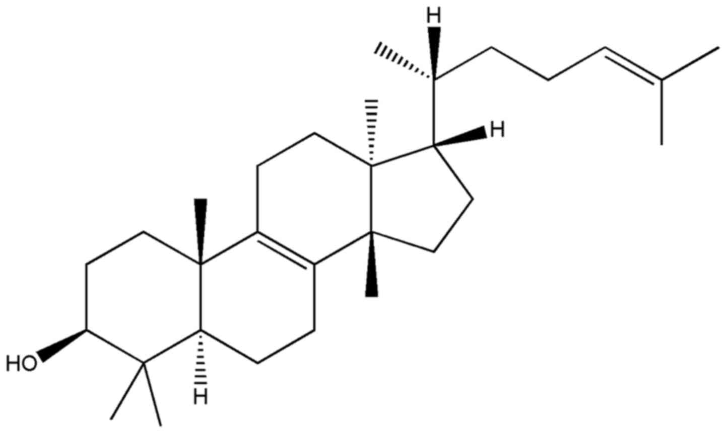
g). The chemical structure of the euphol, representeted in Fig. 1, was determined by elemental analyses
of 1H NMR and 13C NMR spectral data, and by comparison with their
respective authentic compounds using Chemdraw software version 7.0
(22,23) (PubChem CID: 441678). The 1H NMR (500
MHz) and 13C NMR (126 MHz) spectra were recorded on a Bruker 500
MHz instrument. The MS spectra were recorded on a Perkin Elmer
instrument, model API 150 and run in ES-MS positive mode: MH+ 427
m/e, MH+ _H2O 409 m/e. The NMR parameters were 13C NMR (CDCl3):
15.7 (C29), 15.8 (C18), 17.9 (C26), 19.1 (C21), 19.2 (C6), 20.4
(C19), 21.7 (C11), 24.9 (C30), 25.0 (C23), 25.9 (C27), 28.0 (C2,
C7), 28.2 (C28), 28.3 (C16), 30.0 (C15), 31.1 (C12), 35.4 (C1),
35.7 (C22), 36.1 (C20), 37.5 (C10), 39.2 (C4), 44.3 (C13), 49.9
(C17), 50.2 (C14), 51.2 (C5), 79.2 (C3), 125.4 (C24), 131.1 (C25),
133.8 (C9), 134.3 (C8) and 1H NMR (CDCl3): 0.75 (3H, s, H-18), 0.80
(3H, s, H-29), 0.85 (3H, d, H-21), 0.87 (3H, s, H-30), 0.95 (3H, s,
H-19), 1.00 (3H, s, H-28), 1.50 (3H, s, H-26), 1.68 (3H, s, H-27),
3.23 (1H, dd, H-3), 5.09 (1H, bt, H-24). The tetracyclic triterpene
euphol used in this study showed >95% purity.
The extract fraction was initially dissolved in
dimethyl sulfoxide (DMSO) at a concentration of 50 mg/ml and stored
at −20°C. The intermediate dilutions of the experimental compound
were prepared to obtain a concentration of 1% DMSO.
Cell viability assay
The cytotoxicity effect of euphol was assessed by
Cell Titer 96 Aqueous cell proliferation assay (MTS assay; Promega
Corporation, Madison, WI, USA), following the manufacturer's
instructions as previous described (20,24). For
experiments, the cells (third and fifth passage) were plated into
96-well plates (until a maximum 5×10³ cells/well). The
plate was incubated overnight for optimal attachment of adherent
lines, and then placed under low serum-starved conditions for 24 h
(DMEM supplemented with 0.5% of FBS). Subsequently, the cells were
treated with increasing concentrations of the test compound diluted
in DMEM (0.5% FBS) and incubated for 72 h. The control groups
received the same amount of vehicle (1% DMSO, final concentration).
Growth of tumoral cells was quantified by ability of living cells
to reduce the yellow dye MTS to a blue formazan product. Two hours
before the end of incubation, 10 µl of MTS were added to each well,
and the plate further incubated for 2 h at 37°C. Absorbance was
measured in automatic microplate reader Varioskan (Thermo Fisher
Scientific, Inc.) at 490 nm. The response to euphol treatment was
assessed by standardizing treated groups to the untreated control,
and were expressed as a percentage relatively to control cells, in
DMSO alone (considered as 100% viability) ± SD.
IC50 determination
The results of absorbance values of treated cells
were converted to percentage of cell viability in cells in with
presence of the vehicle (DMSO), which were used as control,
corresponding to 100% survival. The analysis of the non-linear
regression curve using GraphPad PRISM version 7 (GraphPad Software,
Inc., La Jolla, CA, USA) the was carried out on results of
viability, yielding an equation used to calculate the concentration
of substance required to produce 50% reduction in cell viability
(IC50) as previous determined (25,26).
Drug combination studies
Combination studies were done with fixed
concentrations (determinated IC50 value) of standard
chemotherapeutic paclitaxel (Sigma-T7402; Sigma-Aldrich; Merck
KGaA, Darmstadt, Germany) and gemcitabine hydrochloride
(Sigma-G6423; Sigma-Aldrich; Merck KGaA, Darmstadt, Germany) (data
not shown), exposed simultaneously to increasing concentrations of
euphol. The drug interactions were evaluated by the combination
index (CI) that was calculated by the Chou-Talalay equation, which
takes into account both the potency (Dm or IC50) and the
shape of the dose-effect curve (27,28),
using CalcuSyn software version 2.0 (Biosoft, Ferguson, MO, USA).
In CI analysis, synergy was defined as CI values significantly
lower than 1.0; antagonism as CI values significantly higher than
1.0; and additivity as CI values equal to 1.0 at drug
IC50 value for each cell line.
Proliferation assay
The proliferation effect of euphol was assessed by
BrdU assay kit (Roche Applied Science, Mannheim, Germany) following
the manufacturer's instructions. In brief, 5×10³
cells/well were seeded in a 96-well, flat-bottom plate. The plate
was incubated overnight for optimal attachment of adherent lines,
and then placed under low serum-starved conditions for 24 h
(DMEM-0.5% FBS). Subsequently, the cells were treated with
increasing concentrations of the test compound diluted in DMEM
(0.5% FBS) and incubated for 72 h. The control groups received the
same amount of vehicle (1% DMSO, final concentration). Therefore,
the proliferation effect was assessed by BrdU assay kit following
the manufacturer's instructions. Experiments were done in
triplicate in three independent experiments for each cell line.
The criterion of GI to ascertain the cell line
sensibility to euphol was previously described (29). Mean GI values was calculated at fixed
dose of 15 µM of euphol (concentration closer to the mean
IC50 value for all cell lines) and established as
100%-percentage of viable cells at this dose. Samples exhibiting
more than 60% GI in the presence of 15 µM euphol were classified as
highly sensitive (HS); moderately sensitive (MS) when located
between 40–60%; and resistant when showing less than 40%. All the
assays were done in triplicate and repeated at least three times
for each cell line.
Wound-healing migration assay
Cell migration properties were evaluated by
wound-healing assay, as previsously described by our group
(26,30). The pancreatic cancer cell lines,
Mia-Pa-Ca-2 and Panc-1, were plated in 6-well plates and grown to
confluence. A sterile tip was used to create a scratch in monolayer
cells. Cells were then incubated with euphol at 8.46 and 21.47 µM.
The ‘wounded’ areas were photographed by phase contrast microscopy
(Model IX71; Olympus Corporation, Tokyo, Japan) to evaluate wound
closure (0, 24, 48 and 72 h). The migration rate of individual
cells was determined by measuring the distances covered from the
initial time to the selected time-points (bar of distance tool,
DP2-BSW Olympus version 2.2). The percentage of the relative
migration distance was calculated as wound area at a given time
compared to the initial wound surface. Pictures shown are
representative of three independent experiments performed in
triplicates.
Colony formation-assay
Inhibition of anchorage-independent was assesed by
soft-type-agar assay as reported (30). We placed 1 ml of acellular solution
of 0.6% agar (combining equal volumes of 1.2% Noble agar with 2×
concentrated DMEM with 20% FBS) into a six-well plate and incubated
at 37°C for 10 min; 2×10⁴ cells of Mia-Pa-Ca-2 and
Panc-1 were homogenized in solution containing DMEM supplemented
with 0.35% agar (upper layer agar; equal volumes of 0.7% Noble agar
and 2X concentrated DMEM with 20% FBS) and seeded onto acellular
coating. After solidification 0.5 ml of DMEM medium + 10% FBS was
added. The medium was changed every two days, and DMEM medium +
0.5% euphol at 3 and 10 µM was added. The cells were incubated at
37°C in a humidified atmosphere of 5% CO2 for 20 days;
colonies formed were stained with 0.05% crystal violet for 15 min.
Photo-documented colonies were analyzed using the Image J Software.
The assay was performed in two biological replicates and the
experiments were done in duplicate.
Statistical analysis
The results of in vitro experiments are
expressed as mean ± standard deviation (SD) of three independent
experiments. IC50 values were obtained by nonlinear
regression. We applied Student's t-test for comparing two different
conditions whereas two-way analysis of variance with Tukey's post
hoc test was used for assessing differences between more groups.
P<0.05 was considered to indicate a statistically significant
difference. All statistical analyzes were performed using GraphPad
PRISM version 7 (GraphPad Software, Inc.).
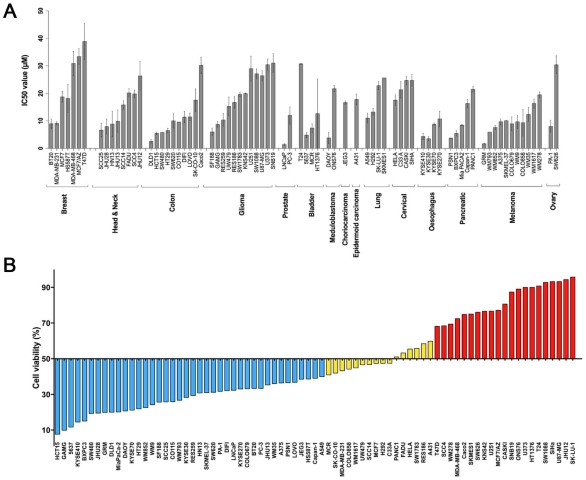
Results
Euphol promotes cytotoxity in human
cancer cell lines
The antitumor effect of euphol in vitro was
assessed using MTS assay on 73 human cancer lines from 15 solid
tumor models (breast, colon, bladder, prostate, lung, pancreas,
esophageal, head and neck, cervical, epidermoid carcinoma,
meduloblastoma, placental choriocarcinoma, ovarian carcinoma,
glioblastoma, and melanoma) (Table
I). We generated complete dose-response curves and
IC50 values for this euphol treatment. Among each tumor
type, the distinct cell lines exhibited a heterogeneous profile of
response to euphol (Fig. 2A). The
mean of IC50 values was 15.14 (6.47 µg/ml), but varied
significantly between individual cell lines with up to a more than
27-fold difference in the IC50 values [IC50
range: 1.41–38.89 µM (0.60–16.62 µg/ml)]. Esophageal squamous cell
carcinoma and pancreatic carcinomas showed the most sensitive
profiles (IC50 mean 11.08 and 6.84 µM, respectively,
Fig. 2A and Table I), followed by prostate, melanoma and
colon cancer cell lines.
To allow a better classification of the cell lines
response to euphol, we determined their GI. We found that 50.68%
(37/77) were classified as HS, 21.92% (16/73) were MS, and 27.4%
(20/77) were resistant (Fig. 2B and
Table I). Esophageal (100%),
prostate (100%) and pancreatic (80%) cancer models showed a higher
percentage of HS cell lines. At variance, glioma (54.5%) followed
by breast tumor type (42%) has the most cancer cell lines scored as
resistant.
Biological effect of euphol on
pancreatic cancer cell lines
We further investigated the biological effect of
euphol on pancreatic cancer cell lines, the most sensitive tumoral
type in our study. To determine whether the effect of euphol on
cancer cells is cytotoxic or cytostatic, its effect was also
evaluated on the proliferation of pancreatic cell lines by BrdU
incorporation. As shown in Fig. 3A,
euphol exhibited dose-dependent effects on proliferation of
pancreatic cancer cell lines (Panc-1 and Mia-Pa-Ca-2) but varied
significantly between individual cell lines. In both cancer cell
lines, low doses of euphol slightly decreased proliferation and the
dose of 17.51 µM was able to inhibit almost 50% of the
proliferation.
Euphol also exhibited dose-dependent effects on cell
viability on pancreatic cancer cell lines (Panc-1 and Mia-Pa-Ca-2).
Although euphol decreased the proliferation, the strongest
inhibition of proliferation was seen at 35.1 µM concentration of
euphol after 72 h, with almost 39.4% for Mia-Pa-Ca-2 cells and 51%
for Panc-1 cells (Fig. 3A), whereas
the same concentration suppressed cell viability of Mia-Pa-Ca-2
cells by 10.7% and Panc-1 cells by 22.4% (Fig. 3B). Thus, comparing the effects on
cell viability in concentrations of the same magnitude, euphol
seemed to inhibit growth through a more cytotoxic than cytostatic
fashion.
Next, we investigated its biological effect on
pancreatic cancer cell lines. In untreated conditions for both cell
lines (Mia-Pa-Ca-2 and Panc-1, complete close up of the wound took
longer than 72 h, suggesting that these cell lines have an
intrinsic low migratory capability. Those two cells lines were
treated with euphol at 8.46 and 21.47 µM. In spite of the low
migratory feature of the cells, euphol treatment significantly
inhibited cell migration in Mia-Pa-Ca-2 at 72 h when compared to
control cells (Fig. 3C). We could
not evaluate the effect of euphol in Panc-1 cell motility, since
this cell present an inherent low motility phenotype hampered an
adequate assessment of any inhibition. The findings suggest that
euphol has a cytotoxic effect but were not able to inhibit the
migration of this cell line (Fig.
3D).
In addition to cell proliferation
(anchorage-dependent growth) and cell migration, colony formation
(anchorage-independent growth) is one of the typical
characteristics of the metastatic potential of cancer cells in
vitro and strongly correlates with tumorigenesis in vivo
(31). Therefore, we evaluated the
effect of euphol in anchorage-independent growth of Panc-1 cancer
cells. Panc-1 cells formed colonies on agar after 20 days of
incubation, and the presence of euphol at IC50 value
resulted in a significant suppression of number and size of
colonies (P<0.05; Fig. 3E). The
ability to form colony was reduced by 90% compared to the untreated
control. Similarly, we proceeded the same way with Mia-Pa-Ca-2 cell
line, however we can not observe the colonies formation even in the
control condition suggesting a low tumorigenic potential of this
cell line (data not shown).
Euphol potentiates
chemotherapeutic-induced decrease in cell viability
We also evaluated the potential combinatorial value
of euphol in the context of standard esophageal and pancreatic
tumor therapy. We found that euphol and gemcitabine combination
treatment showed a synergistic effect (CI<1) in 50% of
pancreatic cells lines investigated (mean CI values, range:
0.76–0.8; Table II), being the
combination more effective than single agents. Likewise, euphol was
able to synergistically sensitize most esophageal cells lines to
paclitaxel treatment (mean CI values, range: 0.37–0.55; Table III).
| Table II.Drug combination studies in
pancreatic cancer cell lines. |
Table II.
Drug combination studies in
pancreatic cancer cell lines.
| Cell line | Euphol Mean
IC50 (µM) | SD | GEM Mean
IC50 (µM) | SD | CI
GEM+Euphola |
|---|
| Mia-PaCa-2 | 8.46 | 0.39 | 1.65 | 0.51 | 5.16 |
| PANC-1 | 21.47 | 1.83 | 10.37 | 2.62 | 1.5 |
| PSN-1 | 3.71 | 0.17 | 0.51 | 0.02 | 0.8 |
| BXPC-3 | 5.47 | 1.64 | 0.61 | 0.05 | 0.76 |
| Table III.Drug combination studies in
Oesophagus cancer cell lines. |
Table III.
Drug combination studies in
Oesophagus cancer cell lines.
| Cell line | Euphol Mean
IC50 (µM) | SD | Paclitaxel Mean
IC50 (µM) | SD | CI
PC+Euphola |
|---|
| KYSE30 | 3.52 | 1.28 | 0.015 | 0.003 | 0.54 |
| KYSE70 | 8.77 | 0.74 | 0.009 | 0.001 | 0.37 |
| KYSE270 | 10.71 | 3.95 | 0.018 | 0.002 | 0.55 |
| KYSE410 | 4.35 | 2.03 | 0.023 | 0.008 | 2.02 |
Discussion
In the present study, we investigated the cytotoxity
effects of euphol on a large panel of 73 human cancer cell lines,
derived from 15 solid tumor models including breast, colon,
bladder, prostate, lung, pancreas, esophagus, head and neck,
cervical cancer as well as glioma and melanoma. We observed that
euphol exhibited dose-dependent cytotoxic effects on all human
cancer cell lines tested with the highest effect (reduced
viability) for pancreas and esophageal cancer lines, followed by
prostate, melanoma and colon cancer cell lines. According to the
American National Cancer Institute criteria, euphol would be a
promising compound for further analysis since its IC50
values were lower than 30 µg/ml (http://www.cancer.gov) (32). The cytotoxic and/or growth-inhibition
effects of euphol were identified at low IC50 values,
being lower than 30 µg/ml for 71 out 73 cancer cell lines tested.
Our results are in agreement with earlier in vitro reports
that suggested E. tirucalli crude extracts or euphol may
have antitumoral effect. The relative toxicity of the E.
tirucalli crude extracts on Mia-Pa-Ca-2 was also evaluated by
Munro et al, which demonstrated that methanol extracts
exerted a significant decrease in cell viability at 25 µg/ml
(33). Also, Choene et al,
investigated E. tirucali crude extracts that contains
different types of secondary metabolites mainly terpenes and
flavonoids, and reported its effect on breast cancer (MCF-7 and
MDA-MB-231) cell cycle arrest (34).
Lin et al, reported the effect of euphol in gastric cancer
cell lines (CSN, CS12, AGS and MKN45) with an IC50
values of 49.6, 12.8, 14.7 and 14.4 (µg/ml), respectively (17). The authors also reported that euphol
induced apoptosis by upregulation of ERK signaling (17). Another study analyzing the T47D
breast cancer cells showed that euphol has an antiproliferative
activity, with IC50 values of 260 µM (16). The results suggest a cytostatic
effect for euphol that induced GI by cell cycle arrest at the G0/G1
phase. In our study we observed a lower IC50 value
(38.89 µM) in the T47D cells, yet, also above the limite considered
effective by NCI. It is worthy of note that although the extract of
E. tirucalli containing 64% euphol (7), in our study breast cancer cell lines
were the less responsive to euphol in which the HPLC purity
revealed above 95%. This finding seems to be in accordance with the
potential of the compound found in phytochemical evaluation, which
indicated that it is a tetracyclic triterpene alcohol (7,35).
To gain more insight into the role played by euphol
in tumorigenesis, we investigated the biological effect of euphol
on pancreatic cancer cell lines. Euphol inhibited cell
proliferation (anchorage-dependent growth) as well as colony
formation (anchorage-independent growth) of pancreatic cancer
cells. We also showed that euphol inhibits cell migration of
Mia-Pa-Ca-2 cancer cell line. One of the suitable molecular cancer
targets is protein kinase (ERK), which is an important factor in
the regulation of cell migration of numerous cell types. The ERK
pathway inhibitors PD98059 and U0126 inhibit the migration of
diverse cell types in response to cell matrix proteins, such as
fibronectin, vitronectin and collagen (36). Supporting a possible role of ERK
inhibition on migration modulation by euphol, Passos et al,
showed that, at the intracellular level, euphol reduced TPA-induced
extracellular signal-regulated ERK activation in skin inflammation
in mice (13). However, these data
are in disagreement with Lin et al, which showed that euphol
selectively induced gastric cancer cells apoptosis by activation of
ERK signaling (17). Taken together,
these findings provided further support that euphol effect may
depend on the cellular context and showed that further
investigation regarding euphol in other cancer cell lines and in
other experimental model are required.
In addition, we investigated the combination of
euphol to chemotherapy in pancreas and esophageal cancer lines and
we found that euphol when combined with a gemcitabine and
paclitaxel treatment seems to have a synergistic effect
(chemo-sensitization) leading to lower doses of therapeutic agents.
This synergy (chemo-sensitization) is of major interest since those
two standard chemotherapy drugs formed the therapy backbone for
those cancers, the level of responses seen in practice is still
suboptimal and there is an urgent need for improvement (37,38).
The present study constitutes, to our knowledge, the
first largest screening of euphol efficacy on human cancer cell
lines. We showed that euphol could be a promising agent on large
number of tumor types, in particularly in esophageal and pancreatic
cancer. One important limitation of the present study is the lack
of normal counterpart cells of the distinct tumor types evaluated.
Therefore, additional studies are warranted to address this topic.
This study also revealed the inhitbition/reduction of some hallmark
events, such as proliferation and migration as part of the
mechanism of action of this compound on pancreatic cancer cells.
Finally, the euphol also showed synergistic interactions with
chemotherapeutic drugs used in clinical practice. Our results
provide insights for further studies suggesting euphol as an
interesting antineoplastic alone or in combination for cancer
treatment.
Acknowledgements
The authors would like to acknowledge the
discussions of Amazônia Fitomedicamentos Scientific Committee, in
particular Dr. Amilcar Tanuri.
Funding
This study was supported by grants from Amazônia
Fitomedicamentos Ltda. (grant no. FITO 05/2012) and Barretos Cancer
Hospital, all from Brazil.
Availability of data and material
All data generated or analyzed during this study are
included in this published article.
Authors' contributions
VAOS designed the experiments, and participated in
data acquisition and interpretation. VAOS and MNR carried out the
studies of cell culture, including cytotoxicity and proliferation
assays, wound healing migration assay, colony formation assay, drug
combination studies and statistical analysis. AT helped to carry
out the cell viability assay. RJSO and OM contributed to the design
of some experiments, interpretation of data and were involved in
critically revising the manuscript. JPL helped to design the drug
combination experiments, and helped to draft and critically revise
the manuscript. LFP was responsible for the preparation of extracts
and contributed to the discussion of cytotoxicity results. RMR
conceived the study, participated in its design and coordination,
interpreted the data, drafted the manuscript and was involved in
revising it critically for important intellectual content. All
authors read and approved the final manuscript.
Ethics approval and consent to
participate
Not applicable.
Consent for publication
Not applicable.
Competing interests
The drug euphol was provided by Amazônia
Fitomedicamentos Ltda. LFP is one of the authors and also one of
the inventors of euphol's patent. The Amazônia Fitomedicamentos
Ltda. is the sole and exclusive owner of the respective
intellectual property rights. This study was supported by grants
from Amazônia Fitomedicamentos Ltda as part of the euphol
pre-clinical studies and VAOS and MNR received a scholarship from
Amazônia Fitomedicamentos Ltda. to conduct of the study.
Glossary
Abbreviations
Abbreviations:
|
ANOVA
|
analysis of variance
|
|
CI
|
combination index
|
|
DNA
|
deoxyribonucleic acid
|
|
DMEM
|
dulbecco's modified eagle's medium
|
|
DMSO
|
dimethyl sulfoxide
|
|
ETHE
|
E. tirucalli hydroalcoholic
extract
|
|
FBS
|
fetal bovine serum
|
|
FDA
|
food and drug administration
|
|
g
|
gram
|
|
GI
|
growth inhibition
|
|
HPLC
|
high performance liquid chromatography
analysis
|
|
HS
|
highly sensitive
|
|
IC50
|
half maximal inhibitory
concentration
|
|
MHz
|
megahertz
|
|
ml
|
milliliter
|
|
MS
|
moderately sensitive
|
|
MTS
|
3-(4,5-dimethylthiazol-2-yl)-5-(3-carboxymethoxyphenyl)-2-(4-sulfophenyl)-2H-tetrazolium
|
|
NCI
|
National Cancer Institute
|
|
NMR
|
nuclear magnetic resonance
|
|
P/S
|
penicillin/streptomycin solution
|
|
R
|
resistant
|
|
RPMI-1640
|
Roswell Park Memorial
Institute-1640
|
|
SD
|
standard deviation
|
|
STR
|
short tandem repeat
|
|
WHO
|
World Health Organization
|
|
uM
|
micromolar
|
References
|
1
|
Lee JA, Uhlik MT, Moxham CM, Tomandl D and
Sall DJ: Modern phenotypic drug discovery is a viable, neoclassic
pharma strategy. J Med Chem. 55:4527–4538. 2012. View Article : Google Scholar : PubMed/NCBI
|
|
2
|
Khazir J, Riley DL, Pilcher LA, De-Maayer
P and Mir BA: Anticancer agents from diverse natural sources. Nat
Prod Commun. 9:1655–1669. 2014.PubMed/NCBI
|
|
3
|
Cragg GM, Newman DJ and Yang SS: Natural
product extracts of plant and marine origin having antileukemia
potential. The NCI experience. J Nat Prod. 69:488–498. 2006.
View Article : Google Scholar
|
|
4
|
WHO Model List of Essential Medicines Home
Page. Aug 4–2016
|
|
5
|
Hopkins AL: Network pharmacology: The next
paradigm in drug discovery. Nat Chem Biol. 4:682–690. 2008.
View Article : Google Scholar : PubMed/NCBI
|
|
6
|
Braz-Filho R: Contribuição da fitoquímica
para o desenvolvimento de um país emergente. Quim Nova. 33:229–239.
2010. View Article : Google Scholar
|
|
7
|
Dutra RC, Campos MM, Santos AR and Calixto
JB: Medicinal plants in Brazil: Pharmacological studies, drug
discovery, challenges and perspectives. Pharmacol Res. 112:4–29.
2016. View Article : Google Scholar : PubMed/NCBI
|
|
8
|
Franco-Salla GB, Prates J, Cardin LT, Dos
Santos AR, Silva WA Jr, da Cunha BR, Tajara EH, Oliani SM and
Rodrigues-Lisoni FC: Euphorbia tirucalli modulates gene expression
in larynx squamous cell carcinoma. BMC Complement Altern Med.
16:1362016. View Article : Google Scholar : PubMed/NCBI
|
|
9
|
Prakash E and Gupta DK: Cytotoxic
activities of extracts of medicinal plants of euphorbiacae family
studied on seven human cancer cell lines. Univers J Plant Sci.
1:113–117. 2013.
|
|
10
|
Keating GM: Ingenol mebutate gel 0.015 and
0.05%: In actinic keratosis. Drugs. 72:2397–2405. 2012. View Article : Google Scholar : PubMed/NCBI
|
|
11
|
Lebwohl M, Swanson N, Anderson LL,
Melgaard A, Xu Z and Berman B: Ingenol mebutate gel for actinic
keratosis. N Engl J Med. 366:1010–1019. 2012. View Article : Google Scholar : PubMed/NCBI
|
|
12
|
Dutra RC, Bicca MA, Segat GC, Silva KA,
Motta EM, Pianowski LF, Costa R and Calixto JB: The antinociceptive
effects of the tetracyclic triterpene euphol in inflammatory and
neuropathic pain models: The potential role of PKCepsilon.
Neuroscience. 303:126–137. 2015. View Article : Google Scholar : PubMed/NCBI
|
|
13
|
Passos GF, Medeiros R, Marcon R,
Nascimento AF, Calixto JB and Pianowski LF: The role of PKC/ERK1/2
signaling in the anti-inflammatory effect of tetracyclic triterpene
euphol on TPA-induced skin inflammation in mice. Eur J Pharmacol.
698:413–420. 2013. View Article : Google Scholar : PubMed/NCBI
|
|
14
|
Bani S, Kaul A, Khan B, Gupta VK, Satti
NK, Suri KA and Qazi GN: Anti-arthritic activity of a biopolymeric
fraction from Euphorbia tirucalli. J Ethnopharmacol. 110:92–98.
2007. View Article : Google Scholar : PubMed/NCBI
|
|
15
|
Dutra RC, de Souza PR, Bento AF, Marcon R,
Bicca MA, Pianowski LF and Calixto JB: Euphol prevents experimental
autoimmune encephalomyelitis in mice: Evidence for the underlying
mechanisms. Biochem Pharmacol. 83:531–542. 2012. View Article : Google Scholar : PubMed/NCBI
|
|
16
|
Wang L, Wang G, Yang D, Guo X, Xu Y, Feng
B and Kang J: Euphol arrests breast cancer cells at the G1 phase
through the modulation of cyclin D1, p21 and p27 expression. Mol
Med Rep. 8:1279–1285. 2013. View Article : Google Scholar : PubMed/NCBI
|
|
17
|
Lin MW, Lin AS, Wu DC, Wang SS, Chang FR,
Wu YC and Huang YB: Euphol from Euphorbia tirucalli selectively
inhibits human gastric cancer cell growth through the induction of
ERK1/2-mediated apoptosis. Food Chem Toxicol. 50:4333–4339. 2012.
View Article : Google Scholar : PubMed/NCBI
|
|
18
|
Santos OJ, Sauaia Filho EN, Nascimento FR,
Júnior FC, Fialho EM, Santos RH, Santos RA and Serra IC: Use of raw
Euphorbia tirucalli extract for inhibition of ascitic Ehrlich
tumor. Rev Col Bras Cir. 43:18–21. 2016.(In English, Portuguese).
View Article : Google Scholar : PubMed/NCBI
|
|
19
|
MacNeil A, Sumba OP, Lutzke ML, Moormann A
and Rochford R: Activation of the Epstein-Barr virus lytic cycle by
the latex of the plant Euphorbia tirucalli. Br J Cancer.
88:1566–1569. 2003. View Article : Google Scholar : PubMed/NCBI
|
|
20
|
Silva-Oliveira RJ, Silva VA, Martinho O,
Cruvinel-Carloni A, Melendez ME, Rosa MN, de Paula FE, de Souza
Viana L, Carvalho AL and Reis RM: Cytotoxicity of allitinib, an
irreversible anti-EGFR agent, in a large panel of human
cancer-derived cell lines: KRAS mutation status as a predictive
biomarker. Cell Oncol(Dordr). 39:253–263. 2016. View Article : Google Scholar : PubMed/NCBI
|
|
21
|
Dirks WG, Faehnrich S, Estella IA and
Drexler HG: Short tandem repeat DNA typing provides an
international reference standard for authentication of human cell
lines. Altex. 22:103–109. 2005.PubMed/NCBI
|
|
22
|
Yasukawa K, Akihisa T, Yoshida ZY and
Takido M: Inhibitory effect of euphol, a triterpene alcohol from
the roots of Euphorbia kansui, on tumour promotion by
12-O-tetradecanoylphorbol-13-acetate in two-stage carcinogenesis in
mouse skin. J Pharm Pharmacol. 52:119–124. 2000. View Article : Google Scholar : PubMed/NCBI
|
|
23
|
Dutra RC, Simao da Silva KA, Bento AF,
Marcon R, Paszcuk AF, Meotti FC, Pianowski LF and Calixto JB:
Euphol, a tetracyclic triterpene produces antinociceptive effects
in inflammatory and neuropathic pain: The involvement of
cannabinoid system. Neuropharmacology. 63:593–605. 2012. View Article : Google Scholar : PubMed/NCBI
|
|
24
|
Teixeira TL, Oliveira Silva VA, da Cunha
DB, Polettini FL, Thomaz CD, Pianca AA, Zambom FL, da Silva Leitão
Mazzi DP, Reis RM and Mazzi MV: Isolation, characterization and
screening of the in vitro cytotoxic activity of a novel L-amino
acid oxidase (LAAOcdt) from Crotalus durissus terrificus venom on
human cancer cell lines. Toxicon. 119:203–217. 2016. View Article : Google Scholar : PubMed/NCBI
|
|
25
|
Martinho O, Zucca LE and Reis RM: AXL as a
modulator of sunitinib response in glioblastoma cell lines. Exp
Cell Res. 332:1–10. 2015. View Article : Google Scholar : PubMed/NCBI
|
|
26
|
Martinho O, Silva-Oliveira R,
Miranda-Goncalves V, Clara C, Almeida JR, Carvalho AL, Barata JT
and Reis RM: In vitro and in vivo analysis of RTK inhibitor
efficacy and identification of its novel targets in glioblastomas.
Transl Oncol. 6:187–196. 2013. View Article : Google Scholar : PubMed/NCBI
|
|
27
|
Chou TC and Talalay P: Quantitative
analysis of dose-effect relationships: The combined effects of
multiple drugs or enzyme inhibitors. Adv Enzyme Regul. 22:27–55.
1984. View Article : Google Scholar : PubMed/NCBI
|
|
28
|
Bruzzese F, Di Gennaro E, Avallone A, Pepe
S, Arra C, Caraglia M, Tagliaferri P and Budillon A: Synergistic
antitumor activity of epidermal growth factor receptor tyrosine
kinase inhibitor gefitinib and IFN-alpha in head and neck cancer
cells in vitro and in vivo. Clin Cancer Res. 12:617–625. 2006.
View Article : Google Scholar : PubMed/NCBI
|
|
29
|
Konecny GE, Glas R, Dering J, Manivong K,
Qi J, Finn RS, Yang GR, Hong KL, Ginther C, Winterhoff B, et al:
Activity of the multikinase inhibitor dasatinib against ovarian
cancer cells. Br J Cancer. 101:1699–1708. 2009. View Article : Google Scholar : PubMed/NCBI
|
|
30
|
Moniz S, Martinho O, Pinto F, Sousa B,
Loureiro C, Oliveira MJ, Moita LF, Honavar M, Pinheiro C, Pires M,
et al: Loss of WNK2 expression by promoter gene methylation occurs
in adult gliomas and triggers Rac1-mediated tumour cell
invasiveness. Hum Mol Genet. 22:84–95. 2013. View Article : Google Scholar : PubMed/NCBI
|
|
31
|
Freedman VH and Shin SI: Cellular
tumorigenicity in nude mice: Correlation with cell growth in
semi-solid medium. Cell. 3:355–359. 1974. View Article : Google Scholar : PubMed/NCBI
|
|
32
|
Trendowski M: Recent advances in the
development of antineoplastic agents derived from natural products.
Drugs. 75:1993–2016. 2015. View Article : Google Scholar : PubMed/NCBI
|
|
33
|
Munro B, Vuong QV, Chalmers AC, Goldsmith
CD, Bowyer MC and Scarlett CJ: Phytochemical, antioxidant and
anti-cancer properties of Euphorbia tirucalli methanolic and
aqueous extracts. Antioxidants (Basel). 4:647–661. 2015. View Article : Google Scholar : PubMed/NCBI
|
|
34
|
Choene M and Motadi L: Validation of the
antiproliferative effects of Euphorbia tirucalli extracts in breast
cancer cell lines. Mol Biol (Mosk). 50:115–127. 2016. View Article : Google Scholar : PubMed/NCBI
|
|
35
|
Silva AC, de Faria DE, Borges NB, de Souza
IA, Peters VM and Guerra Mde O: Toxicological screening of
Euphorbia tirucalli L: Developmental toxicity studies in rats. J
Ethnopharmacol. 110:154–159. 2007. View Article : Google Scholar : PubMed/NCBI
|
|
36
|
Klemke RL, Cai S, Giannini AL, Gallagher
PJ, de Lanerolle P and Cheresh DA: Regulation of cell motility by
mitogen-activated protein kinase. J Cell Biol. 137:481–492. 1997.
View Article : Google Scholar : PubMed/NCBI
|
|
37
|
Voutsadakis IA: Molecular predictors of
gemcitabine response in pancreatic cancer. World J Gastrointest
Oncol. 3:153–164. 2011. View Article : Google Scholar : PubMed/NCBI
|
|
38
|
Wiedmann MW and Mossner J: New and
emerging combination therapies for esophageal cancer. Cancer Manag
Res. 5:133–146. 2013. View Article : Google Scholar : PubMed/NCBI
|