Introduction
Osteoporosis (OP) is a bone disease characterized by
a low bone mass and deterioration of bone tissue, leading to
increased bone fragility and increasing the susceptibility of
patients to fracture (1). OP
commonly affects the elderly and its prevalence and associated
complications are rapidly increasing worldwide (1). It is associated with a reduction in
bone density, which increases bone fragility and the probability of
fracture, particularly of the hip, wrist and spine. It is estimated
that >200 million people have OP and it has been predicted that
the number of those experiencing hip fractures may rise to 6.26
million by 2050 (2,3). Current therapeutic strategies to treat
the bone degradation that occurs as a result of OP include the use
of bisphosphonates that increase bone mass, strength and
turnover.
Zoledronic acid (ZA) is a potent antiresorptive
bisphosphonate that actively inhibits osteoclast proliferation,
reduces the risk of fractures in patients with postmenopausal
conditions and is therefore used to treat OP (4). Bisphosphonates with nitrogen containing
side chains (N-BPs) inhibit a key enzyme in the mevalonate pathway,
farnesyl pyrophosphate synthase, thereby preventing the prenylation
of small GTPases in osteoclasts (5).
The enhanced permeation and retention effect of the nanoparticle
drug delivery system also increases the accumulation of ZA in
osteoclasts and inhibits the disruption of mature osteoclast
activity that causes cellular apoptosis (5,6).
Accumulation of ZA in the bone may pose a serious health risk. It
has been suggested that nanotechnologies and nanomaterials may be
used to formulate alternative treatments of OP and its
complications (7). In addition, it
has been demonstrated that bisphosphonates inhibit bone resorption
at sites of high osteoclast activity and they have therefore been
extensively used to treat patients with OP (8).
Nanoparticles are making a notable impact on the
medical landscape. Numerous examples of therapeutic strategies
involving nanoparticles, including biocompatible drug carriers, are
currently undergoing clinical trials and may be developed as more
beneficial alternatives to traditional therapies (9–11).
Liposomes are artificially constructed nanostructures and have been
extensively studied due to their superior properties over other
nanosystems. These include minimal toxicity, no immunogenicity,
good biodegradability and biocompatibility, making them ideal drug
delivery carriers (12–15). In addition, phospholipid bilayers
allow liposomes to encapsulate hydrophobic and hydrophilic
bioactive molecules. Nanocarriers with bone-targeting ligands may
therefore be developed as a promising solution to deliver drugs to
affected sites (16–18).
Hydroxyapatite (HA) is widely used as a naturally
occurring inorganic bone substitute material due to its properties
of biocompatibility and osteoconductivity. This is due to its
similarity with the mineral components of bone and it is therefore
able to induce local deposition/accumulation in the bone tissue
(19,20). It has been demonstrated that, unlike
bisphosphonates, HA does not stimulate negative osteoclast
activity; however, it stimulates positive bone remodeling and
osteogenic activity (21). Following
administration of such drug-loaded nanocarriers that form strong
interactions with HA, the rapid retention and accumulation of
nanocarriers onto the bone tissue may occur. However, the
construction of synthetic bone graft substitutes to complement the
ability of the bones to self-repair is challenging (22,23).
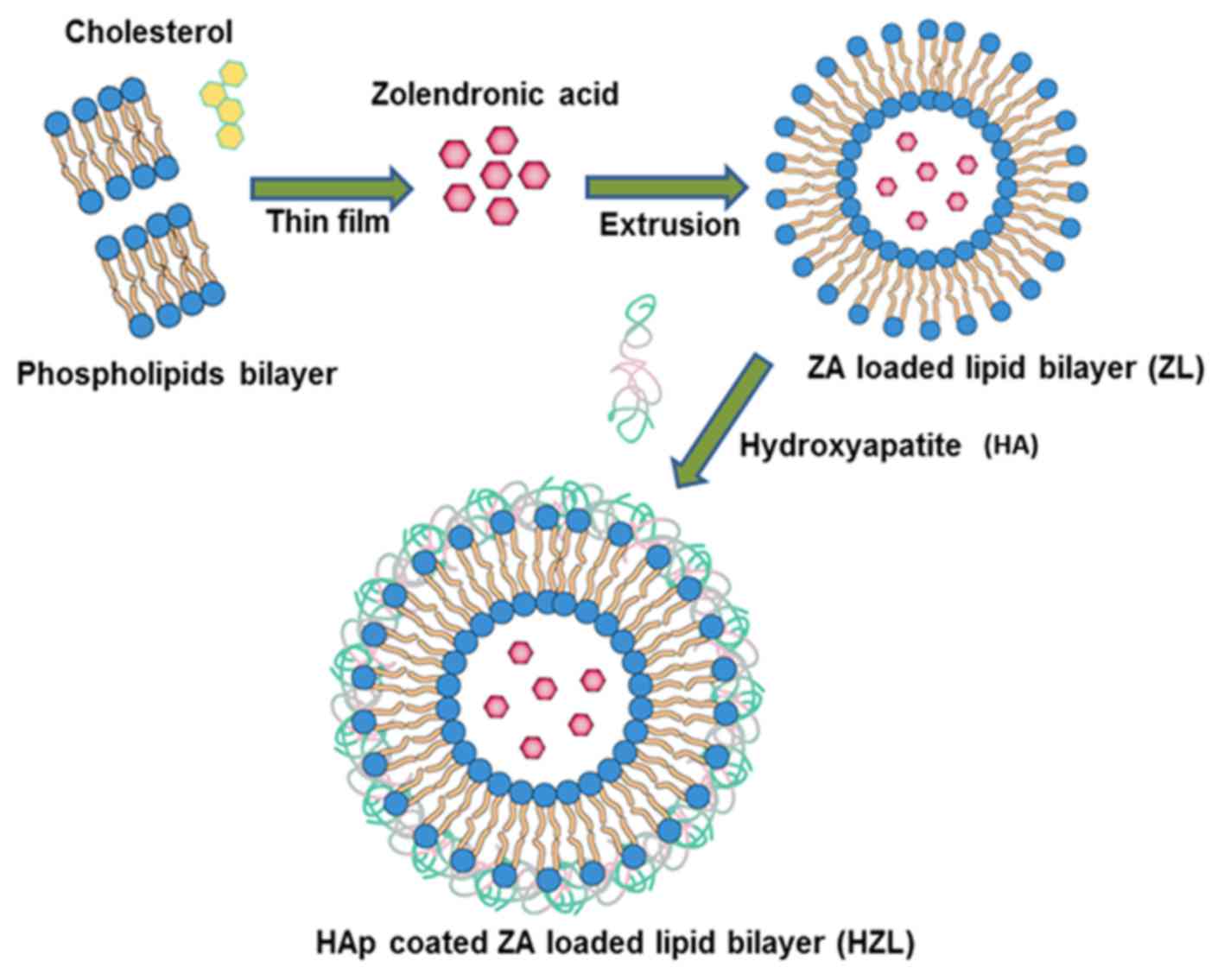
In the present study, HA-coated ZA-loaded lipid
bilayer nanoparticles (HZL NPs) were designed and constructed. ZA
was encapsulated in the hydrophilic cores of nanoparticles and, as
HA is negatively charged, it was able to electrostatically interact
with the surface of the positively charged lipid molecules.
Therefore, HZL had a dual function: ZA was present in the inner
core of the liposome to facilitate sustained drug release and HA
was present in the outer layer to provide osteoconductivity for
bone cells. Therefore, several physicochemical parameters were
characterized in the present study and the affinity of
nanoparticles towards HA was evaluated using the human osteoblast
cell line HFOb 1.19. The results of the present study indicate that
this bone-targeting drug delivery system may be developed as a
novel therapeutic strategy to treat patients with OP.
Materials and methods
Reagents
ZA was purchased from Sigma-Aldrich; Merck KGaA
(Darmstadt, Germany). L-α-phosphatidylcholine (EPC),
1,2-dioleoyl-3-trimethylammonium-propane (DOTAP), and cholesterol
(CHOL) were purchased from Avanti Polar Lipids Inc. (Alabaster, AL,
USA). HA powder with small particle sizes was obtained from
Oerlitken Metco Switzerland (Pfaffikon, Switzerland). The human
osteoblast cell line HFOb 1.19 was purchased from the American Type
Culture Collection (Manassas, VA, USA). All chemicals were of
analytical grade and used without further purification.
Preparation of HZL NPs
Liposomes consisting of EPC/DOTAP/CHOL (3:1:1) were
prepared by hydration of a thin lipid film followed by extrusion.
The lipid mixture was dissolved in 1 ml chloroform/methanol (2:1
v/v) mixture, the resulting solution was added to a round-bottom
flask and the solvent was removed using a rotary evaporator.
Subsequently, the lipid film was hydrated with 3 mg ZA and 1 ml 0.2
µm filtered distilled water, and the resulting suspension was
gently mixed in the presence of glass beads until the lipid layer
was removed from the glass wall. Following this step, the flask was
left at room temperature for a further 2 h. The liposome suspension
was then extruded using an extruder system (Avanti Wind Systems,
Franklin, WI, USA), which repeatedly passed the suspension under a
nitrogen atmosphere through polycarbonate membranes with decreasing
pore sizes.
Briefly, 0.020 M calcium acetate and 0.010 M
phosphoric acid solutions were added to the liposomal solution
(1:1:1 v/v %), followed by the addition of an ammonium solution
(20% v/v) to adjust the pH to 9.0 (19). The mixture was then incubated to form
a HA coating on the liposomal surface which finally resulted in
HA-coated ZA-loaded lipid layer nanoparticles (HZLs) were then
isolated using the Sephadex G-50 column (GE Healthcare, Chicago,
IL, USA). Following preparation, liposomes were stored at 4°C. Each
formulation was prepared in triplicate.
Particle size and measurement of
ζ-potential
The mean diameter and polydispersity of
nanoparticles was measured using a dynamic light scattering
technique. A Zetasizer Nano ZS (Malvern Instruments, Ltd., Malvern,
UK) was used to measure the size at room temperature, with a
detection angle of 90°. ζ-potential measurements of the formulation
were determined using a detection angle of 17°. Each sample was
diluted 10-fold with filtered water prior to each measurement. All
values were calculated as the mean of three separate batches.
Morphological characterization
The morphology of the samples was characterized
using a JSM field emission scanning electron microscope (JEOL Ltd.,
Tokyo, Japan). Powdered nanoparticles were spread onto a carbon
tape over a stub and vacuum-dried. Gold coating was applied using
an ion-sputtering device.
Encapsulation and loading
efficiency
Quantitative analysis of ZA was performed by
high-performance liquid chromatography (HPLC) (Agilent
Technologies, Inc., Santa Clara, CA, USA) using a C18 (1) column (3 µm, 150×4.6 mm) (Agilent). The
injection volume used was 20 µl with a 1 ml/min flow rate using a
mobile phase 20:80 (v/v) of acetonitrile and an aqueous solution (8
mM di-potassium hydrogen orthophosphate, 2 mM di-sodium hydrogen
orthophosphate and 7 mM tetra-n-butyl ammonium hydrogen sulfate,
adjusted at pH 7.0 with sodium hydroxide). Briefly, the drug-loaded
nanocarrier was filtered with an Amicon centrifugal filter
(Sigma-Aldrich; Merck KGaA) by centrifugation at a high speed of
2,571 × g for 20 min at 37°C. The unencapsulated ZA was analyzed at
215 nm to quatify the amount to be loaded in the nanoparticle using
equations 1 and 2. Furthermore, a standard curve of ZA was
individually plotted. The ZA encapsulation efficiency was
calculated using the following equations:
Encapsulation efficiency=Total amount of ZA-Amount of free
ZA×100Total amount of ZA
Loading efficiency=Total amount of ZA-Amount of free ZA×100Total
weight of nanoparticles
In vitro release studies
To investigate the release of ZA from HZL NPs, a
fixed amount (1 mg/ml) of drug-loaded nanoparticles was incubated
in 10 ml phosphate buffer solution [PBS (pH 7.4) ionic strength 0.1
M with 0.1% Tween 80] in a dialysis bag (molecular cut-off, 10 kDa)
at 37°C and under gentle magnetic stirring (10 × g). At fixed time
intervals, 1 ml supernatant was withdrawn and replaced with fresh
buffer solution to maintain a sink condition. Furthermore, the
amount of ZA released in the samples collected was extracted by
acetonitrile and determined by HPLC using the aforementioned
protocol. All experiments were performed in triplicate.
Cell culture
HFOb 1.19 cells were obtained from the American Type
Culture Collection (Manassas, VA, USA) and cells were cultured for
24 h in Dulbecco's Modified Eagle's medium (DMEM) supplemented with
10% fetal bovine serum (Stem Cell Technologies, Seoul, Korea), 2 mM
L-glutamine, 0.3 mg/l G418, 100 U/ml penicillin and 100 mg/ml
streptomycin. Cells were maintained at 37°C with 5% CO2
in a humidified incubator.
MTT assay
The cytotoxic potential of the individual
formulations was evaluated using an MTT assay. Briefly, cells were
seeded into 96-well plates at a density of 0.5×10⁴
cells/well in 0.1 ml growth medium (Stem Cell Technologies) and
incubated for 72 h at 37°C. The following day, the cells were
treated with the respective formulations [free ZA, ZA-loaded lipid
bilayer (ZL) and HZL NPs] at concentrations between 0.001 to 50 µM
and also treated with blank formulation, where the blank liposome
concentration was equivalent to the drug concentration of the
formulation. Respective formulations were incubated for 72 h at
37°C. At each time interval, the cells were washed twice with PBS
and treated with MTT solution (5 mg/ml) and incubated for another 4
h at 37°C. The resultant formazan crystals were then dissolved in
100 µl dimethyl sulfoxide (DMSO). The mixture was gently agitated
in a microplate reader prior to measuring the absorbance at 570 nm
and each experiment was repeated 6 times.
Apoptosis assay
Cell apoptosis was evaluated using the Annexin
V/fluorescein isothiocyanate (FITC) apoptosis detection kit (BD
Biosciences, Franklin Lakes, NJ, USA) and propidium iodide (PI;
Sigma-Aldrich; Merck KGaA). Briefly, cells were harvested using
0.25% trypsin 48 h following treatment with different formulations,
washed twice in cold PBS and re-suspended in binding buffer.
Subsequently, cells were incubated with 5 µl Annexin V/FITC and 5
µl PI for 15 min at room temperature in the dark. A flow cytometer
was used to measure the apoptosis of osteoblasts.
HA affinity test
In total, 3 mg HZL NPs were dissolved in DMSO.
Subsequently, 1 mg nanocarrier in DMSO was added to 15 µl
trimethylamine. This solution was then mixed with 7.5 ml distilled
water, sonicated for 5 min at 37°C and dialyzed for 48 h. A total
of 2 ml dialysis solution was stirred with 500 µl HA suspension (10
mg/ml) in buffer solution. Following incubation, the suspension was
centrifuged for 4 min at 2,571 × g and washed with buffer solution
at room temperature. The supernatant was then collected and
lyophilized and analyzed using a UV spectrophotometer at 280 nm.
HZL NPs without HA coating were used as a control.
Statistical analysis
All results are expressed as the mean ± standard
deviation. Data were analyzed using analysis of variance or the
Student's t-test using IBM SPSS Statistics for Windows (version
14.0; SPSS Inc., Chicago, IL, USA) to determine whether differences
between test groups were statistically significant. P<0.05 was
considered to indicate a statistically significant difference.
Results
Characterization of HZL NPs
Briefly, the schematic illustration depicted in
Fig. 1 demonstrates the successful
loading of hydrophilic ZA onto the aqueous core of the lipid
nanoparticle using the extrusion method. EPC and DOTAP were
selected to prepare stable positively charged liposomes using the
modified thin film method. An increase in the ratio of DOTAP in
liposomes also leads to a more positive z-potential, since DOTAP
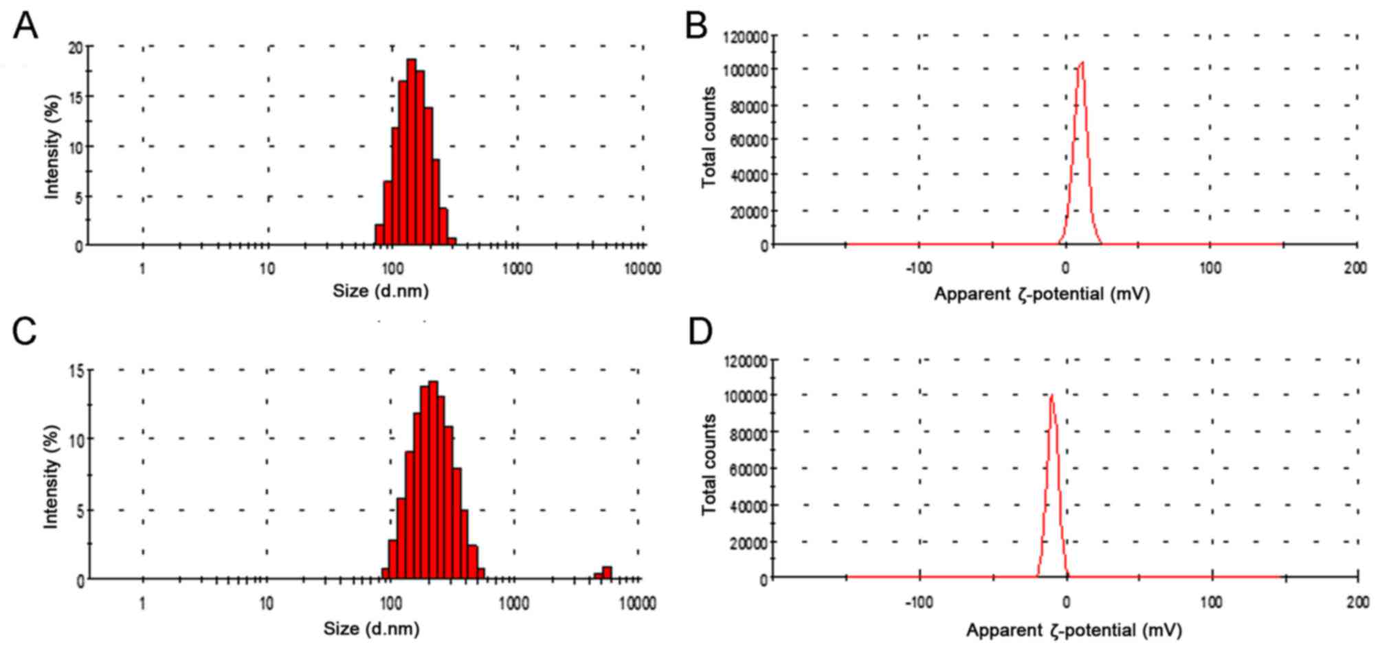
itself is positively charged. Dynamic light scattering analysis is
depicted in Fig. 2. The Z-potential
of the liposome (3:1) was 12.4 mV and the hydrodynamic size of the
ZA loaded liposome was 172.4±3.47 nm (Fig. 2A). HA was fabricated on the surface
of the lipid nanoparticle using the precipitation method and
following HA precipitation, the mean particle size increased from
172.4 to 197.8 nm (Fig. 2C).
Furthermore, following HA coating, the surface charge on the
liposomes decreased from 12.4 to−6.3 mV (Fig. 2B and D). These results indicate that
HA coating screens the negative charge of the liposomes. In
addition, the loading efficiency of ZA was high (~83%)
corresponding to 9.28% drug loading due to the negative charge of
the ZA interacting with the positively charged bilayer. This
indicates a major contribution to the high encapsulation efficiency
of ZA in ZL.
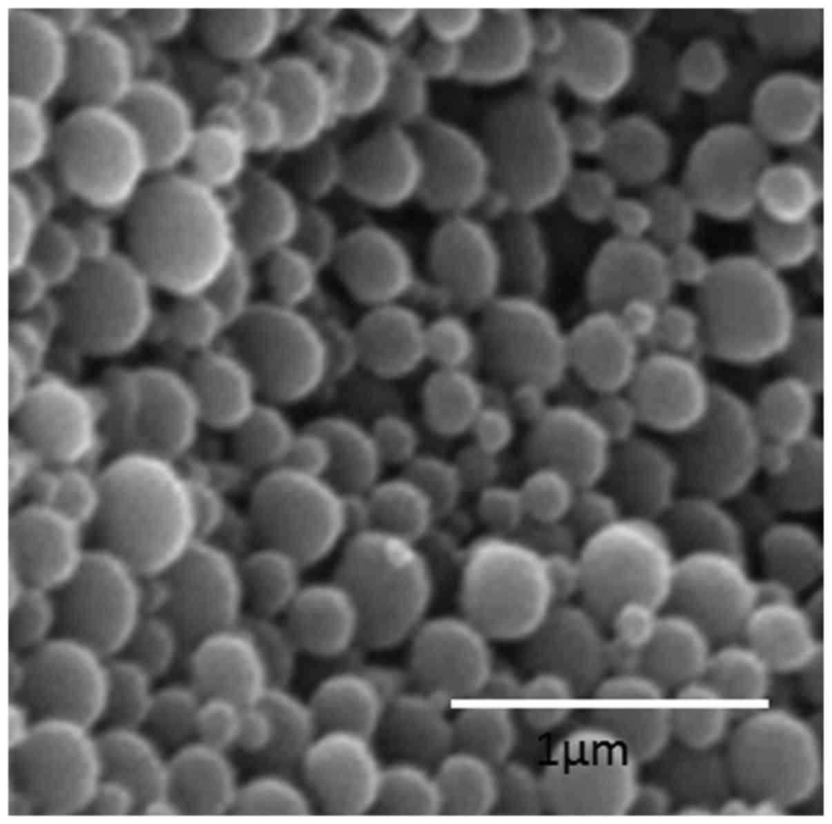
Surface morphology
The nanoparticle surface morphology of HZL was
investigated using scanning electron microscopy (SEM). As observed
in Fig. 3, HZL NPs were spherically
shaped and uniformly distributed on the SEM grid. The SEM images of
HZL revealed well-defined, spherical-shaped particles with a
uniform distribution. The spherical nature of the particle was
attributed to the self-assembly of the lipid bilayer. This figure
reveals the SEM micrographs of particles of the HZL complex
containing particles of various sizes (from a few microns to a few
hundred microns). However, the overall size of the particles was
larger as confirmed by scale bar and consisted of a smooth surface
following coating with lipid as seen in SEM image.
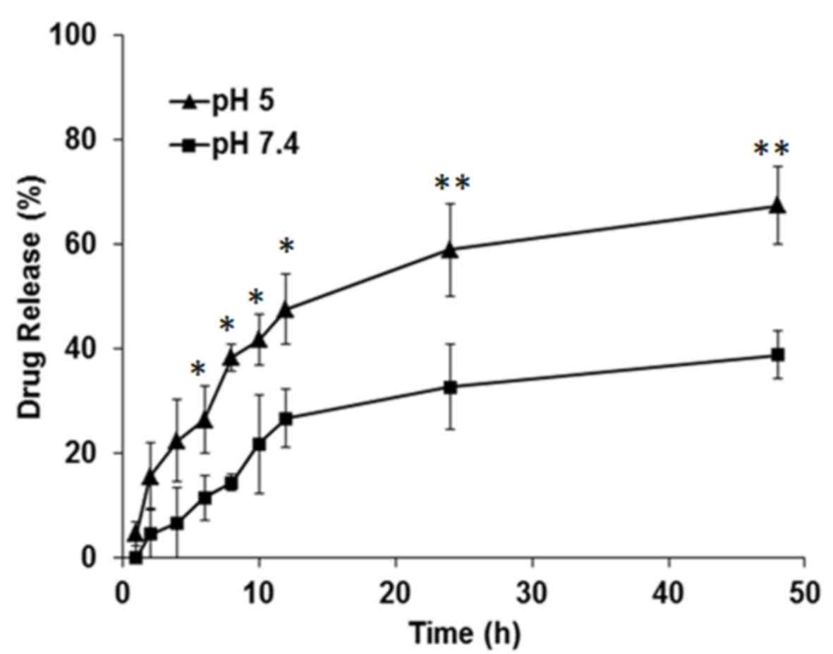
In vitro drug release
As presented in Fig.
4, the amount of the drug cumulatively released from HZL was
higher at a lower pH (5.0) than at physiological pH (7.4). The
difference between the release of the drug at pH 5.0 and 7.4 was
statistically significant at 10 to 50 h (P<0.05 and P<0.01).
ZA-loaded HZL NPs at physiological pH exhibited a biphasic drug
release pattern characterized by an initial rapid release in which
8.42±2.89% ZA was released over 6 h, followed by a sustained
continuous release phase in which 38.17±2.12% ZA was released from
the lipid bilayer nanoparticle over 48 h. The pH around the border
of osteoblasts is ~5.0 during bone remodeling and homeostatic body
fluid is ~7.4. HA more readily dissolves in acidic pH and under
acidic conditions, there was an immediate burst release of
34.1±4.81% drug over 8 h, due to the HA surface layer dissolving
immediately. Consequently, drug release followed a sustained
release profile from the nanoparticles, with 64.2±3.75% of drug
being released following 48 h.
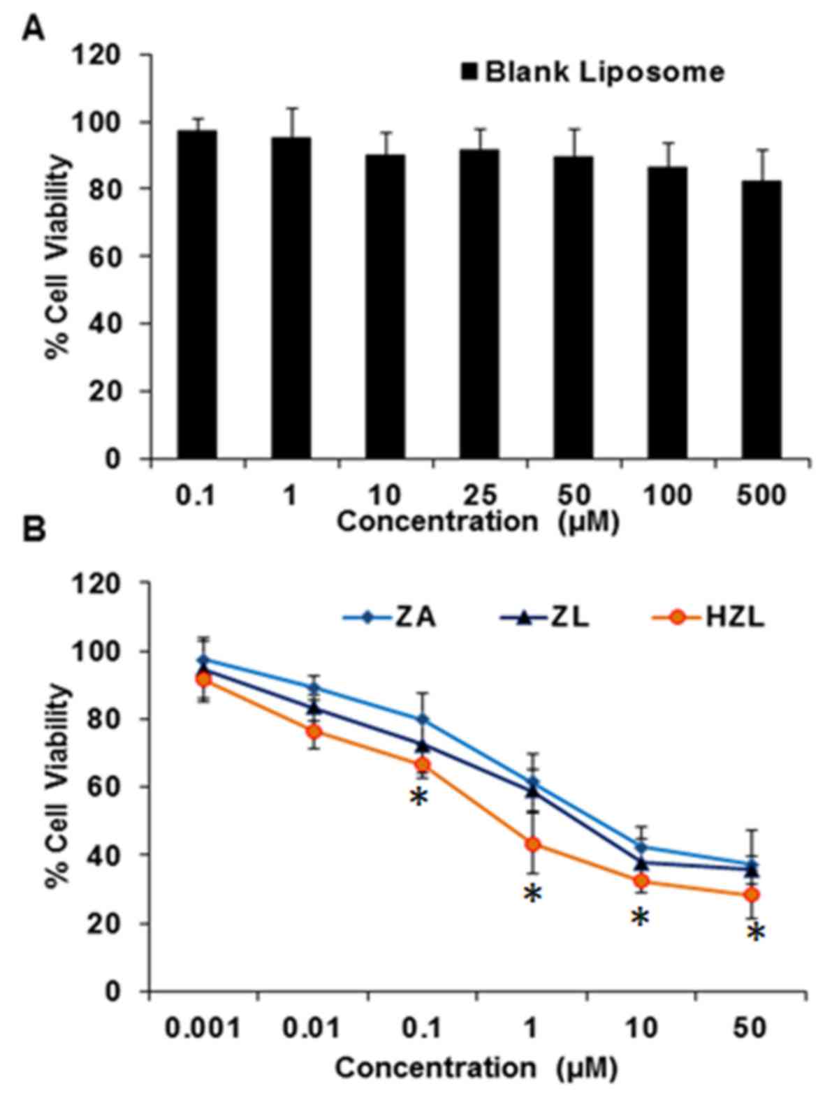
In vitro cytotoxicity
The cytotoxicity of ZA, ZL and HZL NPs was measured
by an MTT assay. The viability of HFOb 1.19 cells remained >80%
when they were treated with different concentrations of a blank
liposome (Fig. 5A). Furthermore, the
cytotoxicity of osteoblasts treated with HZL NPs was higher
compared with cells treated with ZL-viability was 48% following
treatment with 1 µM HZL NP and incubation for 72 h (Fig. 5B). Furthermore, toxicity was
dose-dependent, with higher concentrations of ZA, ZL and HZL NP
inducing higher toxicity. Notably, HZL NP treatment resulted in
significantly decreased viability when compared to that of free ZA
at 0.1 to 50 µM (P<0.05). As expected, the osteoclast viability
and differentiation were greatly affected by the presence of HZL NP
and exhibited significantly higher cytotoxicity compared with the
cytotoxicity induced by ZA and ZL following 48 h incubation. HZL NP
may have induced higher toxicity in osteoblasts due to its higher
cellular release of ZA.
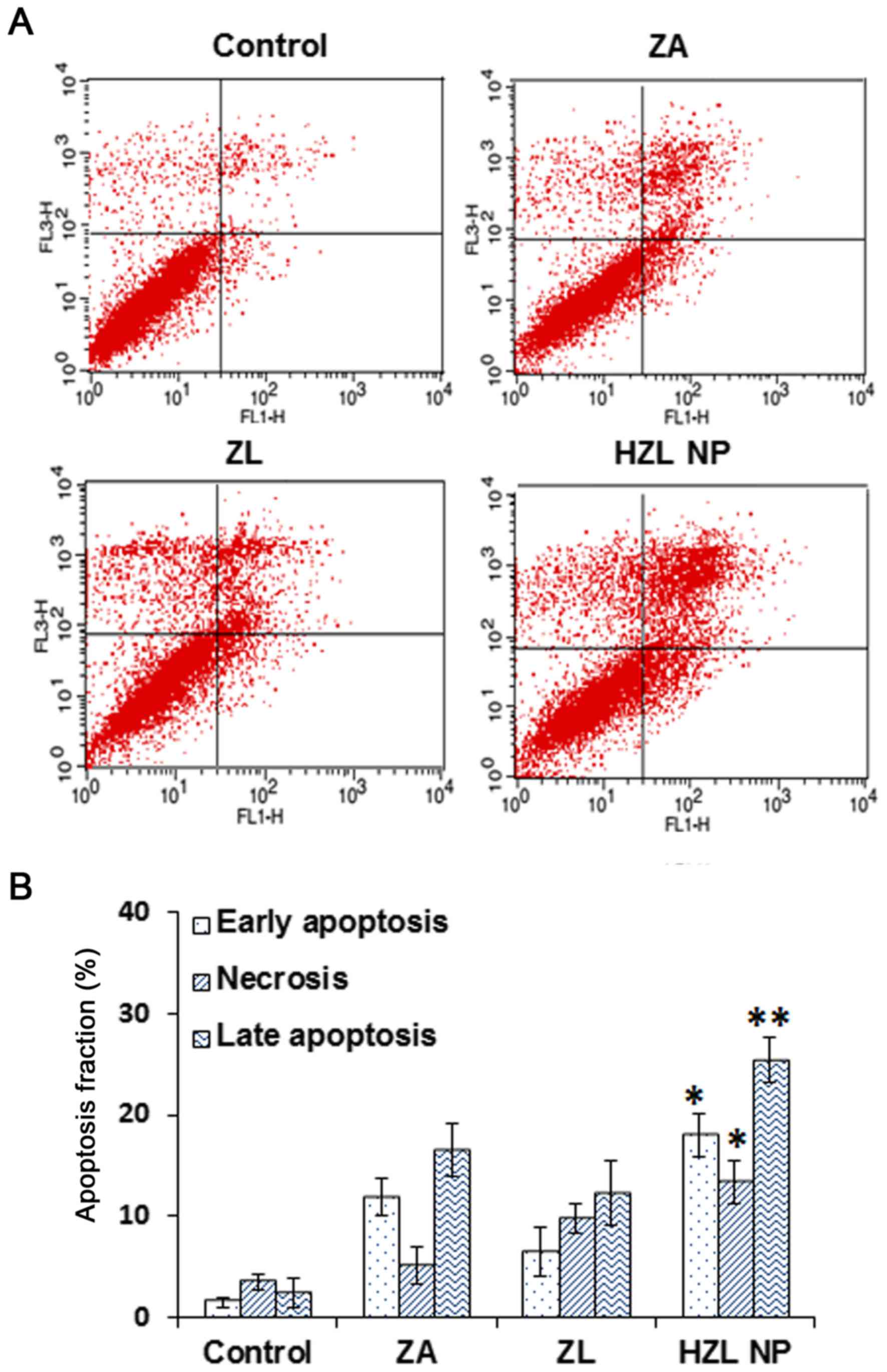
Apoptosis assay
Annexin V/PI flow cytometric analyses were performed
to determine whether the inhibition of cell viability differed
following treatment with ZA, ZL or HZL NP and to distinguish among
healthy, early apoptotic and late apoptotic or necrotic cells in
each group of cells. Qualitative and quantitative results
demonstrated that following treatment with 10 µM ZA for 2 days, the
proportion of apoptotic HFOb 1.19 cells in all treatment groups
with ZA, ZL and HZL NP was significantly higher than that of the
control group (Fig. 6). In cells
cultured with ZA or ZL for 2 days, the percentage of viable cells
decreased and the percentage of early apoptotic cells and late
apoptotic or necrotic cells increased. However, cells treated with
HZL NPs exhibited higher toxicity compared with cells treated with
ZL; the proportion of early apoptotic cells increased from 7.5±2.1
to 18.1±12.4% (P<0.05). Furthermore, the proportion of late
apoptotic and necrotic cells increased significantly to 28.7±3.7
and 16.1±1.2%, respectively (P<0.01 and P<0.05, respectively)
compared with cells treated with free ZA. These data were
consistent with the trends of cell viability and indicate that the
inhibition of cell viability by ZA released by HZL NP is due to the
induction of apoptosis.
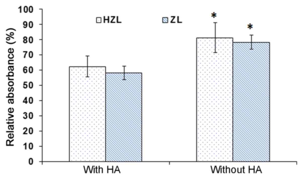
HA affinity test
To confirm the affinity of the nanoparticle to HA, a
quantitative affinity test was performed (Fig. 7). HZL NPs were incubated with and
without HA in buffer maintained at pH 7.4. The absorbance of HZL NP
incubated with HA was 62%. However, the absorbance of HZL NPs
without HA was 82%, resulting in a high amount of nanocarrier
adhering to the surface of HA, due to the presence of HA on the
surface of the NPs. The absorbance of ZL NPs was ~57 and 76% with
and without HA, respectively. The absorbance of NPs was
significantly higher in NPs incubated without HA compared with
those incubated with HA (P<0.05; Fig.
7). ZA exhibited strong affinity with HA and the interactions
between bisphosphates and HA involved the P-C-P backbone and also
the hydroxyl groups (24). Notably,
ZL exhibited affinity in the presence of HA as the ZA interacts
with the HA conjugate on the surface, which may release and help to
induce affinity towards HA whereas in the HZL NPs, the HA along
with the drug may serve a vital role in stimulating adhesion. HZL
NPs bind and accumulate in bone sites where the pores in the bone
are 70–100 nm (25). Therefore, the
NPs prepared with pores ≤100 nm can easily target and deliver drugs
to the bone.
Discussion
OP is a progressive bone disease that makes the bone
more porous (26). Bisphosphonates
inhibiting osteoclast resorption are widely used in the prevention
and treatment of the disease. Risedronate is a nitrogen-containing
bisphosphonate approved by the Food and Drug Administration as a
treatment of postmenopausal OP. Its primary mechanism is the
inhibition of farnesyl diphosphate synthase, which ultimately
results in the inhibition of osteoclast adhesion via osteoclast
apoptosis on the margin of bone where absorption takes place
(27). Lü et al (23) described the synthesis of the
HA-nanofibre composite used to induce osteogenesis by the
employment of mesenchymal stem cells (MSCs). The ability of HA to
induce MSCs to differentiate into osteoblasts may be applicable in
bone tissue engineering.
The present study therefore investigated the ability
of HA-coated ZA-loaded lipid nanoparticle to inhibit the
progression of OP. It has been hypothesized that HA coated
nanoparticles may effectively inhibit osteoclasts and thereby
improve the symptoms of OP. In the present study, liposomal systems
were used, due to their inherent biocompatibility (28,29).
Liposomal systems were coated with HA by loading zoledronate in the
lipid bilayer, the association between zoledronate and HA and the
response of bone cells to these novel biomaterials was determined.
The Z-potential of the DOTAP liposome (3:1) was 12.4 mV and the
hydrodynamic size of ZA-loaded liposome was 172.4±3.47 nm. HA was
then fabricated on the surface of the lipid nanoparticle using the
precipitation method. As the HA was negatively charged, it
electrostatically interacted with the positively charged lipid
layer and formed a coat around the HZL NP. Additionally, the
precipitation of HA on the surface of liposomes resulted in the
formation of a core-shell structure. The negative charge of the
liposomes was screened following HA coating. SEM images of HZL
revealed well-defined, spherical-shaped particles with a uniform
distribution.
ZA-loaded HZL NPs exhibited a biphasic drug release
pattern characterized by an initial rapid release followed by a
sustained continuous release from the lipid bilayer nanoparticle
after 48 h. HZL has numerous potential advantages as a bone drug
delivery system due to its osteoconductivity. A switch from
physiological pH to a lower pH around the inflamed sites may
trigger drug release in these specific areas. Such sustained and
prolonged release behavior may enhance the efficiency of OP
therapy, as continually sustained drug release effectively prevents
bone deformation during long-term treatment. Furthermore, it has
been demonstrated that HZL NPs are optimized and the controlled
release of ZA makes it suitable to treat OP. Blank nanocarriers
exhibit lower cytotoxicity, which proves their biocompatibility and
the nontoxic nature of the carrier used to deliver the drug to the
diseased site. ZA-loaded HZL NPs increased the toxicity on
osteoblasts indicating the potent antiresorptive action of ZA. The
apoptosis rate in cells treated with HZL NPs is increased. ZA
inhibits farnesyl diphosphate synthase, a key enzyme in the
mevalonic acid pathway, and thereby inhibits the prenylation of
small GTPases. This enzyme maintains the osteoclast function, and
is widely used to treat bone disorders, including osteoporosis and
metastatic bone disease. Small GTPases accumulate in the cells,
which erroneously stimulate the downstream pathway, inhibiting
osteoclast formation and inducing apoptosis (30). Thus, bone resorption mediated by
osteoclasts is reduced, lowering the bone turnover rate and leading
to a loss of bone mass.
In conclusion, in the current study HZL NPs were
successfully prepared using a thin film and the extrusion method.
The results demonstrated that the precipitation of HA on the
surface was highly effective at treating the symptoms of OP.
Furthermore, the nanosized hydrodynamic size and SEM image of the
nanoparticle demonstrate that they are suitable for the delivery of
ZA to specific sites in the bone. Additionally, a typical biphasic
release pattern was observed with a sustained prolonged drug
release exhibited throughout. Furthermore, higher concentrations of
ZA induced cytotoxicity and exhibited a significant proportion of
osteoclasts in the early and late phases of apoptosis. Therefore,
HZL NPs exhibited a high selectivity for bone tissues and revealed
a strong affinity towards HA, indicating that they may enhance
therapeutic efficacy and may be developed as a novel method of
treating patients with OP.
Acknowledgements
Not applicable.
Funding
No funding was received.
Availability of data and materials
The datasets used and/or analyzed during the current
study are available from the corresponding author on reasonable
request.
Authors' contributions
TG and SF were responsible for the formulation and
initial characterization of nanoparticles. PS performed all the
biological studies. YC designed the present study and wrote the
manuscript.
Ethics approval and consent to
participate
Not applicable.
Patient consent for publication
Not applicable.
Competing interests
The authors declare that they have no competing
interests.
References
|
1
|
Akesson K: New approaches to
pharmacological treatment of osteoporosis. Bull World Health Organ.
81:657–664. 2003.PubMed/NCBI
|
|
2
|
Dhanwal DK, Dennison EM, Harvey NC and
Cooper C: Epidemiology of hip fracture: Worldwide geographic
variation. Ind J Orthop. 45:15–22. 2011. View Article : Google Scholar
|
|
3
|
Drake MT, Clarke BL and Lewiecki EM: The
pathophysiology and treatment of osteoporosis. Clin Ther.
37:1837–1850. 2015. View Article : Google Scholar : PubMed/NCBI
|
|
4
|
Dalle Carbonare L, Zanatta M, Gasparetto A
and Valenti MT: Safety and tolerability of zoledronic acid and
other bisphosphonates in osteoporosis management. Drug Healthc
Patient Saf. 2:121–137. 2010. View Article : Google Scholar : PubMed/NCBI
|
|
5
|
Rogers MJ, Crockett JC, Coxon FP and
Mönkkönen J: Biochemical and molecular mechanisms of action of
bisphosphonates. Bone. 49:34–41. 2011. View Article : Google Scholar : PubMed/NCBI
|
|
6
|
Dunford JE, Thompson K, Coxon FP, Luckman
SP, Hahn FM, Poulter CD, Ebetino FH and Rogers MJ:
Structure-activity relationships for inhibition of farnesyl
diphosphate synthase in vitro and inhibition of bone resorption in
vivo by nitrogen-containing bisphosphonates. J Pharmacol Exp Ther.
296:235–242. 2001.PubMed/NCBI
|
|
7
|
Salzano G, Marra M, Porru M, Zappavigna S,
Abbruzzese A, La Rotonda MI, Leonetti C, Caraglia M and De Rosa G:
Self-assembly nanoparticles for the delivery of bisphosphonates
into tumors. Int J Pharm. 403:292–297. 2011. View Article : Google Scholar : PubMed/NCBI
|
|
8
|
Bellido T and Plotkin LI: Novel actions of
bisphosphonates in bone: Preservation of osteoblast and osteocyte
viability. Bone. 49:50–55. 2011. View Article : Google Scholar : PubMed/NCBI
|
|
9
|
Wei D, Jung J, Yang H, Stout DA and Yang
L: Nanotechnology treatment options for osteoporosis and its
corresponding consequences. Curr Osteoporos Rep. 14:239–247. 2016.
View Article : Google Scholar : PubMed/NCBI
|
|
10
|
Barry M, Pearce H, Cross L, Tatullo M and
Gaharwar AK: Advances in nanotechnology for the treatment of
osteoporosis. Curr Osteoporos Rep. 14:87–94. 2016. View Article : Google Scholar : PubMed/NCBI
|
|
11
|
Ramasamy T, Ruttala HB, Gupta B, Poudal
BK, Choi HG, Yong CS and Kim JO: Smart chemistry-based nanosized
drug delivery systems for systemic applications: A comprehensive
review. J Control Release. 258:226–253. 2017. View Article : Google Scholar : PubMed/NCBI
|
|
12
|
Peng H, Liu X, Wang G, Li M, Bratlie KM,
Cochran E and Wang Q: Polymeric multifunctional nanomaterials for
theranostics. J Mater Chem B. 3:6856–6870. 2015. View Article : Google Scholar
|
|
13
|
Ikoba U, Peng H, Li H, Miller C, Yu C and
Wang Q: Nanocarriers in therapy of infectious and inflammatory
diseases. Nanoscale. 7:4291–4305. 2015. View Article : Google Scholar : PubMed/NCBI
|
|
14
|
Ruttala HB, Ramasamy T, Poudal BK, Choi Y,
Choi JY, Kim J, Ku Kwang S, Choi HG, Soon Yong C and Kim Oh J:
Molecularly targeted co-delivery of a histone deacetylase inhibitor
and paclitaxel by lipid-protein hybrid nanoparticles for
synergistic combinational chemotherapy. Oncotarget. 8:14925–14940.
2017. View Article : Google Scholar : PubMed/NCBI
|
|
15
|
Ruttala HB, Ramasamy T, Gupta B, Choi HG,
Yong CS and Kim JO: Multiple polysaccharide-drug complex-loaded
liposomes: A unique strategy in drug loading and cancer targeting.
Carbohydr Polym. 173:57–66. 2017. View Article : Google Scholar : PubMed/NCBI
|
|
16
|
Wang Q, Cheng H, Peng H, Zhou H, Li PY and
Langer R: Non-genetic engineering of cells for drug delivery and
cell-based therapy. Adv Drug Deliv Rev. 91:125–140. 2015.
View Article : Google Scholar : PubMed/NCBI
|
|
17
|
Meng S, Su B, Li W, Ding Y, Tang L, Zhou
W, Song Y and Caicun Z: Integrin-targeted paclitaxel nanoliposomes
for tumor therapy. Med Oncol. 28:1180–1187. 2011. View Article : Google Scholar : PubMed/NCBI
|
|
18
|
Ruttala HB, Ramasamy T, Shin BS, Choi HG,
Yong CS and Kim JO: Layer-by-layer assembly of hierarchical
nanoarchitectures to enhance the systemic performance of
nanoparticle albumin-bound paclitaxel. Int J Pharm. 519:11–21.
2017. View Article : Google Scholar : PubMed/NCBI
|
|
19
|
Xu Q, Tanaka Y and Czernuszka JT:
Encapsulation and release of a hydrophobic drug from hydroxyapatite
coated liposomes. Biomaterials. 28:2687–2694. 2007. View Article : Google Scholar : PubMed/NCBI
|
|
20
|
Venkatesan P, Puvvada N, Dash R, Kumar
Prashanth BN, Sarkar D, Azab B, Pathak A, Kundu SC, Fisher PB and
Mandal M: The potential of celecoxib-loaded hydroxyapatite-chitosan
nanocomposite for the treatment of colon cancer. Biomaterials.
32:3794–3806. 2011. View Article : Google Scholar : PubMed/NCBI
|
|
21
|
Iafisco M, Ruffini A, Adamiano A, Sprio S
and Tampieri A: Biomimetic magnesium-carbonate-apatite nanocrystals
endowed with strontium ions as anti-osteoporotic trigger. Mater Sci
Eng C Mater Biol Appl. 35:212–219. 2014. View Article : Google Scholar : PubMed/NCBI
|
|
22
|
Pilia M, Guda T and Appleford M:
Development of composite scaffolds for load-bearing segmental bone
defects. BioMed Res Int. 2013:4582532013. View Article : Google Scholar : PubMed/NCBI
|
|
23
|
Lü LX, Zhang XF, Wang YY, Ortiz L, Mao X,
Jiang ZL, Xiao ZD and Huang NP: Effects of
hydroxyapatite-containing composite nanofibers on osteogenesis of
mesenchymal stem cells in vitro and bone regeneration in vivo. ACS
Appl Mater Interfaces. 5:319–330. 2013. View Article : Google Scholar : PubMed/NCBI
|
|
24
|
Jueng H, Sung KC, Eun SP, Kun H, Hee DH
and Byung CS: Enhanced stability of hydroxyapatite-coated liposomes
for ultrasound-triggered drug release. Bull Korean Chem Soc.
36:83–87. 2015. View Article : Google Scholar
|
|
25
|
Yewle JN, Puleo DA and Bachas LG: Enhanced
affinity bifunctional bisphosphonates for targeted delivery of
therapeutic agents to bone. Bioconjug Chem. 22:2496–2506. 2011.
View Article : Google Scholar : PubMed/NCBI
|
|
26
|
Wang D, Miller S, Sima M, Kopecková P and
Kopecek J: Synthesis and evaluation of water-soluble polymeric
bone-targeted drug delivery systems. Bioconjug Chem. 14:853–859.
2003. View Article : Google Scholar : PubMed/NCBI
|
|
27
|
Cummings SR and Melton LJ: Epidemiology
and outcomes of osteoporotic fractures. Lancet. 359:1761–1767.
2002. View Article : Google Scholar : PubMed/NCBI
|
|
28
|
Nishikawa M, Akatsu T, Katayama Y,
Yasutomo Y, Kado S, Kugal N, Yamamoto M and Nagata N:
Bisphosphonates act on osteoblastic cells and inhibit osteoclast
formation in mouse marrow cultures. Bone. 18:9–14. 1996. View Article : Google Scholar : PubMed/NCBI
|
|
29
|
Gaspar MM, Gobbo O and Ehrhardt C:
Generation of liposome aerosols with the Aeroneb Pro and the
AeroProbe nebulizers. J Liposome Res. 20:55–61. 2010. View Article : Google Scholar : PubMed/NCBI
|
|
30
|
Dunford JE, Rogers MJ, Ebetino FH, Phipps
RJ and Coxon FP: Inhibition of protein prenylation by
bisphosphonates causes sustained activation of Rac, Cdc42, and Rho
GTPases. J Bone Miner Res. 21:684–694. 2006. View Article : Google Scholar : PubMed/NCBI
|