Introduction
In 2012, approximately 239,000 women were diagnosed
with ovarian cancer worldwide, while 152,000 women succumbed to the
disease (1). These data suggest that
mortality is observed in almost 65% of all women with ovarian
cancer, and the mortality rates are even higher in women diagnosed
at an advanced disease stage of the disease (stage IV). Treatment
of ovarian cancer primarily includes cytoreductive surgery and
platinum-based chemotherapy (2).
Although clinical remission is possible, the long-term outcome
remains poor since the majority of patients will relapse and
succumb to the disease within 5 years, with the 5-year survival
being ~30% (3,4). Therefore, novel therapies need to be
integrated into ovarian cancer treatment strategies in order to
achieve durable clinical outcomes.
Recently, it has been reported that lanthanum
complexes were effective candidate drugs in the treatment of
malignant tumors (5). For instance,
lanthanum complex has demonstrated considerable cytotoxicity on
human cervical carcinoma cells in vitro (6,7).
Lanthanum chloride (LaCl3) also significantly inhibited
the growth and induced the apoptosis of leukemia cell lines, HL-60
and NB4 (8). In addition, lanthanum
has been involved in the growth arrest of human gastric cancer
cells (9). All these observations
suggested that lanthanum, a rare earth element, has the capacity to
promote anti-tumor regulation (10).
In the present study, the therapeutic effect of
LaCl3 on ovarian tumors was elucidated, based on the
investigation of ovarian cancer cell lines in vitro and a
xenograft animal model in vivo. It was observed that
LaCl3 was able to enhance the inhibitory effect of
cisplatin on the cell proliferation and carcinogenesis. These data
provided a solid experimental basis for the treatment of ovarian
cancer, revealing a potential strong anti-tumor candidate in the
treatment of this tumor.
Materials and methods
Animals
A total of 60 female BALB/C nude mice (4-6 weeks old
and weighing 13-15 g) were purchased from Shanghai SLAC Laboratory
Animal Co., Ltd. (Shanghai, China). All animals were housed in
micro-isolator cages in a light- and temperature-controlled room
under a 12/12 h light/dark cycle at 23-25°C. All animals had free
access to food and water according to the Guide for the Care and
Use of Laboratory Animals (11). In
particular, any effort was put to avoid unnecessary pain of the
animals. All the animal protocols were approved by the Ethics
Committee of the First Affiliated Hospital of Nanchang University
(Nanchang, China).
Cell culture and treatment
A cisplatin-sensitive human ovarian cancer cell line
COC1 (American Type Culture Collection, Manassas, VA, USA) was
cultured in RPMI-1640 medium (Hyclone; GE Healthcare Life Sciences,
Logan, UT, USA) supplemented with 10% fetal bovine serum (Hyclone;
GE Healthcare Life Sciences) at 37°C and 5% CO2. The
cisplatin-resistant subline COC1/DDP was obtained by culturing COC1
cells in 0.2-0.5 µg/ml cisplatin (Sigma-Aldrich; Merck KGaA,
Darmstadt, Germany) with gradually increasing concentrations for 3
weeks at 37°C and 5% CO2 to finally obtain steady
cisplatin resistant cells. Cells were transferred into
cisplatin-free medium for 48 h prior to experiments.
For in vitro studies, cells were transferred
into cisplatin-free medium for 48 h prior to experiments. COC1 or
COC1/DDP cells were divided into 4 groups: The LaCl3
group in which cells were treated with 20 µg/ml LaCl3
(Sigma-Aldrich; Merck KGaA); the cisplatin group in which cells
were treated with 20 µg/ml cisplatin (Sigma-Aldrich; Merck KGaA);
the LaCl3/cisplatin group in which cells were treated
with 20 µg/ml LaCl3 and 20 µg/ml cisplatin; and the
control group in which the cell were left untreated. Cells were
cultured on 96 well at a density of 1×10⁴
cells/well.
For the cells prepared for injection into mice, the
medium was replaced by fresh medium in order to remove dead and
detached cells at 3-4 h before harvesting. Cells were centrifuged
at 251 × g for 4 min at room temperture, washed twice with
phosphate-buffered saline (PBS) and stored on ice. Cells were
suspended in 300 µl PBS containing 2.0×10⁶ cells per
injection.
Xenograft animal model of ovarian
cancer
The inoculation area of the mice was cleaned and
sterilized with ethanol and iodine solutions. COC1 or COC1/DDP
cells were injected subcutaneously into nude mice at the scapular
region. An evident tumor (200-300 mm3) was observed 4
weeks after cell injection. At that time, 6 mice were anesthetized
using 45 mg/kg pentobarbital by intraperitoneal injection, an
incision was made to expose the abdomen and ovary, then tumor
tissues were dissected from the nude mice and cut into the small
sections (~10 mm3). Then, the tumors were subsequently
transplanted into the ovaries of the other 40 mice. At 3 weeks
after transplantation, the incision was opened again and the tumor
size was measured. Then mice were injected with tumor cells were
further divided into 4 groups with 5 mice in each group. The
LaCl3 group was treated with 10 mg/kg LaCl3
by intraperitoneal injection. The cisplatin group was treated with
10 mg/kg cisplatin by intraperitoneal injection. The
LaCl3/cisplatin group was treated with 10 mg/kg
LaCl3 and 10 mg/kg cisplatin by intraperitoneal
injection. The control group was treated with 0.5 ml 0.9% sodium
chloride solution by intraperitoneal injection. Each treatment was
given once a week for 4 weeks. Mice were sacrificed at 4 weeks
after each corresponding treatments, tumor tissues were then
dissected from the ovaries and the tumor volume was calculated
according to the following formula: Volume (mm3)=length
× width × height × π/6.
Terminal deoxynucleotidyl
transferase-mediated dUTP nick end labeling (TUNEL) assay
TUNEL assay was performed using an in situ
cell death detection kit (Santa Cruz Biotechnology, Inc., Dallas,
TX, USA). Briefly, cells were cultured for 48 h after
LaCl3, cisplatin or combined of LaCl3 and
cisplatin treatment. Then cells were fixed with 4% paraformaldehyde
and permeabilized with 0.2% Triton X-100. Subsequently, fragmented
DNA was labeled with fluorescein-12-dUTP (Thermo Fisher Scientific,
Inc., Waltham, MA, USA) at 37°C for 1 h, and the reaction was
terminated by the addition of saline-sodium citrate buffer,
followed by washing twice with PBS. TUNEL-positive nuclei were
investigated under a fluorescent microscope (Nikon Corp., Tokyo,
Japan) and counted from the entire sample.
Immunohistochemical analysis
The cells were washed in PBS, blocked for 60 min in
0.3% Triton X-100 in PBS and 5% bovine serum albumin. Next, samples
were incubated overnight at 4°C with primary antibodies against:
B-cell lymphoma-2 (BCL-2; cat. no. ab692; 1:500), Ki67 (cat. no.
ab15580, 1:500), breast cancer 1 (BRCA1; cat. no. ab16780; 1:500),
BRCA2 (cat. no. ab27976; 1:500) and excision repair
cross-complementation group 1 (ERCC1; cat. no. ab2356; 1:100; all
purchased from Abcam, Cambridge, UK). Horseradish
peroxidase-conjugated anti-mouse secondary antibodies (cat. no.
ab193651; 1:500; Abcam) was subsequently applied to the slides, and
incubated for 1 h at room temperature. Finally,
DAB/H2O2 was added to the surface of the
slide and the slides were visualized by light microscopy at a
magnification of ×200.
Statistical analysis
The measurement data was expressed by mean ±
standard deviation. Comparisons among groups were conducted using
one-way analysis of variance followed by a Tukey post hoc test.
P<0.05 was considered to indicate differences that were
statistically significant. All analyses were made using SPSS
(version 20.0; IBM Corp., Armonk, NY, USA).
Results
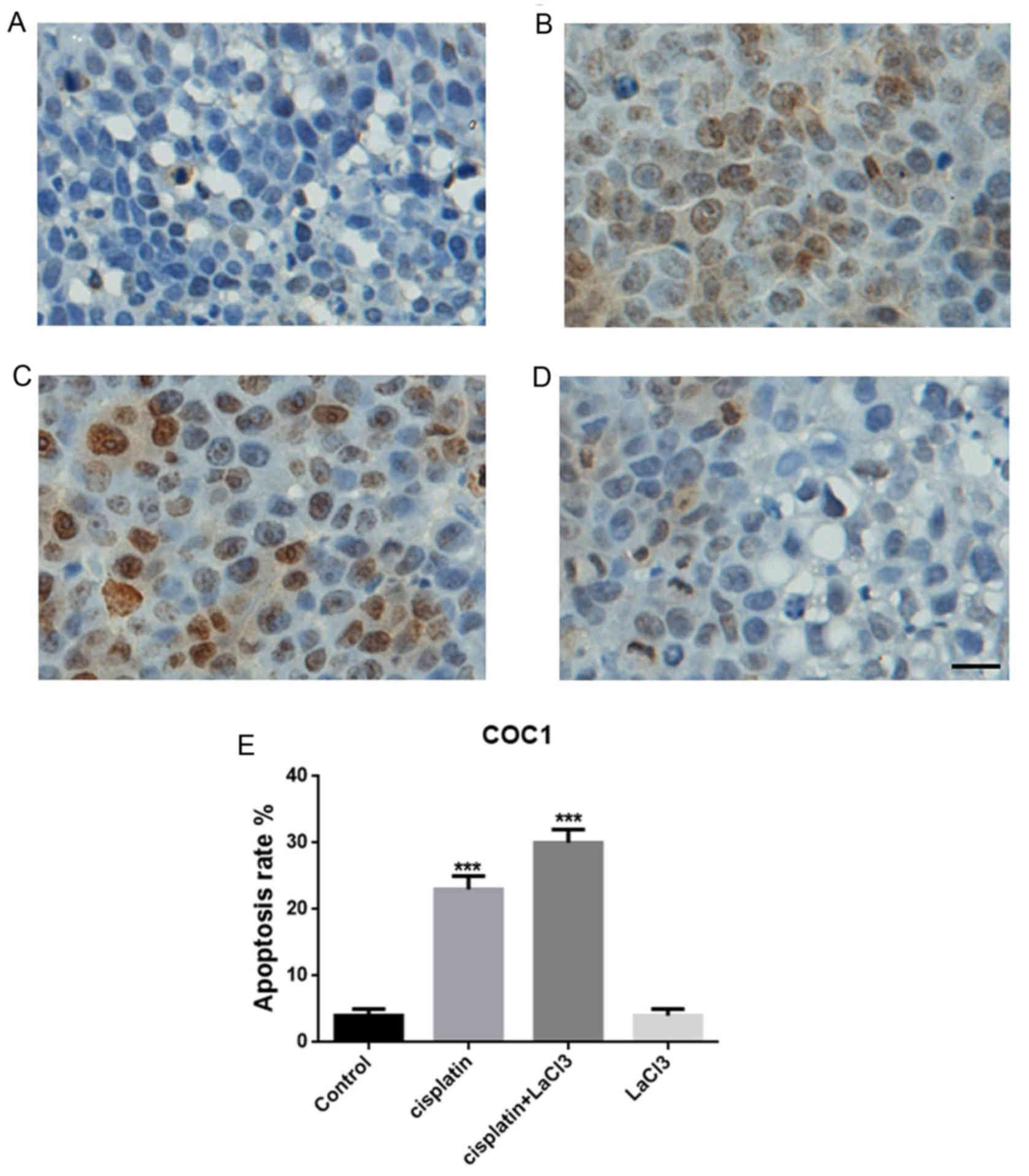
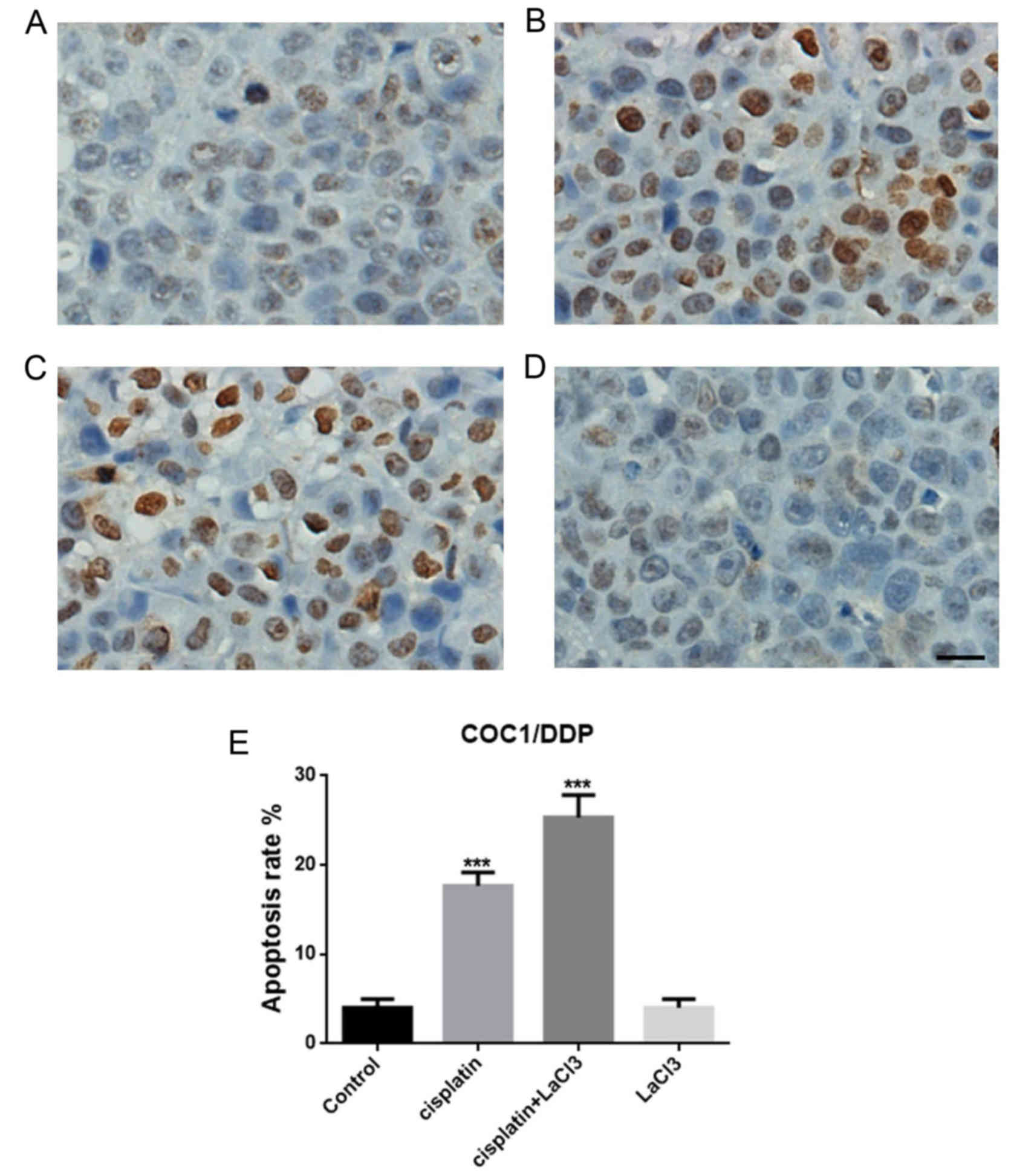
Co-application of LaCl3 and
cisplatin substantially promotes COC1 cell death in vitro
TUNEL assay was conducted to evaluate the cell death
following treatment with cisplatin, LaCl3 or cisplatin +
LaCl3 in the COC1 and COC1/DDP cell lines (Fig. 1A-D). The data indicated that the
percentage of TUNEL-positive cells in the cisplatin-treated COC1
cells were significantly higher compared with that in the control
group (P<0.001; Fig. 1E). By
contrast, the addition of LaCl3 further promoted the
apoptotic effect of cisplatin, leading to a markedly higher
percentage of apoptotic cells in the cisplatin+LaCl3
co-treated group (P<0.001; Fig.
1C). However, application of LaCl3 only did not
evidently affect the cell death or apoptosis (Fig. 1D). Similar results were obtained in
the COC1/DDP cells (Fig. 2). Thus,
it can be concluded that LaCl3 was able to enhance the
apoptotic effect of cisplatin on the ovarian cancer cells.
Combination of LaCl3 and
cisplatin affects the expression of tumor-associated proteins
Next, the present study determined whether the
co-application of LaCl3 is able to alter the expression
of tumor-associated proteins. Firstly, the expression of
proliferative cell marker Ki67 in COC1 cells was decreased in the
presence of cisplatin as compared with the control cells (Fig. 3A). LaCl3 stimulation
together with cisplatin further decreased the expression of Ki67.
By contrast, the effect of LaCl3 on the Ki67 expression
was minor. The expression of BCL-2, an oncogene and anti-apoptotic
protein, was also examined, and presented a trend similar to Ki67
(Fig. 3A and B). On the contrary,
the expression levels of human tumor suppressor genes, BRCA1 and
BRCA2, exhibited the opposite results (Fig. 3C and D, respectively). Namely, the
application of cisplatin alone increased the expression levels of
BRCA1 and BRCA2, while co-treatment with cisplatin and
LaCl3 further increased the expression levels of BRCA1
and BRCA2. Furthermore, the current study also detected the
expression of ERCC1, which is responsible for DNA repair, and
identified a similar pattern to that of Ki67 and BCL-2, with a
decrease observed in ERCC1 expression in the cisplatin and
cisplatin + LaCl3 groups (Fig. 3E). In the COC1/DDP cells, all the
results were similar with those observed in the COC1 cells
(Fig. 4). Collectively, the
immunochemical data indicated that LaCl3 and cisplatin
combined treatment decreased the expression of proteins associated
with proliferation and oncogenesis, and increased the expression of
tumor suppressor protein.
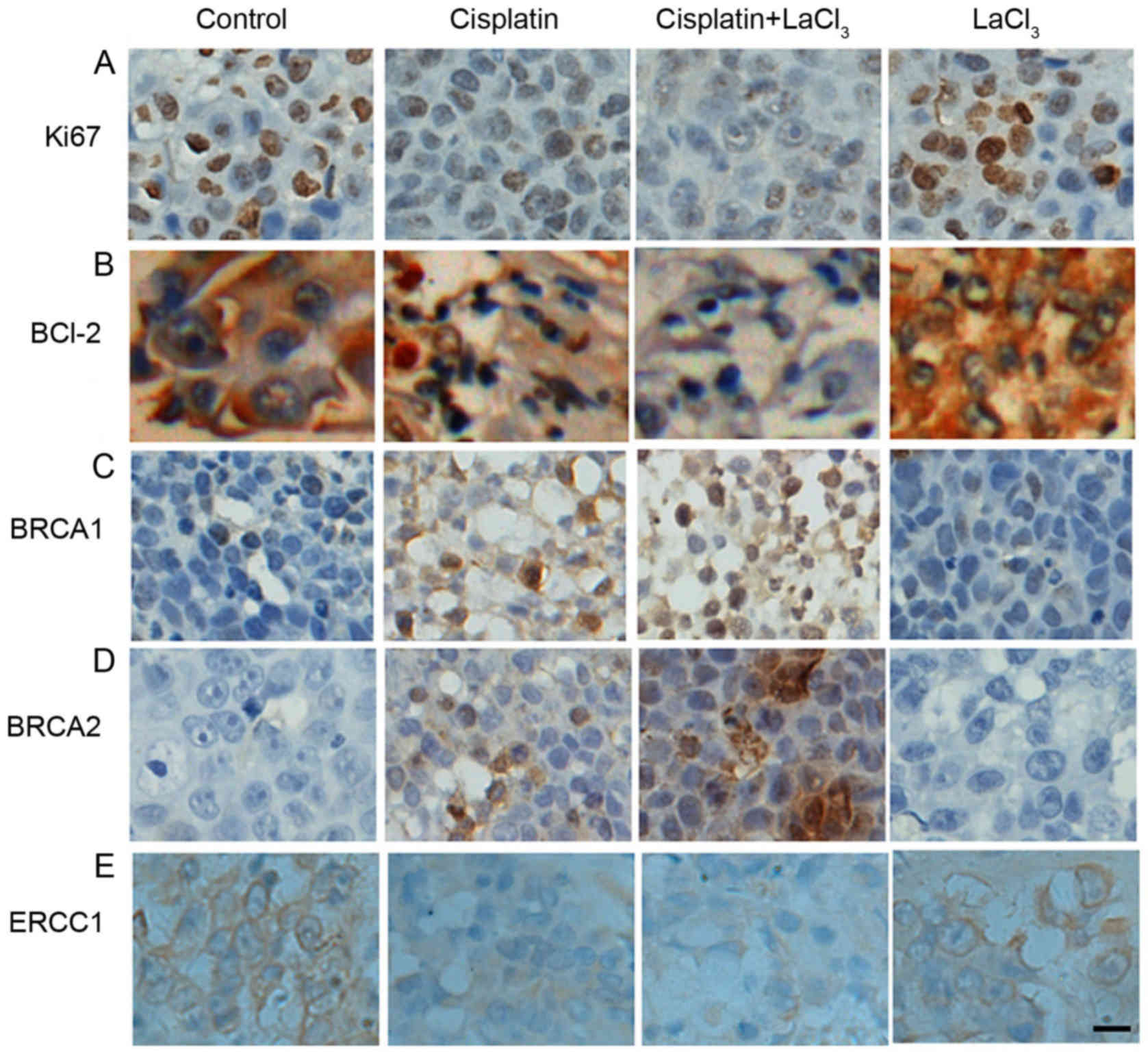 | Figure 3.LaCl3 and cisplatin
co-treatment altered the expression levels of tumor-associated
proteins in COC1 cells. Expression levels of (A) Ki67, (B) BCL-2,
(C) BRCA1, (D) BRCA2, and (E) ERCC1 are demonstrated. Ki67
expression decreased in the presence of cisplatin compared with the
control, while application of cisplatin + LaCl3 further
decreased Ki67 expression, and LaCl3 alone had only a
minor effect. Similar results were obtained for BCL-2 and ERCC1
expression levels. By contrast, cisplatin increased the expression
levels of BRCA1 and BRCA2 compared with the control cells, while
co-application of LaCl3 and cisplatin further increased
the levels, and LaCl3 alone did not have a marked
effect. Scale bar, 20 µm. Magnification, ×200. LaCl3,
lanthanum chloride; BCL-2, B-cell lymphoma-2; BRCA, breast cancer;
ERCC1, excision repair cross-complementation group 1. |
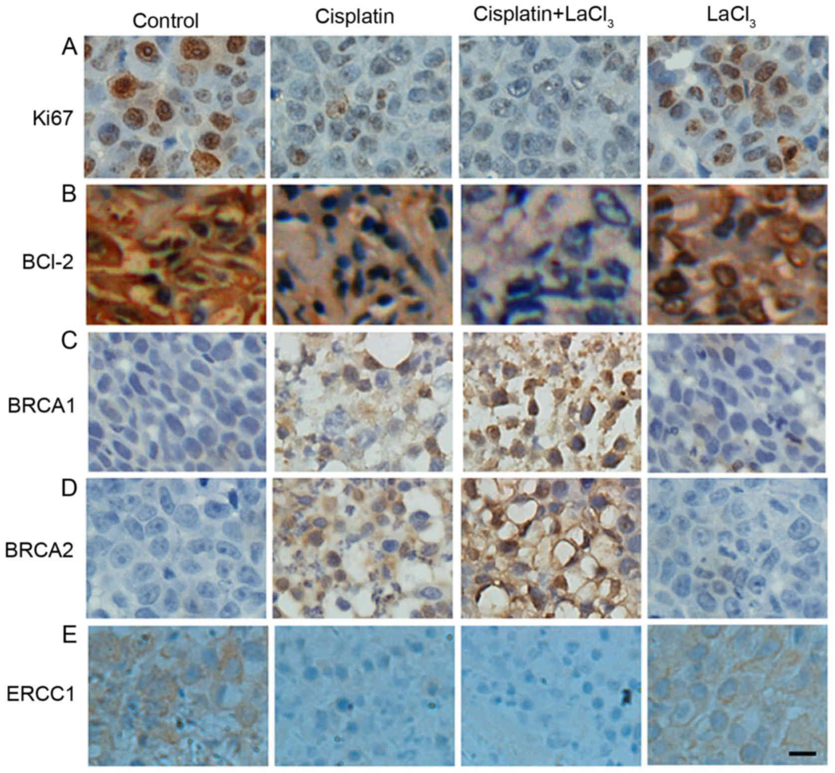 | Figure 4.Co-treatment with LaCl3
with cisplatin altered the expression levels of (A) Ki67, (B)
BCL-2, (C) BRCA1, (D) BRCA2, and (E) ERCC1 in COC1/DPP cells. The
effects of different treatments were similar with those in COC1
cells. Scale bar, 20 µm. Magnification, ×200. LaCl3,
lanthanum chloride; BCL-2, B-cell lymphoma-2; BRCA, breast cancer;
ERCC1, excision repair cross-complementation group 1. |
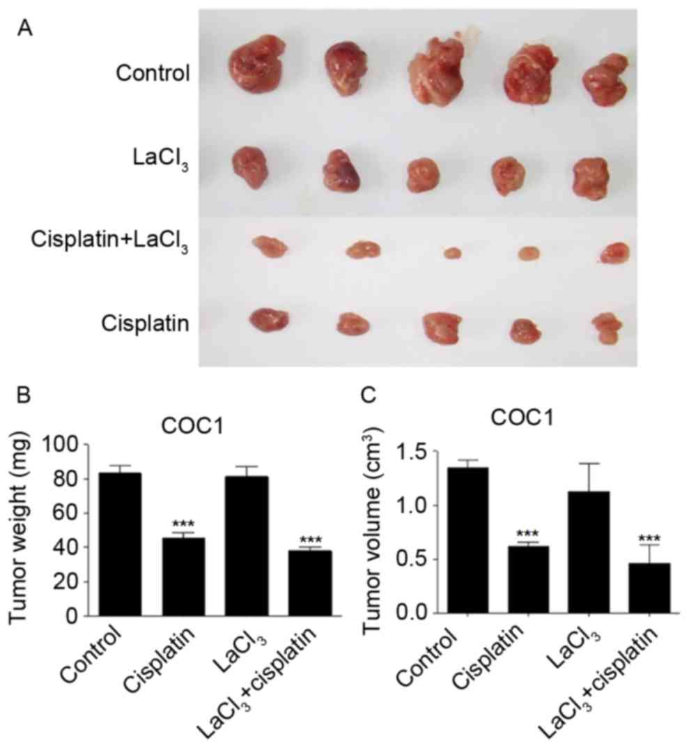
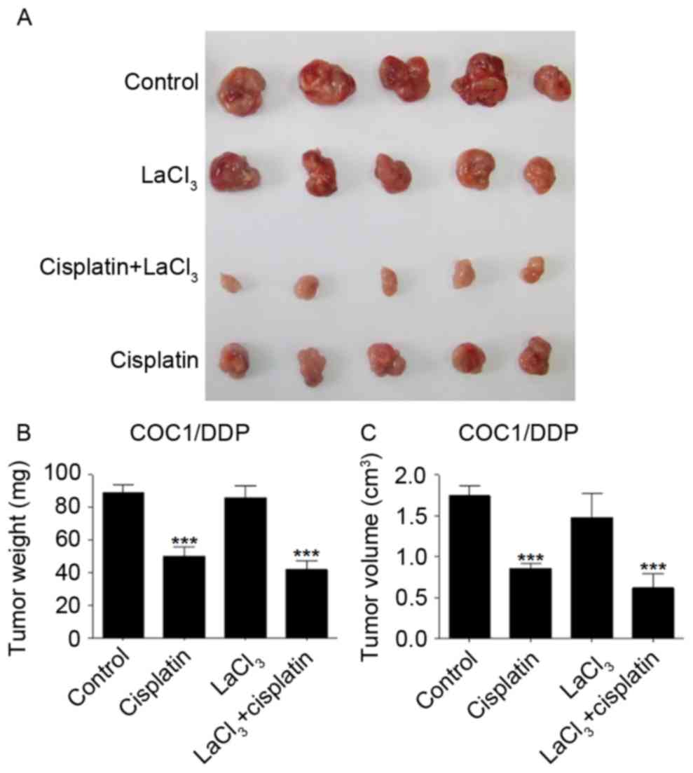
LaCl3 and cisplatin
co-treatment inhibits the tumor growth in vivo
The present study also evaluated whether the
application of LaCl3 and cisplatin affected the tumor
growth in vivo. The tumor xenograft animal model,
established using COC1 cells, demonstrated that cisplatin and
LaCl3 co-incubation was able to inhibit the tumor growth
in vivo (Fig. 5A). Cisplatin
treatment significantly reduced the tumor weight and size by 47 and
54%, respectively (P<0.001; Fig. 5B
and C). Co-application of cisplatin and LaCl3
further and significantly decreased the tumor weight and size by 56
and 66%, respectively, compared with those in the control animals
(P<0.001). By contrast, LaCl3 alone did not affect
the tumor growth. The xenograft of COC1/DDP cells demonstrated
similar results (Fig. 6). These
findings indicated that LaCl3 exerted an inhibitory
effect on the in vivo growth of ovarian tumor.
Discussion
In the present study, it was observed that
LaCl3 promoted the cisplatin-induced inhibition of
ovarian cancer cell growth and tumorigenesis in a mouse model of
ovarian cancer. In addition, this process was associated with the
alteration of the expression levels of specific tumor proteins. The
current research provides novel in vitro and in vivo
evidence to support the potential clinical application of
LaCl3 in the treatment of ovarian cancer.
Although it has been previously reported that
LaCl3 affects multiple tumor growth based on cell-based
assays and animal experiments (10,12–14), its
precise biological role remains largely unclear. Therefore, further
investigations on the underlying mechanisms are required. The
lanthanum compound KP772 has anticancer properties associated with
p53-independent G0/G1 arrest and apoptosis induction (12). Apoptosis is a critical stage in the
cell cycle, maintaining the balance between proliferation and cell
death. In the present study, it was demonstrated that the
application of LaCl3 increased the percentage of
TUNEL-positive COC1 or COC1/DDP cells induced by cisplatin,
indicating that LaCl3 was able to interfere with the
cell cycle progression and promote apoptosis. This increased ratio
of apoptotic cells was possibly due to a decrease in the percentage
of cells in S and G2/M phases and increase in the percentage of
cells in G0/G1 phase. However, the cell cycle status requires
further investigation in the future work.
Xenograft models, which are more biologically
representative of patient tumors, are valuable models in predicting
the clinical outcome (15). In the
present study, using a xenograft model in nude mice, it was
observed that the tumorigenesis ability of COC1 and COC1/DDP cells
was reduced in the presence of LaCl3 and cisplatin, as
determined by the decrease in tumor size and weight in vivo.
This finding provides strong evidence on the anticancer effect of
cisplatin in combination with LaCl3, though
LaCl3 itself did not demonstrate an anticancer effect,
it can enhance the anticancer ability of cisplatin. However, one
limitation of the present study is that the phenotypic instability
of tumor cells in long-term culture was not considered. A previous
study has reported that xenografts derived from cell lines
generally presented a more homogeneous, undifferentiated histology
and thus may be different from in vitro conditions that
occur during extensive culturing (16). This affects the outcome and needs to
be considered in future experiments.
Through immunohistochemical analysis, the
co-application of LaCl3 and cisplatin was observed to
result in further downregulation of Ki67, BCL-2 and ERCC1, as well
as in the upregulation of BRCA1 and BRCA2. Ki67 is a proliferative
cell marker that is used as a biomarker in cancer (17), while BCL-2 is an oncogene with
anti-apoptotic function (18). In
addition, ERCC1 participates in DNA repair in lesions (19). Upregulation of these proteins
demonstrates the weakened ability of tumor cell repair,
anti-apoptosis and proliferation. By contrast, BRCA1 and BRCA2 are
human tumor suppressor genes (20),
and the increased expression of these two proteins indicates
enhanced tumor growth suppression. Thus, in the present study,
downregulation of Ki67, BCL-2 and ERCC1, as well as upregulation of
BRCA1 and BRCA2, induced by LaCl3 and cisplatin
indicated the suppression of cancer growth, which is direct
evidence supporting the antitumor application of LaCl3
in the treatment of ovarian cancer.
In conclusion, the results of the present study
suggested that the combination of cisplatin with LaCl3
was capable of inducing ovarian cancer cell apoptosis in
vitro and inhibiting tumor growth in vivo. The current
study provides novel evidence to establish an experimental basis
for the clinical application of LaCl3 as an anticancer
drug. Further studies are required to reveal the mechanism involved
in the synergistic role of LaCl3 and cisplatin.
Acknowledgements
Not applicable.
Funding
No funding was received.
Availability of data and materials
The datasets used and/or analyzed during the current
study are available from the corresponding author on reasonable
request.
Authors' contributions
FW and YZ conducted the experiments and wrote the
manuscript. SF and ShL analyzed the data. SiL designed the
study.
Ethics approval and consent to
participate
All the animal protocols were approved by the Ethics
Committee of the First Affiliated Hospital of Nanchang University
(Nanchang, China).
Patient consent for publication
Not applicable.
Competing interests
The authors declare that they have no competing
interests.
References
|
1
|
Cress RD, Chen YS, Morris CR, Petersen M
and Leiserowitz GS: Characteristics of long-term survivors of
epithelial ovarian cancer. Obstet Gynecol. 126:491–497. 2015.
View Article : Google Scholar : PubMed/NCBI
|
|
2
|
Petrillo M, Anchora LP, Scambia G and
Fagotti A: Cytoreductive surgery plus platinum-based hyperthermic
intraperitoneal chemotherapy in epithelial ovarian cancer: A
promising integrated approach to improve locoregional control.
Oncologist. 21:532–534. 2016. View Article : Google Scholar : PubMed/NCBI
|
|
3
|
Baldwin LA, Huang B, Miller RW, Tucker T,
Goodrich ST, Podzielinski I, DeSimone CP, Ueland FR, van Nagell JR
and Seamon LG: Ten-year relative survival for epithelial ovarian
cancer. Obstet Gynecol. 120:612–618. 2012. View Article : Google Scholar : PubMed/NCBI
|
|
4
|
Baldwin L, Ware R, Huang B, Tucker T,
Goodrich S, Podzielinski I, DeSimone C, Ueland F, van Nagell J and
Seamon L: Ten-year relative survival for epithelial ovarian cancer.
Gynecol Oncol. 120 Suppl 1:S34–S35. 2011. View Article : Google Scholar
|
|
5
|
Kapoor S: Lanthanum and its rapidly
emerging role as an anti-carcinogenic agent. J Cell Biochem.
106:1932009. View Article : Google Scholar : PubMed/NCBI
|
|
6
|
Durgo K, Halec I, Sola I and Franekić J:
Cytotoxic and genotoxic effects of the quercetin/lanthanum complex
on human cervical carcinoma cells in vitro. Arh Hig Rada Toksiko.
62:221–227. 2011. View Article : Google Scholar
|
|
7
|
Shen L1, Lan Z, Sun X, Shi L, Liu Q and Ni
J: Proteomic analysis of lanthanum citrate-induced apoptosis in
human cervical carcinoma SiHa cells. Biometals. 23:1179–1189. 2010.
View Article : Google Scholar : PubMed/NCBI
|
|
8
|
Dai Y, Li J, Li J, Yu L, Dai G, Hu A, Yuan
L and Wen Z: Effects of rare earth compounds on growth and
apoptosis of leukemic cell lines. In Vitro Cell Dev Biol Anim.
38:373–375. 2002. View Article : Google Scholar : PubMed/NCBI
|
|
9
|
Shi P and Huang ZW: Proteomic detection of
changes in protein synthesis induced by lanthanum in BGC-823 human
gastric cancer cells. Biometals. 18:89–95. 2005. View Article : Google Scholar : PubMed/NCBI
|
|
10
|
Zhang J, Li Y, Hao X, Zhang Q, Yang K, Li
L, Ma L, Wang S and Li X: Recent progress in therapeutic and
diagnostic applications of lanthanides. Mini Rev Med Chem.
11:678–694. 2011. View Article : Google Scholar : PubMed/NCBI
|
|
11
|
The guide for the care and use of
laboratory animals. Ilar J. 56:2015.
|
|
12
|
Heffeter P, Jakupec MA, Körner W, Chiba P,
Pirker C, Dornetshuber R, Elbling L, Sutterlüty H, Micksche M,
Keppler BK and Berger W: Multidrug-resistant cancer cells are
preferential targets of the new antineoplastic lanthanum compound
KP772 (FFC24). Biochem Pharmacol. 73:1873–1886. 2007. View Article : Google Scholar : PubMed/NCBI
|
|
13
|
Heffeter P, Jakupec MA, Körner W, Wild S,
von Keyserlingk NG, Elbling L, Zorbas H, Korynevska A, Knasmüller
S, Sutterlüty H, et al: Anticancer activity of the lanthanum
compound [tris(1,10-phenanthroline)lanthanum(III)]trithiocyanate
(KP772; FFC24). Biochem Pharmacol. 71:426–440. 2006. View Article : Google Scholar : PubMed/NCBI
|
|
14
|
Zhang Z, Wang J, Li J and Xu S:
Telomerase-mediated apoptosis of chicken lymphoblastoid tumor cell
line by lanthanum chloride. Biol Trace Elem Res. 144:657–667. 2011.
View Article : Google Scholar : PubMed/NCBI
|
|
15
|
Rubio-Viqueira B, Jimeno A, Cusatis G,
Zhang X, Iacobuzio-Donahue C, Karikari C, Shi C, Danenberg K,
Danenberg PV, Kuramochi H, et al: An in vivo platform for
translational drug development in pancreatic cancer. Clin Cancer
Res. 12:4652–4661. 2006. View Article : Google Scholar : PubMed/NCBI
|
|
16
|
Hausser HJ and Brenner RE: Phenotypic
instability of Saos-2 cells in long-term culture. Biochem Biophys
Res Commun. 333:216–222. 2005. View Article : Google Scholar : PubMed/NCBI
|
|
17
|
Kos Z and Dabbs DJ: Biomarker assessment
and molecular testing for prognostication in breast cancer.
Histopathology. 68:70–85. 2016. View Article : Google Scholar : PubMed/NCBI
|
|
18
|
Cleary ML, Smith SD and Sklar J: Cloning
and structural analysis of cDNAs for bcl-2 and a hybrid
bcl-2/immunoglobulin transcript resulting from the t(14;18)
translocation. Cell. 47:19–28. 1986. View Article : Google Scholar : PubMed/NCBI
|
|
19
|
van Duin M, de Wit J, Odijk H, Westerveld
A, Yasui A, Koken MH, Hoeijmakers JH and Bootsma D: Molecular
characterization of the human excision repair gene ERCC-1: cDNA
cloning and amino acid homology with the yeast DNA repair gene
RAD10. Cell. 44:913–923. 1986. View Article : Google Scholar : PubMed/NCBI
|
|
20
|
Duncan JA, Reeves JR and Cooke TG: BRCA1
and BRCA2 proteins: Roles in health and disease. Mol Pathol.
51:237–247. 1998. View Article : Google Scholar : PubMed/NCBI
|