Introduction
Breast cancer is one of the most important
malignancies that threaten the life of women worldwide (1,2). In
recent years, advances in early diagnosis and systematic treatment
of breast cancer have led to a significant reduction in mortality
(1,2). However, thus far, breast cancer remains
difficult to cure due to the recurrence, metastasis and tolerance
to radiotherapy and chemotherapy. According to statistics, breast
cancer accounts for ~23% of all cancer cases in women, and
mortality due to breast cancer accounts for ~14% of all
cancer-associated mortalities in women, while the incidence of
breast cancer patients are increasing at an annual rate of >1.3
million (1). Therefore, identifying
novel effective diagnostic and treatment methods is currently
urgent in the field of breast cancer research.
MicroRNAs (miRNAs), a class of endogenous non-coding
single-stranded small RNAs with a length of approximately 18–24
nucleotides, regulate the expression of the endogenous genes at the
transcriptional or post-transcriptional levels via specific binding
to the 3′-untranslated region (UTR) of target mRNA (3–5).
Currently, abnormal expression of miRNAs is observed in almost all
the malignant tumors, indicating that miRNAs are closely associated
with the occurrence and development of tumors. miR-34a is one of
the earliest detectable tumor suppressor miRNAs, located on
chromosome 1p36.23 and widely found in all normal tissues, with the
exception of the lungs (6). miR-34a
has been reported to be significantly decreased in colon cancer,
prostate cancer, pancreatic cancer and other tumor tissues
(7–9), and to be involved in the regulation of
tumor cell proliferation, invasion and apoptosis among other
activities (10). As a downstream
molecule of p53, miR-34a is considered to be an important tumor
suppressor molecule that regulates the expression of multiple
target genes and is involved in tumor suppression via forming a
network with the relevant regulatory molecules (11).
The Notch pathway is a highly conserved signaling
pathway widely expressed in vertebrates and invertebrates, serving
an important regulatory role in embryonic development, organ
maturation and tumor progression (12). In mammals, the Notch signaling
pathway includes four transmembrane receptors (namely Notch1-4) and
five ligands (Delta-like 1, 3 and 4; Jagged-1 and −2) (13). Studies have demonstrated that
activation of Notch1 or Notch4 in mice induces the formation of
spontaneous breast tumors, demonstrating that activation of the
Notch pathway is an important cause of breast cancer (14–16).
In the present study, the aim was to investigate the
expression and role of miR-34a in breast cancer tissues and cells,
as well as to explore the underlying molecular mechanism
involved.
Materials and methods
Human tissue specimens
The study was approved by the Human Ethics Committee
Review Board of Wuxi Taihu Hospital (Wuxi, China), and informed
consent was obtained from each patient. In total, primary breast
cancer tissues and paired adjacent normal tissues were obtained
from 30 females aged between 35 to 65 years (mean age, 50 years)
who underwent resection surgery of breast tumors in the Wuxi Taihu
Hospital between September 2015 and June 2016. None of the patients
had received treatment prior to surgery. All patients recruited for
the current study were diagnosed with invasive ductal carcinoma
according to the morphological criteria detailed in the World
Health Organization histologic classification of breast tumors
(17). Of the 30 patients, 3, 7 and
20 patients were diagnosed with stage III, II and I invasive ductal
carcinoma, respectively. All tissue samples were assessed and
diagnosis was confirmed by pathologists. Tumor and adjacent normal
tissues were snap-frozen in liquid nitrogen and then stored at
−80°C until further use.
Cell culture
The MCF-7 cell line was obtained from American Type
Culture Collection (Manassas, VA, USA) and cultured in Dulbecco's
modified Eagle's medium (Gibco; Thermo Fisher Scientific, Inc.,
Waltham, MA, USA) containing 10% fetal bovine serum (FBS). Cells
were incubated at 37°C in a humidified atmosphere of 5%
CO2, and passaged every 2–3 days.
Cell transfection
At 24 h before cell transfection, MCF-7 cells
(5×10⁴ cells per well) were plated into 6-well plates
and incubated at 37°C with 5% CO2. Next, miR-34a mimic
(item no. P4185-250UCI), mimic control (cat. no. M-03-S), miR-34a
inhibitor (item no. P4185-250UCI) or inhibitor control (cat. no.
M-04; GenePharma Co., Ltd., Shanghai, China) were transfected into
the MCF-7 cells using 30 µl Lipofectamine® 2000 reagent
(Invitrogen; Thermo Fisher Scientific, Inc.) as per the
manufacturer's protocol. Cells without any treatment were
considered as the negative control (NC) group. After incubation for
48 h, these cells were subjected to subsequent experiments. The
transfection efficiency was assessed by detecting miR-34a
expression using reverse transcription-quantitative polymerase
chain reaction (RT-qPCR).
MTT assay
At 48 h after transfection, cell viability was
detected using an MTT assay. Briefly, MCF-7 cells were harvested,
reseeded into a 96-well culture plate (5,000 cells per well) and
cultured at 37°C for 24, 48 or 72 h. Subsequently, MTT solution
(Amresco, LLC, Solon, OH, USA) was added into each culture well and
incubated for another 4 h. Finally, the optical density value was
measured at 570 nm by using a SynergyTM 2 Multi-function Microplate
Reader (BioTek Instruments, Inc., Winooski, VT, USA). Each
experiment was repeated at least three times.
Cell cycle distribution assay
At 48 h after transfection, flow cytometry was
applied to analyze the MCF-7 cell cycle distribution. Briefly, the
transfected MCF-7 cells were collected by trypsinization and then
fixed with ethanol overnight at 4°C. Subsequently, the MCF-7 cells
were washed by PBS and stained with ribonuclease A (100 µg/ml) and
propidium iodide (PI; 50 µg/ml; both Beyotime Institute of
Biotechnology, Shanghai, China) in the dark at 4°C for 30 min.
Finally, a flow cytometer (FACSCanto II; BD Biosciences, Franklin
Lakes, NJ, USA) was used to analyze the cell cycle distribution.
Tests were repeated at least three times.
Apoptosis analysis assay
MCF-7 cells were transfected with miR-34a mimic,
mimic control, miR-34a inhibitor or inhibitor control, and at 48 h
after the transfection, cells were labeled with Annexin V-FITC and
PI (Cell Signaling Technology, Inc., Danvers, MA, USA) as per the
manufacturer's protocol. Flow cytometry (BD Biosciences) was then
applied to analyze cell apoptosis. Tests were repeated three
times.
Cell invasion assay
Transwell assay was used to detect the cell invasion
ability. Briefly, MCF-7 cells were transfected with miR-34a mimic,
mimic control, miR-34a inhibitor or inhibitor control. After 48 h,
the cells were harvested, re-suspended in serum-free medium, and
then seeded into the upper chamber with a Matrigel-coated membrane
matrix. Cell culture medium containing 20% FBS was added to the
lower chamber as a chemoattractant, and the cells were incubated
for 48 h at 37°C with 5% CO2. Finally, the non-invading
cells on the upper surface were removed, while the invasive cells
on the bottom surface of the membrane were fixed with methanol for
30 min at room temperature and then stained with hematoxylin for 5
min at room temperature. A microscope was then used to observe the
stained cells. Tests were repeated three times.
Western blot analysis
At 48 h after transfection, cells were harvested by
cell scrapers and washed with PBS. The total cell proteins were
extracted using a lysis buffer (Cell Signaling Technology, Inc.)
according to the manufacturer's protocol. The concentration of
protein samples was determined with a BCA Protein Assay kit
(Pierce; Thermo Fisher Scientific, Inc.). Equal amount of the
protein samples (30 µg/lane) were resolved by 12% SDS-PAGE and then
transferred onto polyvinylidene difluoride membranes. Subsequent to
blocking with 5% skim milk at room temperature for 2 h, the
membranes were blotted overnight at 4°C with primary antibody
against Notch1 (cat no. 3608; Cell Signaling Technology, Inc.;
dilution, 1:1,000), and then incubated with anti-rabbit horseradish
peroxidase-linked IgG secondary antibody at room temperature for
2–3 h (cat no. 7074; Cell Signaling Technology, Inc.; dilution,
1:2,000). GAPDH antibody (cat no. 5174; Cell Signaling Technology,
Inc.; dilution, 1:5,000) was also used as the internal control and
incubated at room temperature for 2–3 h. Finally, the protein bands
were observed using an enhanced chemiluminescence detection system
(Super Signal West Dura Extended Duration Substrate; Thermo Fisher
Scientific, Inc.).
RT-qPCR analysis
The breast tissues (cancer and adjacent normal),
MCF-10A cells (Type Culture Collection of the Chinese Academy of
Sciences, Shanghai, China) as normal breast cells and MCF-7 cells
were analyzed by PCR to examine miRNA expression. Breast tissues
(50–100 mg) cut into pieces were homogenized prior to total RNA
extraction. Total RNA from breast tissue homogenates, and MCF-10A
and MCF-7 cells was extracted using TRIzol reagent (Invitrogen;
Thermo Fisher Scientific, Inc.) in line with the manufacturer's
protocol. RT was then performed to generate cDNA using a
PrimeScript Reverse Transcription Reagent kit (Takara Biotechnology
Co., Ltd., Beijing, China) according to the manufacturer's
protocol. Subsequently, qPCR was conducted to analyze the cDNA
using a TaqMan Universal PCR Master Mix kit (Thermo Fisher
Scientific, Inc.). The amplification conditions were as follows:
95°C for 10 min, followed by 38 cycles of 95°C for 10 sec and 58°C
for 60 sec. miR-34a expression was normalized to U6 and Notch1 mRNA
expression was normalized to GAPDH. The primer sequences for qPCR
were as follows: U6 forward, 5′-CTCGCTTCGGCAGCACA-3′ and reverse,
5′-AACGCTTCACGAATTTGCGT-3′; GAPDH forward,
5′-GAAGGTGAAGGTCGGAGTC-3′ and reverse, 5′-GAAGATGGTGATGGGATTTC-3′;
miR-34a forward, 5′-CGTCACCTCTTAGGCTTGGA-3′ and reverse,
5′-CATTGGTGTCGTTGTGCTCT-3; and Notch1 forward,
5′-GAGGCGTGGCAGACTATGC-3′ and reverse, 5′-CTTGTACTCCGTCAGCGTGA-3′.
The 2−ΔΔCq method was used to quantify the relative gene
expression levels (18).
Dual-luciferase reporter assay
Bioinformatics analysis was initially conducted with
TargetScan software (http://www.targetscan.org) in order to predict the
potential targets of miR-34a, and the findings revealed that Notch1
was a potential target of miR-34a. To confirm this prediction,
MCF-7 cells were seeded into each well of a 24-well plate
(5×10⁴ cells/well). At 24 h later, the cells were
co-transfected with Notch1 3′UTR pmirGLO plasmid (containing mutant
Notch1 3′UTR or wild-type Notch1 3′UTR) and a miR-34a mimic or
mimic control (NC) vector using Lipofectamine® 2000
reagent according to the manufacturer's protocol. Following
incubation for another 48 h, the luciferase activity was assessed
using the dual-luciferase reporter assay system (Promega
Corporation, Madison, WI, USA).
Statistical analysis
Data are presented as the mean ± standard deviation.
SPSS statistical software (version 17.0; SPSS, Inc., Chicago, IL,
USA) was used for all statistical analyses. Student's t-test or
one-way analysis of variance followed by Tukey's test was used to
compare the data between groups. P<0.05 was considered to denote
a statistically significant difference.
Results
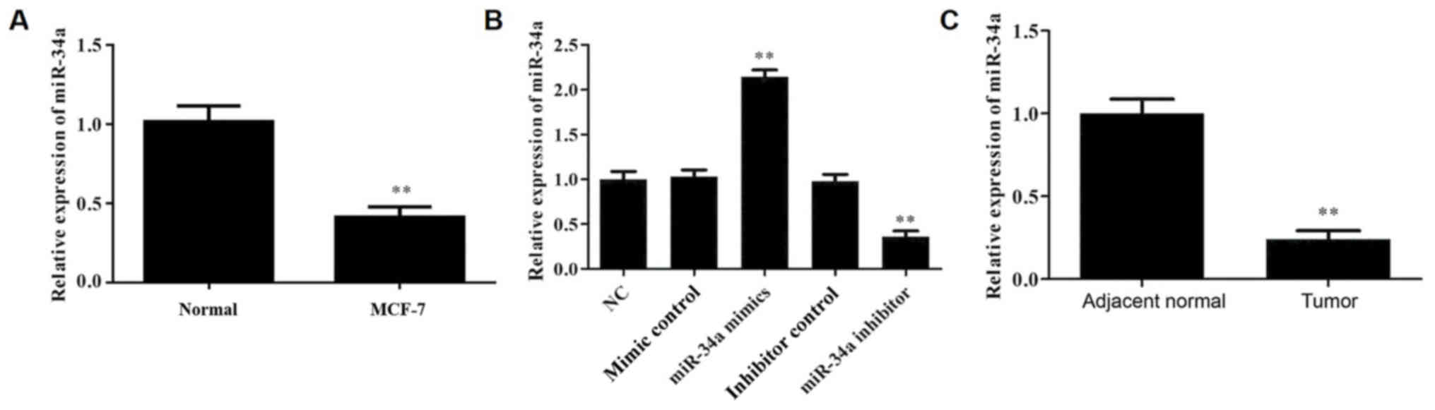
miR-34a is downregulated in breast
cancer
To determine the expression levels of miR-34a in
MCF-10A cells and MCF-7 cells, RT-qPCR was performed. The results
demonstrated that miR-34a expression was significantly lower in
MCF-7 cells compared with normal cells (Fig. 1A). Next, in order to investigate the
role of miR-34a in breast cancer, the MCF-7 cell line was used.
miR-34a was overexpressed or downregulated in MCF-7 cells by
transfections with miR-34a mimic or miR-34a inhibitor,
respectively, and the transfection efficiency was detected by
RT-qPCR (Fig. 1B). Subsequently, the
expression of miR-34a was analyzed by RT-qPCR in breast cancer and
adjacent normal breast tissues. As shown in Fig. 1C, compared with the adjacent normal
tissues, the level of miRNA-34a in breast cancer tissues was
significantly reduced, indicating that miRNA-34a may be involved in
breast cancer progression.
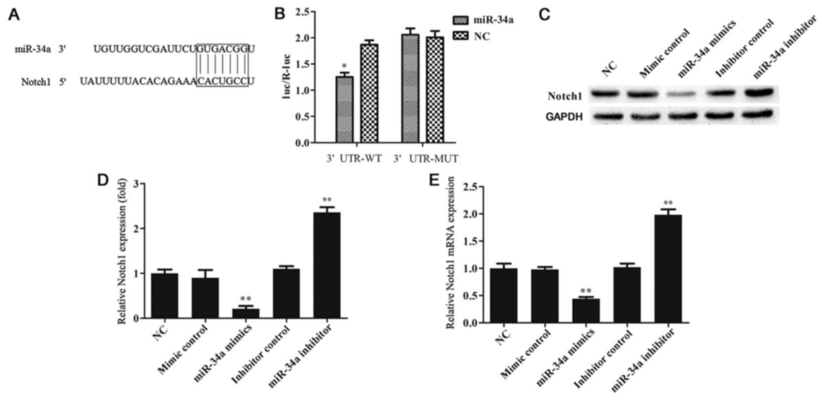
miR-34a directly targets Notch1
Bioinformatics analysis was conducted with the
TargetScan tool to predict the potential targets of miR-34a, and
the findings revealed that Notch1 is one of the target genes of
miR-34a (Fig. 2A). Previous studies
have demonstrated that activation of Notch1 or Notch4 in mice
induced the formation of spontaneous breast tumors, demonstrating
that activation of the Notch signaling pathway is an important
cause of breast cancer (14–16); thus, Notch1 was selected for further
investigation in the present study. To confirm this prediction, a
dual-luciferase reporter assay was used, and the findings indicated
that miR-34a directly targets Notch1 (Fig. 2B). To further reveal whether miR-34a
was able to regulate Notch1 expression in MCF-7 cells, the protein
and mRNA levels of Notch1 were detected by western blotting and
RT-qPCR, respectively. The results indicated that miR-34a
negatively regulated Notch1 expression in MCF-7 cells (Fig. 2C-E).
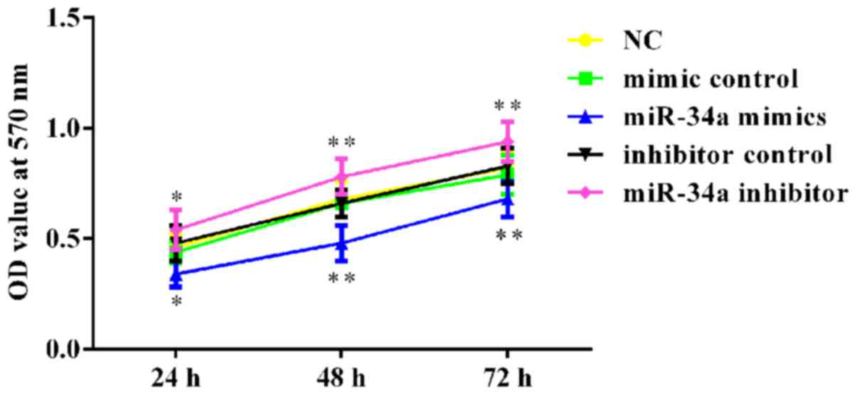
Effect of miR-34a on MCF-7 cell
viability
To investigate the effects of miR-34a on breast
cancer cell viability, an MTT assay was performed. As shown in
Fig. 3, the viability was
significantly reduced in miR-34a mimic-transfected cells, while the
viability of miR-34a inhibitor-transfected cells was notably
increased following transfection at three time points. No
statistically significant differences were detected between the NC
and the mimic control or inhibitor control groups (Fig. 3).
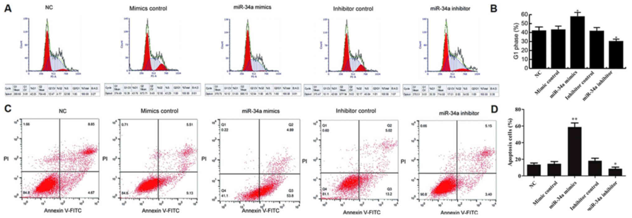
Effect of miR-34a on MCF-7 cell cycle
distribution and apoptosis
To investigate the effects of miR-34a on breast
cancer cell cycle distribution and apoptosis, flow cytometry was
applied. The results demonstrated that, at 48 h after transfection
with miR-34a mimic, MCF-7 cells were significantly arrested in G1
phase when compared with the NC group. In addition, miR-34a
inhibitor markedly declined the G1 phase arrest in MCF-7 cells as
compared with that observed in the NC group. No significant
difference was identified between the NC and the mimic control or
inhibitor control groups (Fig. 4A and
B).
Furthermore, the data revealed that, compared with
the NC group, miR-34a mimic significantly induced MCF-7 cell
apoptosis (early and late apoptosis). By contrast, cell apoptosis
was notably inhibited by miR-34a inhibitor. No significant
differences were detected between the NC and the mimic control or
inhibitor control groups (Fig. 4C and
D).
Effect of miR-34a on MCF-7 cell
invasion
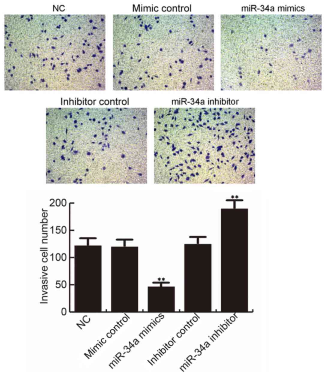
At 48 h after cell transfection, the MCF-7 cell
invasion ability was determined. The findings suggested that,
compared with the NC group, MCF-7 cell invasion ability was
significantly inhibited by miR-34a mimic, while miR-34a inhibitor
notably promoted the MCF-7 cell invasion ability. However, no
significant differences were observed between the NC and the mimic
control or inhibitor control groups (Fig. 5).
Discussion
In recent years, great progress has been made in the
investigation of miR-34a in breast cancer (19–21).
Previous studies have demonstrated that multiple miRNAs are
abnormally expressed in breast cancer and are closely associated
with the development of this tumor (22,23). The
study of miRNAs is important for the diagnosis, treatment and
prognosis prediction of breast cancer (24). Therefore, in the present study, the
expression and role of miR-34a in breast cancer were
investigated.
Initially, the level of miR-34a in human breast
cancer and adjacent normal tissues was detected in the present
study, and the results revealed that miR-34a was significantly
decreased in breast cancer tissues. Next, to further investigate
the role of miR-34a in the development of breast cancer, the MCF-7
cell line was used to perform in vitro investigation. The
bioinformatics prediction tool TargetScan was used to predict the
potential targets of miR-34a, and the results indicated that Notch1
is a target of miR-34a. Notch1 pathway is considered to be an
important pathway regulating the proliferation of breast cancer
cells. Inhibition of Notch1 pathway in breast cancer cells and stem
cells has been reported to effectively inhibit cell proliferation
and promote apoptosis (25). The
underlying mechanism may be associated with the decrease of the
expression of nuclear factor-κB, cyclin B1 and B-cell lymphoma-2,
which are located downstream of Notch1 (26). It has also been reported that miR-34a
is able to inhibit the proliferation of a variety of tumor cells.
For instance, Ma et al (27)
found that miR-34a inhibited the proliferation and promoted the
apoptosis of H1299 cells by targeting transforming growth factor β
receptor II. Gougelet et al (28) also observed that miR-34a reduced the
proliferation ability of primary cultured hepatocarcinoma cells by
inhibiting cyclin D1 and hepatocyte nuclear factor-4α expression
levels. The current study demonstrated that miR-34a was able to
negatively regulate Notch1 expression in MCF-7 cells, and miR-34a
mimic transfection inhibited MCF-7 cell viability, and induced cell
apoptosis and G1 phase arrest.
Notch1 pathway is considered to be involved in the
regulation of breast cancer cell invasion and migration. Recently,
Zhang et al (29) reported
that upregulation of Notch1 in the breast cancer cell line MCF-7
induced epithelial-mesenchymal transition, and promoted cell
invasion and migration. In addition, Notch signaling activation
promoted the ability of cell migration and growth (30). miR-34a is also an important
regulatory factor involved in tumor invasion and metastasis. Lai
et al (31) reported that
miR-34a was able to inhibit colon cancer cell migration through
regulating the sirtuin 1/p53 pathway. Liang et al (32) also indicated that the inhibitory
effect of miR-34a on the invasion and migration of prostate cancer
cells was associated with the targeted regulation of lymphoid
enhancer binding factor 1. Finally, Yu et al (33) demonstrated that miR-34a inhibited the
invasion, migration and angiogenesis of bladder cancer by targeting
CD44 in vitro and in vivo. In the present study, the
results revealed that miR-34a mimic prevented MCF-7 cell invasion,
which is consistent with the findings of the aforementioned
previous studies.
In conclusion, the data of the current study proved
the low expression level of miR-34a in human breast cancer tissues
as compared with the adjacent normal tissues. In addition,
overexpression of miR-34a was found to inhibit breast cancer cell
viability and invasion, as well as induce cell apoptosis and G1
phase arrest. miR-34a may exerts its role, at least partly, by
targeting Notch1, and this miRNA may be a promising therapeutic
target for the treatment of breast cancer. However, only MCF7 cells
were examined in current study; therefore, similar results in other
breast cancer cells would be required to establish the role of
miR-34a in the suppression of breast cancer cell proliferation and
invasion.
Acknowledgements
Not applicable.
Funding
No funding was received.
Availability of data and materials
The datasets used and/or analyzed during the current
study are available from the corresponding author on reasonable
request.
Authors' contributions
XR conceptualized and developed the study design,
and performed most of the experiments. LW and LM cultured the cells
and performed the cell transfections. XR, HZ and YY participated in
western blot analysis. LW, HZ and YY acquired and analyzed the
data. XR, XX, LW and LM conducted the statistical analysis,
interpreted the data and wrote the manuscript. XR and XX made
comments, suggested appropriate modifications and made corrections.
All authors read and approved the final manuscript.
Ethics approval and consent to
participate
The present study was approved by the Human Ethics
Committee Review Board of Wuxi Taihu Hospital, and informed consent
was obtained from each patient.
Patient consent for publication
Not applicable.
Competing interests
The authors declare that they have no competing
interests.
References
|
1
|
Jemal A, Bray F, Center MM, Ferlay J, Ward
E and Forman D: Global cancer statistics. CA Cancer J Clin.
61:69–90. 2011. View Article : Google Scholar : PubMed/NCBI
|
|
2
|
Siegel R, Naishadham D and Jemal A: Cancer
statistics, 2013. CA Cancer J Clin. 63:11–30. 2013. View Article : Google Scholar : PubMed/NCBI
|
|
3
|
Kim VN, Han J and Siomi MC: Biogenesis of
small RNAs in animals. Nat Rev Mol Cell Biol. 10:126–139. 2009.
View Article : Google Scholar : PubMed/NCBI
|
|
4
|
Bartel DP: MicroRNAs: Target recognition
and regulatory functions. Cell. 136:215–233. 2009. View Article : Google Scholar : PubMed/NCBI
|
|
5
|
Valencia-Sanchez MA, Liu J, Hannon GJ and
Parker R: Control of translation and mRNA degradation by miRNAs and
siRNAs. Genes Dev. 20:515–524. 2006. View Article : Google Scholar : PubMed/NCBI
|
|
6
|
Pang RT, Leung CO, Lee CL, Lam KK, Ye TM,
Chiu PC and Yeung WS: MicroRNA-34a is a tumor suppressor in
choriocarcinoma via regulation of Delta-like1. BMC Cancer.
13:252013. View Article : Google Scholar : PubMed/NCBI
|
|
7
|
Fang CY, Qiu SL, Sun F, Li W, Wang Z, Yue
B, Wu X and Yan D: Long non-coding RNA HNF1A-AS1 mediated
repression of miR-34a/SIRT1/p53 feedback loop promotes the
metastatic progression of colon cancer by functioning as a
competing endogenous RNA. Cancer Lett. 410:50–62. 2017. View Article : Google Scholar : PubMed/NCBI
|
|
8
|
Corcoran C, Rani S and O'Driscoll L:
miR-34a is an Intracellular and exosomal predictive biomarker for
response to docetaxelwith clinical relevance to prostate cancer
progression. Prostate. 74:1320–1334. 2014. View Article : Google Scholar : PubMed/NCBI
|
|
9
|
Nalls D, Tang SN, Rodova M, Srivastava RK
and Shankar S: Targeting epigenetic regulation of miR-34a for
treatment of pancreatic cancer by inhibition of pancreatic cancer
stem cells. PLoS One. 6:e240992011. View Article : Google Scholar : PubMed/NCBI
|
|
10
|
Misso G, Di Martino MT, De Rosa G, Farooqi
AA, Lombardi A, Campani V, Zarone MR, Gullà A, Tagliaferri P,
Tassone P and Caraglia M: Mir-34: A new weapon against cancer? Mol
Ther Nucleic Acids. 3:e1942014. View Article : Google Scholar : PubMed/NCBI
|
|
11
|
Hermeking H: The miR-34 family in cancer
and apoptosis. Cell Death Differ. 17:193–199. 2010. View Article : Google Scholar : PubMed/NCBI
|
|
12
|
Yin L, Velazquez OC and Liu ZJ: Notch
signaling: Emerging molecular targets for cancer therapy. Biochem
Pharmacol. 80:690–701. 2010. View Article : Google Scholar : PubMed/NCBI
|
|
13
|
Artavanis-Tsakonas S, Matsuno K and
Fortini ME: Notch signaling. Science. 268:225–232. 1995. View Article : Google Scholar : PubMed/NCBI
|
|
14
|
Shao S and Zhao X, Zhang X, Luo M, Zuo X,
Huang S, Wang Y, Gu S and Zhao X: Notch1 signaling regulates the
epithelial inverted question markmesenchymal transition and
invasion of breast cancer in a Slug-dependent manner. Mol Cancer.
14:282015. View Article : Google Scholar : PubMed/NCBI
|
|
15
|
Naik S, MacFarlane M and Sarin A: Notch4
signaling confers susceptibility to TRAIL-induced apoptosis in
breast cancer cells. J Cell Biochem. 116:1371–1380. 2015.
View Article : Google Scholar : PubMed/NCBI
|
|
16
|
Bakrania AK, Variya BC, Rathod LV and
Patel SS: DEAE-Dextran coated paclitaxel nanoparticles act as
multifunctional nano system for intranuclear delivery to triple
negative breast cancer through VEGF and NOTCH1 inhibition. Eur J
PharmBiopharm. 122:37–38. 2018. View Article : Google Scholar
|
|
17
|
Lakhani SR, Ellis IO, Schnitt SJ, Tan PH
and van de Vijver MJ: WHO Classification of Tumours of the Breast.
4th. Lyon: IARC Press; 2012
|
|
18
|
Livak KJ and Schmittgen TD: Analysis of
relative gene expression data using real-time quantitative PCR and
the 2(-Delta Delta C(T)) method. Methods. 25:402–408. 2001.
View Article : Google Scholar : PubMed/NCBI
|
|
19
|
Xu M, Li D, Yang C and Ji JS: MicroRNA-34a
Inhibition of the TLR signaling pathway via CXCL10 suppresses
breast cancer cell invasion and migration. Cell Physiol Biochem.
46:1286–1304. 2018. View Article : Google Scholar : PubMed/NCBI
|
|
20
|
He R, Liu P, Xie X, Zhou Y, Liao Q, Xiong
W, Li X, Li G, Zeng Z and Tang H: circGFRA1 and GFRA1 act as ceRNAs
in triple negative breast cancer by regulating miR-34a. J Exp Clin
Canc Res. 36:1452017. View Article : Google Scholar
|
|
21
|
Xia Y, Deng XW, Cao MJ, Liu S, Zhang X,
Xiao X, Shen S, Hu Q and Sheng W: Nanodiamond-based layer-by-layer
nanohybrids mediate targeted delivery of miR-34a for triple
negative breast cancer therapy. RSC Adv. 8:13789–13797. 2018.
View Article : Google Scholar
|
|
22
|
Iorio MV, Ferracin M, Liu CG, Veronese A,
Spizzo R, Sabbioni S, Magri E, Pedriali M, Fabbri M, Campiglio M,
et al: MicroRNA gene expression deregulationin human breast cancer.
Cancer Res. 65:7065–7070. 2015. View Article : Google Scholar
|
|
23
|
Volinia S, Galasso M, Sana ME, Wise TF,
Palatini J, Huebner K and Croce CM: Breast cancer signatures for
invasiveness and prognosis defined by deep sequencing of microRNA.
Proc Natl Acad Sci USA. 109:3024–3029. 2012. View Article : Google Scholar : PubMed/NCBI
|
|
24
|
Hoshino I and Matsubara H: MicroRNAs in
cancer diagnosis and therapy: From bench to bedside. Surg Today.
43:467–478. 2013. View Article : Google Scholar : PubMed/NCBI
|
|
25
|
Suman S, Das TP and Damodaran C: Silencing
NOTCH signaling causes growth arrest in both breast cancer stem
cells and breast cancer cells. Br J Cancer. 109:2587–2596. 2013.
View Article : Google Scholar : PubMed/NCBI
|
|
26
|
Pan H, Zhou W, He W, Liu X, Ding Q, Ling
L, Zha X and Wang S: Genistein inhibits MDA-MB-231 triple-negative
breast cancer cell growth by inhibiting NF-κB activity via the
Notch-1 pathway. Int J Mol Med. 30:337–343. 2012. View Article : Google Scholar : PubMed/NCBI
|
|
27
|
Ma ZL, Hou PP, Li YL, Wang DT, Yuan TW,
Wei JL, Zhao BT, Lou JT, Zhao XT, Jin Y and Jin YX: MicroRNA-34a
inhibits the proliferation and promotes the apoptosis of non-small
cell lung cancer H1299 cell line by targeting TGFβR2. Tumour Biol.
36:2481–2490. 2015. View Article : Google Scholar : PubMed/NCBI
|
|
28
|
Gougelet A, Sartor C, Bachelot L, Godard
C, Marchiol C, Renault G, Tores F, Nitschke P, Cavard C, Terris B,
et al: Antitumour activity of an inhibitor of miR-34a in liver
cancer with beta-catenin-mutations. Gut. 65:1024–1034. 2016.
View Article : Google Scholar : PubMed/NCBI
|
|
29
|
Zhang X and Zhao X, Shao S, Zuo X, Ning Q,
Luo M, Gu S and Zhao X: Notch1 induces epithelial-mesenchymal
transition and the cancer stem cell phenotype in breast cancer
cells and STAT3 plays a key role. Int J Oncol. 46:1141–1148. 2015.
View Article : Google Scholar : PubMed/NCBI
|
|
30
|
Bolos V, Mira E, Martinez-Poveda B, Luxán
G, Cañamero M, Martínez-A C, Mañes S and de la Pompa JL: Notch
activation stimulates migration of breast cancer cells and promotes
tumor growth. Breast Cancer Res. 15:R542013. View Article : Google Scholar : PubMed/NCBI
|
|
31
|
Lai M, Du G, Shi R, Yao J, Yang G, Wei Y,
Zhang D, Xu Z, Zhang R, Li Y, et al: miR-34a inhibits migration and
invasion by regulating the SIRT1/p53 pathway in human SW480 cells.
Mol Med Rep. 11:3301–3307. 2015. View Article : Google Scholar : PubMed/NCBI
|
|
32
|
Liang J, Li Y, Daniels G, Sfanos K, De
Marzo A, Wei J, Li X, Chen W, Wang J, Zhong X, et al: LEF1
targeting EMT in prostate cancer invasion is regulated by miR-34a.
Mol Cancer Res. 13:681–688. 2015. View Article : Google Scholar : PubMed/NCBI
|
|
33
|
Yu G, Yao W, Xiao W, Li H, Xu H and Lang
B: MicroRNA-34a functions as an anti-metastatic microRNA and
suppresses angiogenesis in bladder cancer by directly targeting
CD44. J Exp Clin Cancer Res. 33:7792014. View Article : Google Scholar : PubMed/NCBI
|