Introduction
Articular cartilage is avascular and aneural, and
has limited capacity for self-regeneration when injured. Early
intervention is essential to prevent further damage from spreading
to peripheral healthy cartilage and joint surfaces, as
osteoarthritis may occur in the injured joint, which eventually
requires invasive treatment, for example, knee replacement
(1).
Several clinical treatments for cartilage injuries
have been developed, including microfracture (2), abrasion arthroplasty (3), autologous osteochondral transplant
(4,5), autologous chondrocyte implantation
(5,6)
and prosthetic joint replacement (7). Autologous osteochondral transplant
(mosaicplasty) is a technique that involves the transplantation of
one or more cylindric osteochondral plugs harvested from the
non-weight-bearing periphery of the femoral condyles towards the
damaged cartilage site (8).
Mosaicplasty has several advantages in terms of it being a
single-stage and simple procedure, and being cost-effective.
Mosaicplasty provides integration of subchondral bone into the host
tissue and has been satisfied in numerous studies (9–14).
Tissue engineering has emerged as a promising
approach in repairing cartilage defects as it provides an optimal
strategy to develop a scaffold as a native cartilage-bone plug,
which has similar structural and mechanical features as
osteochondral tissue to facilitate the integration with host
cartilage and underlying subchondral bone. The principles of tissue
engineering involve three major components, including biocompatible
scaffolds, the utilization of cell sources, and cell signaling
molecules (15).
Chitosan (CS), silk fibroin (SF), and
nanohydroxyapatite particles (nHA) are commonly used biomaterials
due to their safety and biological compatibility (16–18). It
has been found that, compared with bi-component scaffolds, the
CS/SF/nHA tri-component scaffold has higher compressive strength
and efficiency for inducing cell proliferation, which makes it a
favorable scaffold for tissue engineering and regeneration
(19).
Mesenchymal stem cells (MSCs) have been widely used
as a potential cell source in several studies and clinical trials
(20,21). The high capacity of in vitro
expansion of bone marrow-derived stromal cells (BMSCs) is an
attractive source of cells for cartilage tissue engineering
(22,23). In addition, BMSCs are progenitor
cells of osteoblasts and are essential for the maintenance of bone
quality and quantity (24). It has
been increasingly suggested that BMSCs contribute to rapid healing
in a variety of reconstructive and restorative surgical procedures
(25).
Bone morphogenetic protein (BMP) is an important
molecule for facilitating the recovery of bone and cartilage by
inducing the differentiation of mesenchymal osteoprogenitors and
promoting osteoblastic maturation and function (26,27). Of
all BMPs, BMP-2 is the most potent bone growth factor and has been
widely used for inducing BMSC differentiation into osteoblastic
cells (28–30). BMP-2 protein therapy is a useful
therapy that has been used in the treatment of various animal bony
defect models (31–35). However, the limitation of a short
half-life and high cost associated with large dose requirements in
clinical situations cannot fully satisfy clinical requirements,
which restricts the application of these exogenous proteins
(36). Various vectors have been
established for delivering genes in stem cells efficiently
(37–39). Among these, lentivirus mediated gene
transduction has been found to be effective for the long-term and
stable expression of BMP-2 (31,32,40,41).
A novel treatment, platelet-rich plasma (PRP), is a
safe solution for simulating a natural repair process which
contains a high concentration of platelets and a variety of growth
factors (42). PRP has been used on
BMSCs to assist in bone tissue regeneration. The combination of PRP
and BMSCs has shown promising results on cartilage bone recovery in
animal experiments (43,44).
To optimize the treatment of cartilage defects, the
present study investigated the effect of the combination of
lentivirus-mediated BMP-2-modified BMSCs and PRP on articular
cartilage injury of the rabbit knee following treatment with
mosaicplasty. It was found that cartilage defects were successfully
repaired with BMP-2-transduced BMSCs/PRP at 16 weeks post-surgery.
This combination may provide a promising therapy for cartilage and
bone healing.
Materials and methods
Rabbit BMSC culture
The animal experiments were performed at The Third
Military Medical University (Chongqing, China), and were approved
by the Laboratory Animal Welfare and Ethics Committee of the Third
Military Medical University. The BMSCs were purchased from Cyagen
Biosciences, Inc. (Santa Clara, CA, USA; cat. no. RBXMX-01001). The
cells were plated with the initial seeding in a 100-mm dish and
cultured with Dulbecco's modified Eagle's medium (DMEM, Gibco,
Thermo Fisher Scientific, Inc., Waltham, MA, USA) supplemented with
10% fetal bovine serum (Thermo Fisher Scientific, Inc.) in standard
culture conditions of 37°C and 5% humidified CO2. The
cells were maintained for 3 days prior to the first medium change.
When the cells reached 90% confluency on days 7–9, the adherent
cells were trypsinized and passaged approximately at a 1:3 split.
The identity and mulitpotent differentiation ability along
osteogenic, chondrogenic and adipogenic lineages was confirmed by
Cyagen Biosciences, Inc. Cells in passage three were used for
transduction with lentivirus.
Construction of Lv-BMP-2 and
lentiviral infection of BMSCs
The BMP-2 gene was amplified from Rabbit cDNA with
primers containing NotI and BamHI restriction sites:
Bmp-2-NotI, forward
5′-aaggaaaaaagcggccgcATGGTGGCCGGGACCCGCTG-3′, Bmp-2-BamHI,
reverse 5′-cgcggatccCTAGCGACACCCACAACCCTCCAC-3′. Lv-BMP-2 was
constructed by inserting the BMP-2 gene fragment into the
pLV5-EF1A-GFP plasmid (Western Biotechnology, Inc., Chongqing,
China). The lentivirus was produced by co-transfecting the
pLV5-EF1A-GFP (Lv-control or Lv-BMP-2) and helper plasmid
PG-P1-VSVG, PG-P2-REV and PG-P3-RRE into 293T cells (Western
Biotechnology, Inc.). The rabbit BMSCs at passage three were
transduced with Lv-BMP-2 or Lv-control virus in the presence of 5
µg/ml polybrene. At 24 h post-transduction, the medium was replaced
with fresh DMEM with 10% FBS. The BMSCs were cultured for 72 h.
GFP-positive cells were observed and infection efficiency was
determined using an inverted fluorescence microscope.
PRP preparation (45)
A 10-ml blood sample was harvested from the central
auricular vein of rabbits from each group. The blood was
transferred into an ethylene diamine tetraacetic acid vacutainers
(BD Biosciences, Franklin Lakes, NJ, USA), and plasma was harvested
by centrifugation for 10 min at 1,000 × g at 22°C. The supernatant
was isolated and centrifuged for 20 min at 1,500 × g at 22°C. The
top layer was removed and the platelet-rich lower layer was
collected for future use.
Construction of the osteochondral
defect model
A total of 48 female New Zealand white rabbits
(Jiangsu Zhenlin Biological Technology Co., Ltd., Jiangsu, China)
aged 2–3 months old with a mean weight of 2.5 kg were housed in
room at 26±3°C with a 12 h light/dark cycle. The rabbits were
allowed ad libitum access to food and water. A
full-thickness segment femoral cartilage defect (diameter 5 mm,
thickness 4 mm) was generated through the articular cartilage and
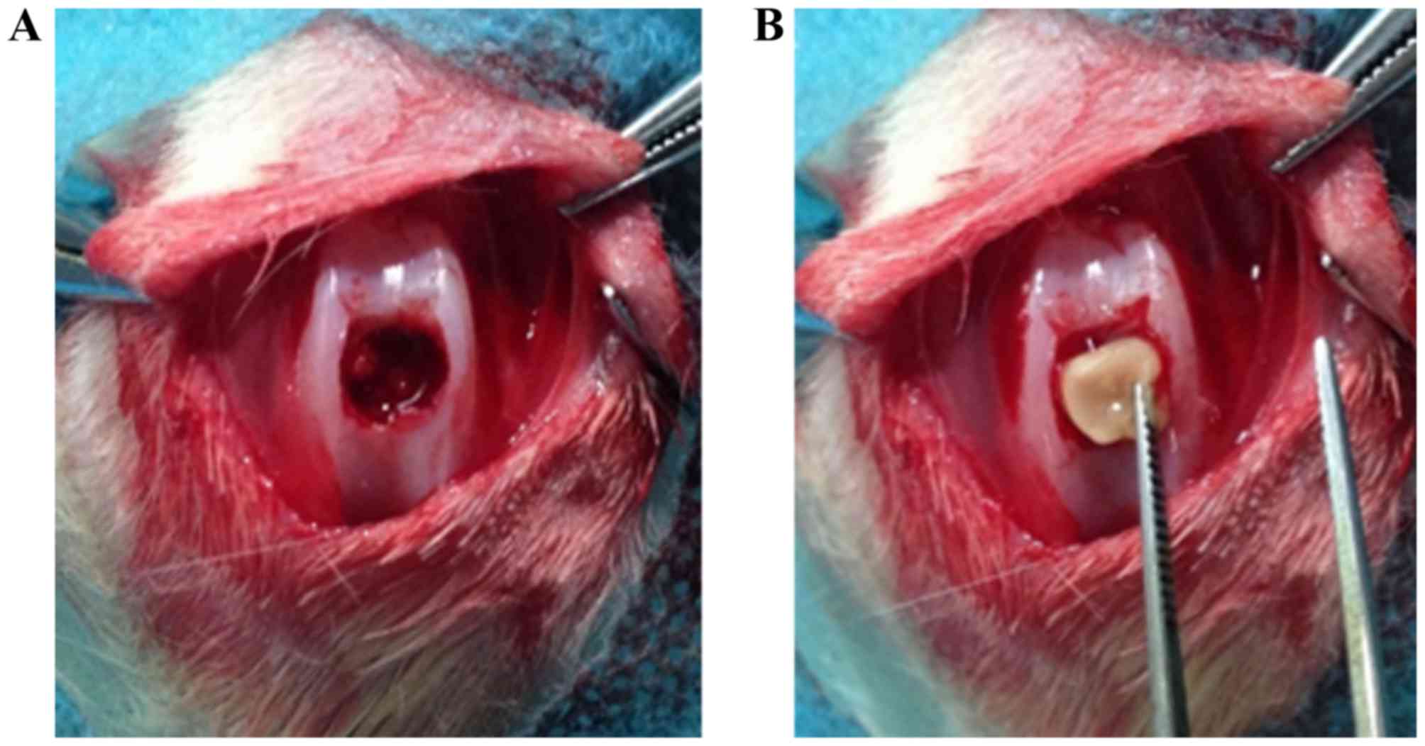
subchondral bone of the patellar groove (Fig. 1A) using a drill equipped with a 5-mm
diameter drill bit. The CS/SF/nHA scaffold treated with or without
PRP and BMSCs was then implanted to fill the cartilage defect on
one knee (Fig. 1B). The rabbits were
divided randomly into four groups: i) Untreated, ii) treated with
BMSCs, iii) treated with Lv-control-BMSCs+PRP; iv) treated with
Lv-BMP-2-BMSCs+PRP. A total of 107 BMSCs and/or 0.2 ml
PRP were used to treat different knees according to the grouping.
The wound was then closed in layers. The animals were returned to
their cages and allowed to move freely without joint
immobilization. The rabbits were sacrificed at 16 weeks. Each
cartilage defect area was evaluated macroscopically.
Scaffold preparation
The biphysic scaffold osteochondral defect repair
were prepared according to our previously described procedures
(19).
Reverse transcription-quantitative
polymerase chain reaction (RT-qPCR) analysis
The cells from each group were collected and total
RNA was extracted using TRIzol reagent (Invitrogen; Thermo Fisher
Scientific, Inc.). The cDNA was then synthesized using a
PrimeScript® 1st Strand cDNA Synthesis kit (Takara
Biotechnology Co., Ltd., Dalian, China). A total of 1 mg cDNA was
used and RT-qPCR analysis was then performed using SYBR®
Premix Ex Taq™ (Takara Biotechnology Co., Ltd.)
according to the manufacturer's protocol. The PCR conditions were
as follows: Stage 1, pre-denaturation: 95°C for 4 min; stage 2, PCR
amplification: 95°C for 10 sec, 60°C for 20 sec, 72°C for 30 sec
for a total of 40 cycles. Each experiment was performed in
triplicate. The primers sequences for alkaline phosphatase (ALP),
type I collagen and type II collagen are presented in Table I (46,47).
Relative mRNA expression was calculated using the 2−ΔΔCt
method (48).
 | Table I.Reverse transcription-quantitative
polymerase chain reaction primer sequences. |
Table I.
Reverse transcription-quantitative
polymerase chain reaction primer sequences.
|
| Primer (5′-3′) |
|---|
|
|
|
|---|
| Gene | Forward | Reverse |
|---|
| ALP (46) |
GCACGACAATCGGGATGAAC |
TCCAGCAGGACGGTCATCA |
| COL I (47) |
TGGCAAGAACGGAGATGACG |
GCACCATCCAAACCACTGAA |
| COL II (47) |
CCACGCTCAAGTCCCTCAAC |
AGTCACCGCTCTTCCACTCG |
| GAPDH |
GGGTGGTGGACCTCATGGT |
CGGTGGTTTGAGGGCTCTTA |
Western blot analysis
The cells from each group were washed with ice-cold
phosphate-buffered saline (PBS) and lysed with lysis buffer
containing 50 mM Tris-HCL (pH 7.4), 0.1% sodium dodecyl sulfate
(SDS), 150 mM NaCl, and protease inhibitor cocktail (Sigma-Aldrich;
Merck KGaA, Darmstadt, Germany). The protein concentration was
determined using Pierce™ BCA Protein Assay kit (Thermo
Fisher Scientific, Inc.) according to the manufacturer's potocol.
The sample was suspended in SDS buffer and boiled for 6 min. A
total of 30 µg/lane proteins were separated onto 30% SDS-PAGE gels
for electrophoresis and then transferred onto a nitrocellulose
membrane. The membranes were blocked in non-fat milk in
Tris-buffered saline with Tween 20 (TBST) for 1 h, and incubated
with primary antibodies for BMP-2 (1:500; cat. no. bs-1012R; BIOSS,
Beijing, China), SRY-box 9 (SOX9; 1:500; cat. no. HPA001758;
Sigma-Aldrich; Merck KGaA) or β-actin (1:1,000; cat. no. MA5-15739;
Thermo Fisher Scientific, Inc.) overnight at 4°C. The membranes
were washed with TBST three times and were incubated with goat
anti-rabbit (cat. no. ab6721) and rabbit anti-mouse (cat. no.
ab97046) secondary antibodies (both 1:1,000; Abcam, Cambridge, UK)
for 1 h at room temperature. The bands were visualized by enhanced
chemiluminescence detection reagents (Bio-Rad Laboratories, Inc.,
Hercules, CA, USA) according to the manufacturer's protocol. The
bands of western blot were quantified with ImageJ (ImageJ bundled
with Java 1.8.0; National Institutes of Health, Bethesda, MD, USA)
(49).
MTT assay
The cells were seeded 2×10³ cells into
96-well plates in triplicate. After 24 h, the cells were washed
twice with PBS and the MTT (Sigma; EMD Millipore) assays were
performed. A total of 20 µl MTT (5 g/l) was added to each well, and
the cells were cultured for an additional 4 h. The absorbance (A)
values at 490 nm were determined with a microplate reader to
determine cell viability.
Micro-computed tomography (CT)
imaging
Specimens obtained at 16 weeks (three animals in
each group) were examined with micro-CT (GE micro-CT system; GE
Healthcare Life Sciences, Logan, UT, USA) for the analyses of bone
and cartilage growth and regeneration.
Alizarin red staining
The cells were cultivated with osteogenetic
induction solution (0.1 µmol/l dexamethasone, 50 mg/l ascorbic acid
and 10 mmol/l β-glycerophosphate) and cultured at 37°C and 5%
CO2. The medium was replaced every 3 days. At 14 days
post-induction, the cells were fixed with 95% ethanol for 10 min,
and incubated in 2% Alizarin red staining solution (Sigma-Aldrich;
Merck KGaA) for 5 min. The staining buffer was removed and the
cells were washed twice with PBS. Calcification deposits were
identified under light microscopy.
Statistical analysis
The data were processed using SPSS statistical
processing software (ver. 21.0; IBM SPSS, Armonk, NY, USA). The
results are expressed as the mean ± standard deviation. A unpaired
Student's t-test was applied for statistical comparisons between
two groups and one-way analysis of variance was applied among
multiple groups. P<0.05 was considered to indicate a
statistically significant difference.
Results
Lentivirus mediated expression of
BMP-2 in BMSCs
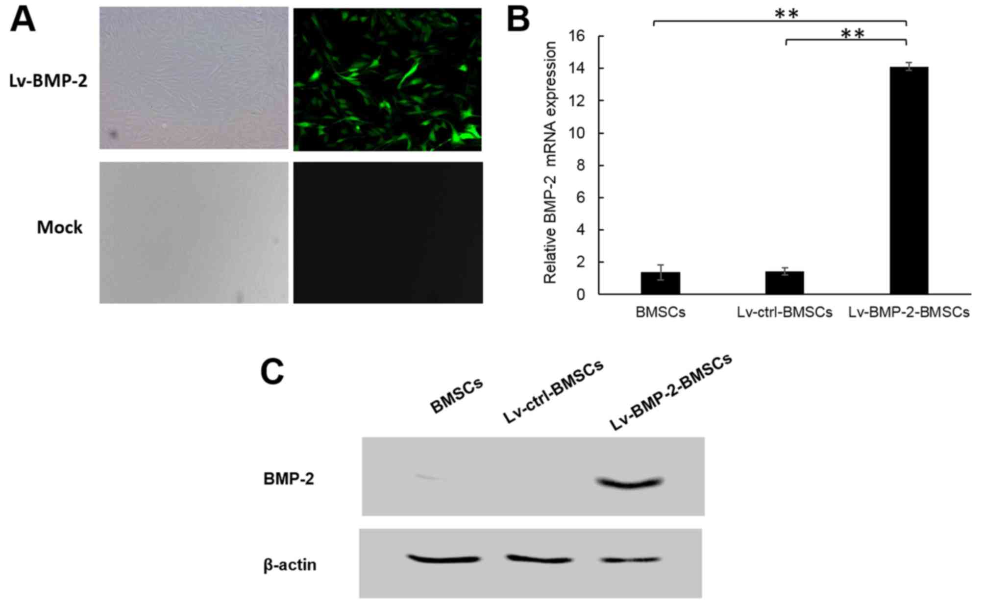
The BMSCs were transduced with lentivirus Lv-BMP-2
or Lv-control at an MOI of 50. According to fluorescence microscopy
observation, the infection efficiency was 90% at 72 h
post-transduction (Fig. 2A). To
confirm the expression of BMP-2, RT-qPCR and western blot analyses
were applied to detect the mRNA and protein levels of BMP-2 in
BMSCs. As shown in Fig. 2B and C,
the endogenous expression of BMP-2 in BMSCs was low, whereas the
Lv-BMP-2 BMSCs expressed significantly higher BMP-2 compared with
the Lv-control BMSCs and non-infected BMSCs at the mRNA and protein
levels. This suggested that the Lv-BMP-2-modified BMSCs had been
successfully established.
Combination of BMP-2 and PRP enhances
the cell viability of BMSCs
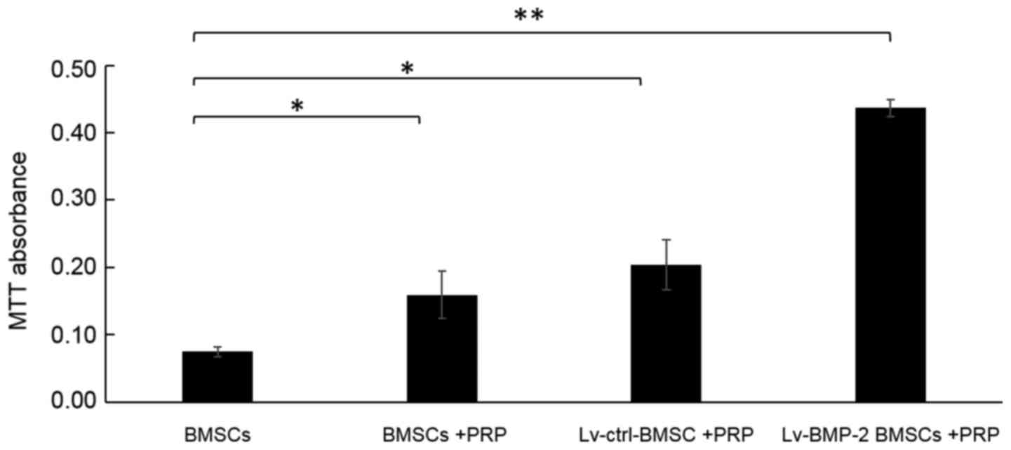
The cell viability of cells was examined using an
MTT assay. The absorbance value at 490 nm was significantly
increased by infection with Lv-BMP-2 in BMSCs with PRP, compared
with that in other groups (Fig. 3).
It was also noted that PRP alone enhanced the cell viability of
BMSCs, although the enhancement was not as marked as for the
combination of BMP-2 and PRP.
Combination of BMP-2 and PRP results
in enhanced expression of osteogenic-related genes in BMSCs
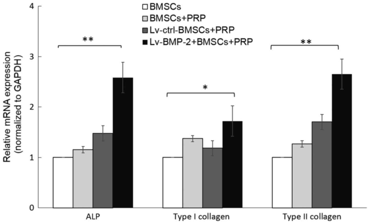
The impact of BMP-2 and PRP on the expression of
osteogenic-related genes was then examined. ALP, and type I
collagen are markers of osteogenic differentiation. In addition,
type II collagen is known to modulate the chondrogenesis of MSCs
and is important in the osteogenic differentiation of BMSCs
(50). To investigate the capacity
of osteogenic differentiation of BMP-2 BMSCs, the cells were
cultured with osteogenesis induction medium, and total mRNA was
isolated from the BMSCs from each group at day 7. The expression
levels of ALP, type I collagen and type II collagen were determined
by RT-qPCR analysis. The data showed that the lentivirus-mediated
expression of BMP-2 in BMSCs upregulated the expression of ALP,
type I collagen and type II collagen, compared with expression in
the Lv-control group. Although incubation with PRP marginally
increased the expression of these genes, the result was not
statistically significant (Fig.
4).
Combination of BMP-2 and PRP induces
higher osteogenic differentiation in BMSCs
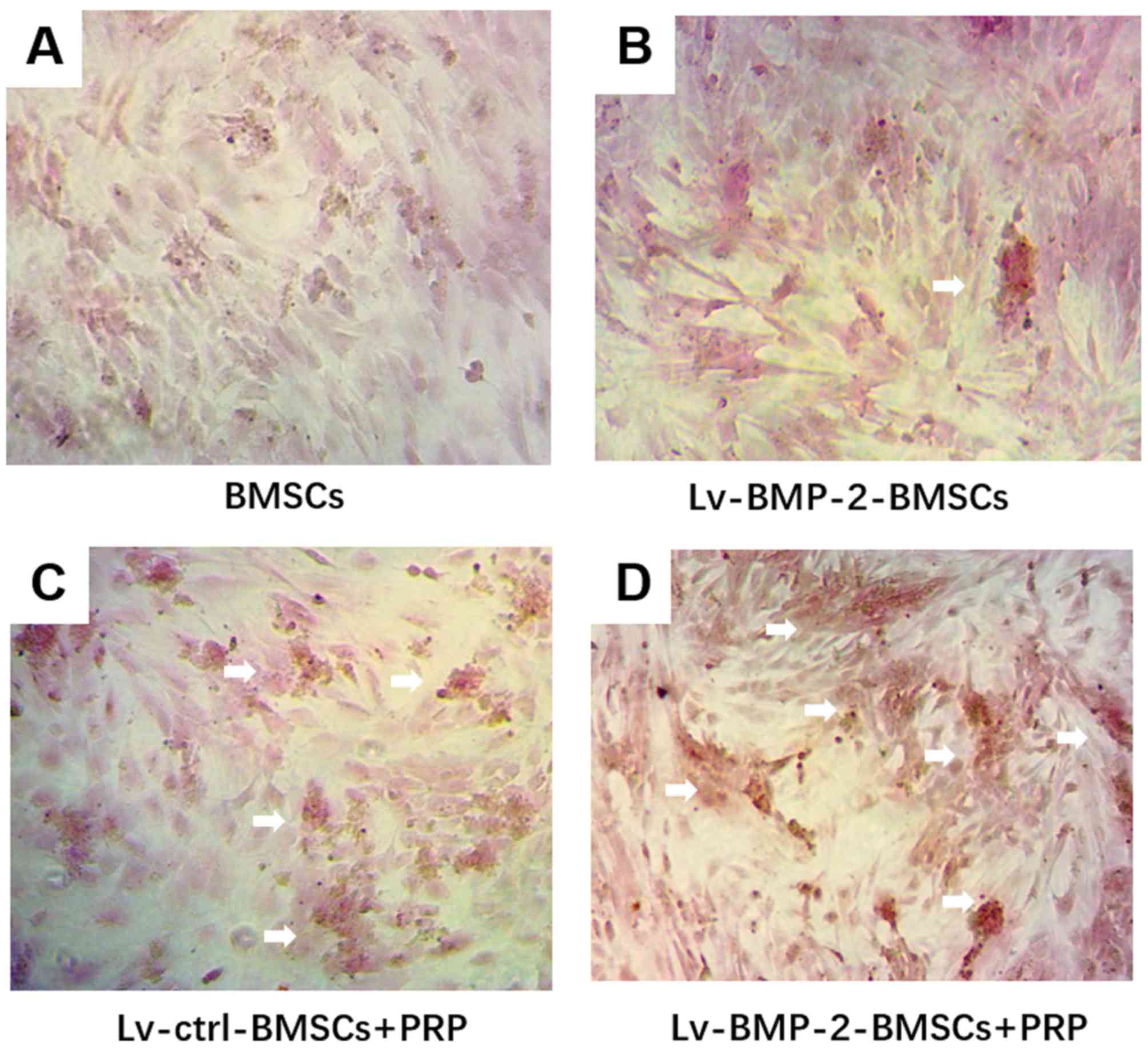
Following 14 days of culture in osteogenesis
induction medium, followed by Alizarin red staining, mineralised
nodules were observed under the microscope. The mineralized nodules
were more intense in the LvBMP-2-modified BMSCs + PRP group,
compared with those in the other groups (Fig. 5A-D). Although BMP-2 or PRP alone
marginally increased the osteogenic differentiation,
LvBMP-2-modified BMSCs+PRP exerted the most marked enhancement of
osteogenesis.
Combination of BMP-2 and PRP recovers
cartilage defects completely
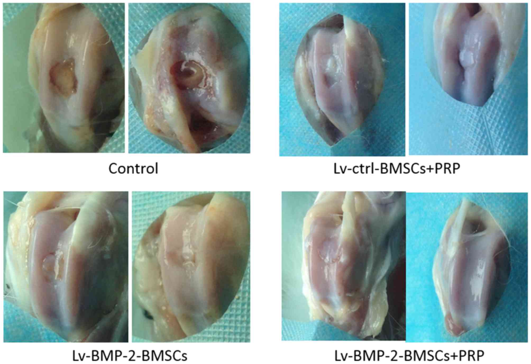
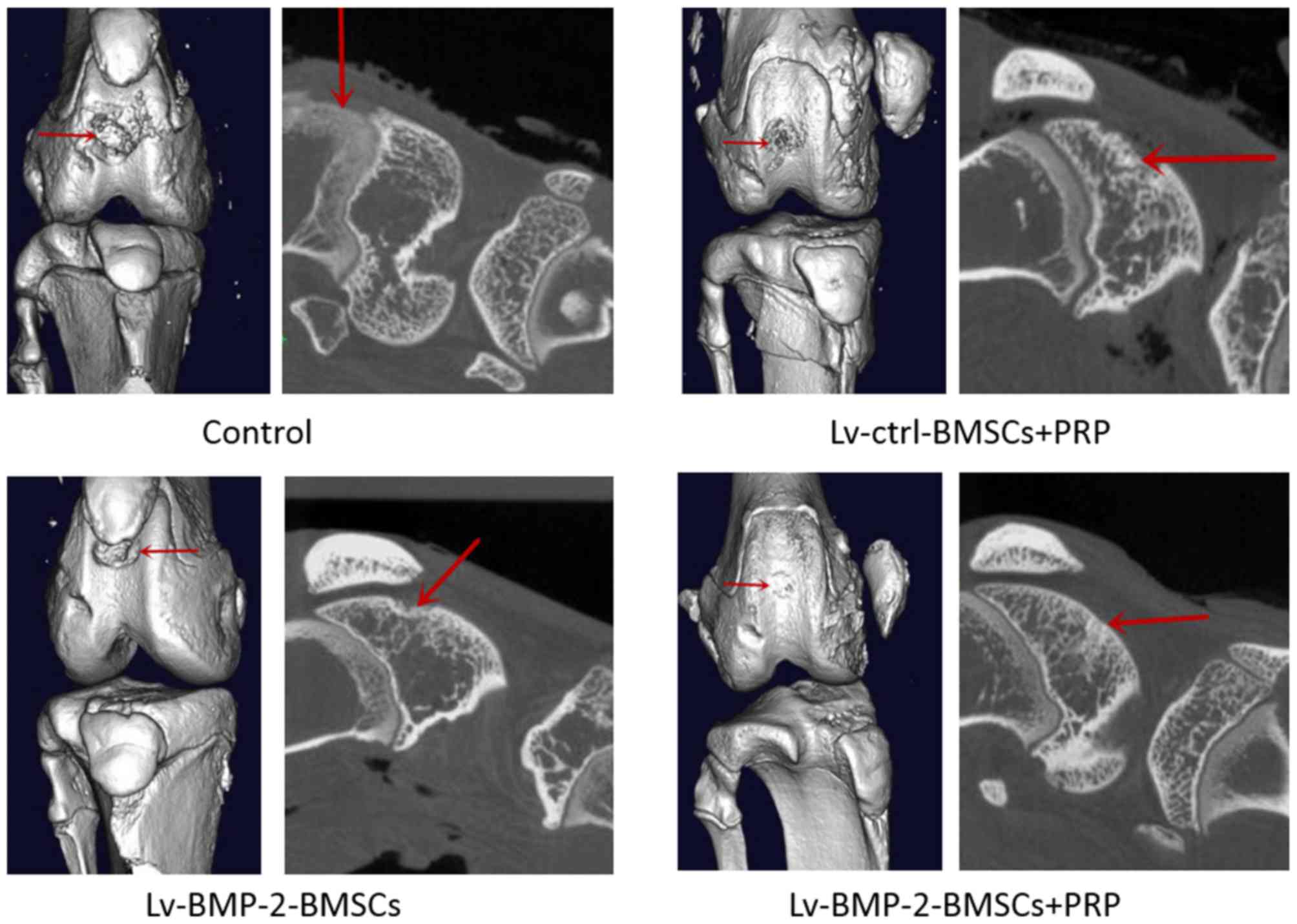
At day 0, a full-thickness defect was created
through the articular cartilage and subchondral bone of the
patellar groove using a drill bit. The CS/SF/nHA scaffold was
implanted to fill the cartilage defect. Subsequently,
107 BMSCs and/or 0.2 ml PRP were used to treated
different knees according to grouping. The wound was then closed in
layers. At 16 weeks, the cartilage defects were examined by
macroscopic observation and micro-CT.
In the control group, the central regions of the
defect sites were filled with a thin, rough reddish-brown tissue
(Fig. 6), the patellar fossa was
significantly larger than that of other groups (Figs. 6 and 7), indicating that cartilage repair
remained limited and incomplete at week 16. The defects in the
Lv-BMP-2 BMSCs and Lv-control BMSCs+PRP groups were similar on
gross observation (Figs. 6 and
7). Although a concavity was
observed in the peripheral region of defects, the surface of
cartilage and bone was shiny and smooth. In the BMP-2 BMSCs+PRP
group, it was found that the surface region of the recovered
cartilage was smooth and shiny, and covered with hyaline-like
cartilage tissue (Fig. 6). No
significant fossa was observed, suggesting that articular cartilage
repair was complete. Taken together, the data showed that the
combination of BMP-2-modified BMSCs with PRP had optimal effects on
the recovery of cartilage and bone defects.
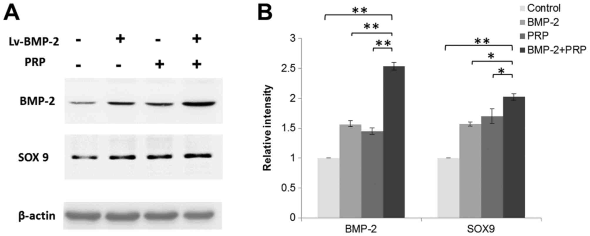
Lentivirus-mediated expression of
BMP-2 is observed in BMSCs for at least 16 weeks
To confirm the expression of BMP-2 in cartilage
defect region in rabbits, the proteins were isolated from articular
cartilage and subchondral bone regions from each group at week 16.
There was an increase of the overall expression of BMP-2 in tissues
around the cartilage defect sites in the Lv-BMP-2+PRP group. It was
also observed that PRP or Lv-BMP-2 alone enhanced the expression of
BMP-2. SRY-box 9 (SOX9), a transcription factor that is important
in cartilage development and formation was also detected. As shown
in Fig. 8A and B, BMP-2 and PRP
increased the expression of SOX9, which contributed to the recovery
of the cartilage defect in vivo. The data indicated that
lentivirus-mediated expression of BMP-2 was present in BMSCs for at
least 16 weeks, and the enhanced expression of BMP-2 combined with
PRP treatment induced the expression of genes for cartilage and
bone formation, which facilitated tissue regeneration in the rabbit
knees.
Discussion
Articular cartilage has limited ability for
self-regeneration, therefore, early intervention is important
(1). MSCs have been widely used for
tissue regeneration, particularly cartilage repair, owing to their
potential for osteoregeneration and osteogenic differentiation
(20,21). There are three important components
for successful tissue regeneration, namely, biocompatible
scaffolds, cell sources, and cell signaling molecules (15). In the present study, the effect of
the combination of a CS/SF/nHA tri-component scaffold,
BMP-2-modified BMSCs and PRP on a rabbit cartilage defect model was
investigated; the results showed that this combination provided
promising results.
CS, SF and nHA have been applied in various tissue
engineering studies due to favourable biological compatibility
(16–18). It has been found that a combination
of two of these improved biocompatibility, enhanced mechanical
strength, or reduced degradation (16,17,51,52). Qi
et al (19), found that the
CS/nHA/SF tri-component scaffold had all of the above advantages,
making it suitable for the aim of the present study.
BMP-2 has been found to be important in bone
formation and BMSC differentiation, compared with other BMP family
members (28–30). However, its limitation of a short
half-life and high cost prevents it from further application. With
the assistance of lentivirus-mediated gene transduction, the
present study showed that the expression of BMP-2 in BMSCs was
sustained over at least 16 weeks, suggesting that it was effective
for long-term and stable expression of BMP-2 (40).
PRP is another important factor for cartilage
defects used in the present study. PRP provides various cell
signaling molecules, which contribute to healing and growth,
including platelet-derived growth factor, vascular endothelial
growth factor, and transforming growth factor-β1 (53). By using PRP, BMP-2-modified BMSCs
exerted beneficial results on repairing cartilage defects in rabbit
knees. Previously Bahmanpour et al (54), found that platelet-rich fibrin (PRF)
had higher evaluation scores of full-thickness lesions in rabbits
than PRP. The use of PRF, rather than PRP, to treat the cartilage
defect in the model used in the present study may assist in
identifying a more effective therapy.
This combination strategy has provided beneficial
outcomes for tissue regeneration in several studies. For example,
Lian et al (55), found that
the combination of BMSCs and PRP assists in diabetic wound repair
and regeneration. BMSCs transfected with BMP-2 also enhanced
tendon-bone healing and improved bone regeneration in canine
segmental ulnar defects (56,57). The
present study combined the advantages of several components for
tissue regeneration and optimized the cartilage defect therapy in a
rabbit model.
Taken together, the data obtained in the present
study suggested that the combination of BMP-2 and PRP enhanced the
efficiency of cartilage and bone repair and provided a novel
promising therapy for tissue engineering.
Acknowledgements
Not applicable.
Funding
The present study was financially supported by the
Science and Technology Plan Projects in Guizhou Province [grant no.
(2016)1420] and the National Natural Science Foundation of China
(grant no. 81660367).
Availability of data and materials
The datasets generated and analysed during the
present study are available from the corresponding author on
reasonable request.
Authors' contributions
JD was involved in the design of the current study.
SR and LY performed all experiments. WH performed the statistical
analyses. All authors participated in manuscript preparation and
were involved in the discussion of the results. All authors have
read and approved the final manuscript.
Ethics approval and consent to
participate
The animal experiments were approved by the
Laboratory Animal Welfare and Ethics Committee of the Third
Military Medical University.
Patient consent for publication
Not applicable.
Competing interests
The authors confirm that they have no competing
interests.
References
|
1
|
Nehrer S, Chiari C, Domayer S, Barkay H
and Yayon A: Results of chondrocyte implantation with a
fibrin-hyaluronan matrix: A preliminary study. Clin Orthop Relat
Res. 466:1849–1855. 2008. View Article : Google Scholar : PubMed/NCBI
|
|
2
|
Steadman JR, Rodkey WG and Rodrigo JJ:
Microfracture: Surgical technique and rehabilitation to treat
chondral defects. Clin Orthop Relat Re S362-S369. 2001. View Article : Google Scholar
|
|
3
|
Bittberg M: Articular cartilage repair: An
update on different clinical repair methods. Ortop Traumatol
Rehabil. 3:235–243. 2001.PubMed/NCBI
|
|
4
|
Mendicino RW, Catanzariti AR and Hallivis
R: Mosaicplasty for the treatment of osteochondral defects of the
ankle joint. Clin Podiatr Med Surg. 18:495–513. 2001.PubMed/NCBI
|
|
5
|
Bentley G, Biant LC, Carrington RW, Akmal
M, Goldberg A, Williams AM, Skinner JA and Pringle J: A
prospective, randomised comparison of autologous chondrocyte
implantation versus mosaicplasty for osteochondral defects in the
knee. J Bone Joint Surg Br. 85:223–230. 2003. View Article : Google Scholar : PubMed/NCBI
|
|
6
|
Brittberg M, Tallheden T, Sjogren-Jansson
B, Lindahl A and Peterson L: Autologous chondrocytes used for
articular cartilage repair: An update. Clin Orthop Relat Res. 391
Suppl:S337–S348. 2001. View Article : Google Scholar : PubMed/NCBI
|
|
7
|
Hunziker EB: Articular cartilage repair:
Basic science and clinical progress. A review of the current status
and prospects. Osteoarthritis Cartilage. 10:432–463. 2002.
View Article : Google Scholar : PubMed/NCBI
|
|
8
|
Hangody L, Rathonyi GK, Duska Z,
Vasarhelyi G, Fules P and Módis L: Autologous osteochondral
mosaicplasty. Surgical technique. J Bone Joint Surg Am. 86-A Suppl
1:S65–S72. 2004. View Article : Google Scholar
|
|
9
|
Ozturk A, Ozdemir MR and Ozkan Y:
Osteochondral autografting (mosaicplasty) in grade IV cartilage
defects in the knee joint: 2- to 7-year results. Int Orthop.
30:200–204. 2006. View Article : Google Scholar : PubMed/NCBI
|
|
10
|
Haklar U, Tuzuner T, Kocaoglu B and Guven
O: Mosaicplasty technique in the treatment of osteochondral lesions
of the knee. Acta Orthop Traumatol Turc. 42:344–349. 2008.(In
Turkish). View Article : Google Scholar : PubMed/NCBI
|
|
11
|
Rose T, Craatz S, Hepp P, Raczynski C,
Weiss J, Josten C and Lill H: The autologous osteochondral
transplantation of the knee: Clinical results, radiographic
findings and histological aspects. Arch Orthop Trauma Surg.
125:628–637. 2005. View Article : Google Scholar : PubMed/NCBI
|
|
12
|
Hangody L, Vasarhelyi G, Hangody LR,
Sukosd Z, Tibay G, Bartha L and Bodó G: Autologous osteochondral
grafting-technique and long-term results. Injury. 39 Suppl
1:S32–S39. 2008. View Article : Google Scholar : PubMed/NCBI
|
|
13
|
Emre TY, Ege T, Kose O, Demircioglu Tekdos
D, Seyhan B and Uzun M: Factors affecting the outcome of
osteochondral autografting (mosaicplasty) in articular cartilage
defects of the knee joint: Retrospective analysis of 152 cases.
Arch Orthop Trauma Surg. 133:531–536. 2013. View Article : Google Scholar : PubMed/NCBI
|
|
14
|
Hangody L, Dobos J, Balo E, Panics G,
Hangody LR and Berkes I: Clinical experiences with autologous
osteochondral mosaicplasty in an athletic population: A 17-year
prospective multicenter study. Am J Sports Med. 38:1125–1133. 2010.
View Article : Google Scholar : PubMed/NCBI
|
|
15
|
Langer R and Vacanti JP: Tissue
engineering. Science. 260:920–926. 1993. View Article : Google Scholar : PubMed/NCBI
|
|
16
|
Bhardwaj N and Kundu SC: Silk fibroin
protein and chitosan polyelectrolyte complex porous scaffolds for
tissue engineering applications. Carbohydrate Polymers. 85:325–333.
2011. View Article : Google Scholar
|
|
17
|
Bhardwaj N, Nguyen QT, Chen AC, Kaplan DL,
Sah RL and Kundu SC: Potential of 3-D tissue constructs engineered
from bovine chondrocytes/silk fibroin-chitosan for in vitro
cartilage tissue engineering. Biomaterials. 32:5773–5781. 2011.
View Article : Google Scholar : PubMed/NCBI
|
|
18
|
Ji WC, Zhang XW and Qiu YS: Selected
suitable seed cell, scaffold and growth factor could maximize the
repair effect using tissue engineering method in spinal cord
injury. World J Exp Med. 6:58–62. 2016. View Article : Google Scholar : PubMed/NCBI
|
|
19
|
Qi XN, Mou ZL, Zhang J and Zhang ZQ:
Preparation of chitosan/silk fibroin/hydroxyapatite porous scaffold
and its characteristics in comparison to bi-component scaffolds. J
Biomed Mater Res A. 102:366–372. 2014. View Article : Google Scholar : PubMed/NCBI
|
|
20
|
Garg P, Mazur MM, Buck AC, Wandtke ME, Liu
J and Ebraheim NA: Prospective Review of Mesenchymal Stem Cells
Differentiation into Osteoblasts. Orthop Surg. 9:13–19. 2017.
View Article : Google Scholar : PubMed/NCBI
|
|
21
|
Fellows CR, Matta C, Zakany R, Khan IM and
Mobasheri A: Adipose, bone marrow and synovial joint-derived
mesenchymal stem cells for cartilage repair. Front Genet.
7:2132016. View Article : Google Scholar : PubMed/NCBI
|
|
22
|
Yamasaki S, Mera H, Itokazu M, Hashimoto Y
and Wakitani S: Cartilage repair with autologous bone marrow
mesenchymal stem cell transplantation: Review of preclinical and
clinical studies. Cartilage. 5:196–202. 2014. View Article : Google Scholar : PubMed/NCBI
|
|
23
|
Potier E, Noailly J and Ito K: Directing
bone marrow-derived stromal cell function with mechanics. J
Biomech. 43:807–817. 2010. View Article : Google Scholar : PubMed/NCBI
|
|
24
|
Chen Q, Shou P, Zheng C, Jiang M, Cao G,
Yang Q, Cao J, Xie N, Velletri T, Zhang X, et al: Fate decision of
mesenchymal stem cells: adipocytes or osteoblasts? Cell Death
Differ. 23:1128–1139. 2016. View Article : Google Scholar : PubMed/NCBI
|
|
25
|
Schneider RK, Anraths J, Kramann R,
Bornemann J, Bovi M, Knüchel R and Neuss S: The role of
biomaterials in the direction of mesenchymal stem cell properties
and extracellular matrix remodelling in dermal tissue engineering.
Biomaterials. 31:7948–7959. 2010. View Article : Google Scholar : PubMed/NCBI
|
|
26
|
Abe E: Function of BMPs and BMP
antagonists in adult bone. Ann N Y Acad Sci. 1068:41–53. 2006.
View Article : Google Scholar : PubMed/NCBI
|
|
27
|
Lochab AK and Extavour CG: Bone
morphogenetic protein (BMP) signaling in animal reproductive system
development and function. Dev Biol. 427:258–269. 2017. View Article : Google Scholar : PubMed/NCBI
|
|
28
|
Zhang Q, He QF, Zhang TH, Yu XL, Liu Q and
Deng FL: Improvement in the delivery system of bone morphogenetic
protein-2: A new approach to promote bone formation. Biomed Mater.
7:0450022012. View Article : Google Scholar : PubMed/NCBI
|
|
29
|
Starman JS, Bosse MJ, Cates CA and Norton
HJ: Recombinant human bone morphogenetic protein-2 use in the
off-label treatment of nonunions and acute fractures: A
retrospective review. J Trauma Acute Care Surg. 72:676–681. 2012.
View Article : Google Scholar : PubMed/NCBI
|
|
30
|
Angle SR, Sena K, Sumner DR, Virkus WW and
Virdi AS: Healing of rat femoral segmental defect with bone
morphogenetic protein-2: A dose response study. J Musculoskelet
Neuronal Interact. 12:28–37. 2012.PubMed/NCBI
|
|
31
|
Hsu WK, Sugiyama O, Park SH, Conduah A,
Feeley BT, Liu NQ, Krenek L, Virk MS, An DS, Chen IS and Lieberman
JR: Lentiviral-mediated BMP-2 gene transfer enhances healing of
segmental femoral defects in rats. Bone. 40:931–938. 2007.
View Article : Google Scholar : PubMed/NCBI
|
|
32
|
Miyazaki M, Sugiyama O, Tow B, Zou J,
Morishita Y, Wei F, Napoli A, Sintuu C, Lieberman JR and Wang JC:
The effects of lentiviral gene therapy with bone morphogenetic
protein-2-producing bone marrow cells on spinal fusion in rats. J
Spinal Disord Tech. 21:372–379. 2008. View Article : Google Scholar : PubMed/NCBI
|
|
33
|
Jiang X, Zhao J, Wang S, Sun X, Zhang X,
Chen J, Kaplan DL and Zhang Z: Mandibular repair in rats with
premineralized silk scaffolds and BMP-2-modified bMSCs.
Biomaterials. 30:4522–4532. 2009. View Article : Google Scholar : PubMed/NCBI
|
|
34
|
Xu XL, Tang T, Dai K, Zhu Z, Guo XE, Yu C
and Lou J: Immune response and effect of adenovirus-mediated human
BMP-2 gene transfer on the repair of segmental tibial bone defects
in goats. Acta Orthop. 76:637–646. 2005. View Article : Google Scholar : PubMed/NCBI
|
|
35
|
Xiao C, Zhou H, Ge S, Tang T, Hou H, Luo M
and Fan X: Repair of orbital wall defects using biocoral scaffolds
combined with bone marrow stem cells enhanced by human bone
morphogenetic protein-2 in a canine model. Int J Mol Med.
26:517–525. 2010.PubMed/NCBI
|
|
36
|
Wozney JM and Rosen V: Bone morphogenetic
protein and bone morphogenetic protein gene family in bone
formation and repair. Clin Orthop Relat Res. 1–37. 1998.PubMed/NCBI
|
|
37
|
Chen Y, Luk KD, Cheung KM, Xu R, Lin MC,
Lu WW, Leong JC and Kung HF: Gene therapy for new bone formation
using adeno-associated viral bone morphogenetic protein-2 vectors.
Gene Ther. 10:1345–1353. 2003. View Article : Google Scholar : PubMed/NCBI
|
|
38
|
Park J, Ries J, Gelse K, Kloss F, von der
Mark K, Wiltfang J, Neukam FW and Schneider H: Bone regeneration in
critical size defects by cell-mediated BMP-2 gene transfer: A
comparison of adenoviral vectors and liposomes. Gene Ther.
10:1089–1098. 2003. View Article : Google Scholar : PubMed/NCBI
|
|
39
|
Ramezani A, Hawley TS and Hawley RG:
Lentiviral vectors for enhanced gene expression in human
hematopoietic cells. Mol Ther. 2:458–469. 2000. View Article : Google Scholar : PubMed/NCBI
|
|
40
|
Sugiyama O, An DS, Kung SP, Feeley BT,
Gamradt S, Liu NQ, Chen IS and Lieberman JR: Lentivirus-mediated
gene transfer induces long-term transgene expression of BMP-2 in
vitro and new bone formation in vivo. Mol Ther. 11:390–398. 2005.
View Article : Google Scholar : PubMed/NCBI
|
|
41
|
Virk MS, Conduah A, Park SH, Liu N,
Sugiyama O, Cuomo A, Kang C and Lieberman JR: Influence of
short-term adenoviral vector and prolonged lentiviral vector
mediated bone morphogenetic protein-2 expression on the quality of
bone repair in a rat femoral defect model. Bone. 42:921–931. 2008.
View Article : Google Scholar : PubMed/NCBI
|
|
42
|
Mehta S and Watson JT: Platelet rich
concentrate: Basic science and current clinical applications. J
Orthop Trauma. 22:432–438. 2008. View Article : Google Scholar : PubMed/NCBI
|
|
43
|
Yamada Y, Ueda M, Naiki T, Takahashi M,
Hata K and Nagasaka T: Autogenous injectable bone for regeneration
with mesenchymal stem cells and platelet-rich plasma:
Tissue-engineered bone regeneration. Tissue Eng. 10:955–964. 2004.
View Article : Google Scholar : PubMed/NCBI
|
|
44
|
Oliva A, Passaro I, Di Pasquale R, Di Feo
A, Criscuolo M, Zappia V, Della Ragione F, D'Amato S, Annunziata M
and Guida L: Ex vivo expansion of bone marrow stromal cells by
platelet-rich plasma: A promising strategy in maxillo-facial
surgery. Int J Immunopathol Pharmacol. 18:47–53. 2005.PubMed/NCBI
|
|
45
|
Park CG, Joo MW, Jeong J, Kang YK and Lee
DR: Evaluation of the effects of the combination of autologous
mesenchymal stem cells and platelet-rich plasma on structural bone
allograft healing. Cell Tissue Bank. 18:229–238. 2017. View Article : Google Scholar : PubMed/NCBI
|
|
46
|
Du X, Huang F, Zhang S, Yao Y, Chen Y,
Huang H and Bai B: Carboxymethylcellulose with phenolic hydroxyl
microcapsules enclosinggene-modified BMSCs for controlled BMP-2
release in vitro. Artif Cells Nanomed Biotechnol. 45:1–14. 2017.
View Article : Google Scholar
|
|
47
|
Wang SJ, Jiang D, Zhang ZZ, Huang AB, Qi
YS, Wang HJ, Zhang JY and Yu JK: Chondrogenic potential of
peripheral blood derived mesenchymal stem cells seeded on
demineralized cancellous bone scaffolds. Sci Rep. 6:364002016.
View Article : Google Scholar : PubMed/NCBI
|
|
48
|
Livak KJ and Schmittgen TD: Analysis of
relative gene expression data using real-time quantitative PCR and
the 2(-Delta Delta C(T)) method. Methods. 25:402–408. 2001.
View Article : Google Scholar : PubMed/NCBI
|
|
49
|
Miller L: Analyzing gels and western blots
with Image. J. Lukemiller. org. Miscellaneous Topics Vaguely
Related to Science¸. 2010.
|
|
50
|
Chiu LH, Lai WF, Chang SF, Wong CC, Fan
CY, Fang CL and Tsai YH: The effect of type II collagen on MSC
osteogenic differentiation and bone defect repair. Biomaterials.
35:2680–2691. 2014. View Article : Google Scholar : PubMed/NCBI
|
|
51
|
Hu Q, Li B, Wang M and Shen J: Preparation
and characterization of biodegradable chitosan/hydroxyapatite
nanocomposite rods via in situ hybridization: A potential material
as internal fixation of bone fracture. Biomaterials. 25:779–785.
2004. View Article : Google Scholar : PubMed/NCBI
|
|
52
|
Frohbergh ME, Katsman A, Botta GP,
Lazarovici P, Schauer CL, Wegst UG and Lelkes PI: Electrospun
hydroxyapatite-containing chitosan nanofibers crosslinked with
genipin for bone tissue engineering. Biomaterials. 33:9167–9178.
2012. View Article : Google Scholar : PubMed/NCBI
|
|
53
|
Eppley BL, Woodell JE and Higgins J:
Platelet quantification and growth factor analysis from
platelet-rich plasma: Implications for wound healing. Plast
Reconstr Surg. 114:1502–1508. 2004. View Article : Google Scholar : PubMed/NCBI
|
|
54
|
Bahmanpour SP, Ghasemi MP, Sadeghi-Naini
MM and Kashani IRP: Effects of platelet-rich plasma &
platelet-rich fibrin with and without stromal cell-derived factor-1
on repairing full-thickness cartilage defects in knees of rabbits.
Iran J Med Sci. 41:507–517. 2016.PubMed/NCBI
|
|
55
|
Lian Z, Yin X, Li H, Jia L, He X, Yan Y,
Liu N, Wan K, Li X and Lin S: Synergistic effect of bone
marrow-derived mesenchymal stem cells and platelet-rich plasma in
streptozotocin-induced diabetic rats. Ann Dermatol. 26:1–10. 2014.
View Article : Google Scholar : PubMed/NCBI
|
|
56
|
Dong Y, Zhang Q, Li Y, Jiang J and Chen S:
Enhancement of tendon-bone healing for anterior cruciate ligament
(ACL) reconstruction using bone marrow-derived mesenchymal stem
cells infected with BMP-2. Int J Mol Sci. 13:13605–13620. 2012.
View Article : Google Scholar : PubMed/NCBI
|
|
57
|
Itoi T, Harada Y, Irie H, Sakamoto M,
Tamura K, Yogo T, Soeta S, Amasaki H, Hara Y and Tagawa M:
Escherichia coli-derived recombinant human bone morphogenetic
protein-2 combined with bone marrow-derived mesenchymal stromal
cells improves bone regeneration in canine segmental ulnar defects.
BMC Vet Res. 12:2012016. View Article : Google Scholar : PubMed/NCBI
|