Introduction
Clinical statistics have shown that chronic kidney
disease (CKD) ranks third in the world, in terms of incidence rate
and number of infected individuals, next to cancer and heart
diseases (1,2). It is caused by interaction of several
kinds of nephropathy, so patients with renal diseases may develop
CKD if they don't receive timely diagnosis and treatment (3,4).
Clinical treatment can lead CKD in remission, but this disease may
cause complications such as the frequent occurrence of
inflammation, decrease in serum calcium, osteoporosis and vitamin D
deficiency. Therefore, attention should be paid to the prevention
of the above complications and finding the successful treatment in
clinic (5–8). In this study, the correlation of CKD
with inflammatory factors, osteoporosis and vitamin D deficiency
were analyzed, providing the basis for the follow-up clinical
treatment.
Patients and methods
General data
A total of 78 patients with CKD presented to the
Union Hospital (Wuhan, China) from December 2015 to December 2017
were selected and divided into three groups according to the
severity of the disease: CKD I–II stage group, CKD III–IV stage
group and CKD V stage group. Among patients in CKD I–II stage
group, there were 12 males and 13 females with body mass index
(BMI) of 25.5±2.76 kg/m2, height of 169.54±17.54 cm and
average age of 58.43±5.65 years. In CKD III–IV stage group, there
were 14 males and 12 females with BMI of 24.9±3.09
kg/m2, height of 170.01±17.09 cm and average age of
59.65±6.09 years. In CKD V stage group, there were 12 males and 15
females with BMI of 25.2±2.76 kg/m2, height of
167.76±16.43 cm and average age of 58.21±5.32 years.
Inclusion criteria: patients who had good adherence
and no mental diseases and whose main organs such as heart, liver
and kidney were not injured.
Exclusion criteria: patients who recently had
infection symptoms or were suffering from cancer or
osteoporosis.
The study was approved by the Ethics Committee of
the Union Hospital and informed consents were signed by the
patients or the guardians.
Methods
Venous blood samples (8 ml) were collected from all
patients on an empty stomach. After leaving the samples to stand
for 30 min, serum was separated through centrifugation at 8,000 × g
for 15 min at 4°C. Inflammatory factors interleukin-6 (IL-6) and
tumor necrosis factor-α (TNF-α) were detected by enzyme-linked
immunoassay. C-reactive protein (CRP) was measured by an
auto-chemistry analyzer (SmartChem; Row2 Technologies, Inc.,
Parsippany, NJ, USA).
Indicators of osteoporosis: serum phosphate and
serum calcium were both measured by an automatic biochemical
analyzer (Mindray Bio-Medical Electronics Co., Ltd., Shenzhen,
China). When serum albumin level of the patient was <40 g/l, the
corrected blood calcium level was calculated. Bone mineral density
(BMD) was measured by a bone density meter (MetriScan; Alara, Inc.,
San Jose, CA, USA) (9).
Determination of 25(OH)D: vitamin D deficiency was
defined as 25(OH)D was <15 ng/ml, and the detection of this
factor was carried out by magnetic particle-based chemiluminescence
(Roche Pharma AG, Grenzach-Wyhlen, Germany) (10). Determination of serum sodium and
serum potassium: ion selective electrode method and the
auto-chemistry analyzer were used. Serum creatinine (Cr): It was
analyzed using the auto-chemistry analyzer after enzymatic assay.
Blood urea nitrogen (BUN): it was determined by
ultraviolet-glutamic acid dehydrogenase assay and analyzed by the
auto-chemistry analyzer.
Statistical analysis
The statistical software, Statistical Product and
Service Solutions (SPSS; SPSS, Inc., Chicago, IL, USA) 17.0, was
used for the statistical analysis of the data. t-test was used for
enumeration data which were expressed as mean ± standard deviation
(SD). ANOVA was used for comparison between multiple groups and the
post hoc test was the Least Significant Difference test. Pearson's
analysis was employed for correlation analysis between variables.
P<0.05 was considered to indicate a statistically significant
difference.
Results
Comparison of general data of patients
among different groups
It was found that there were no differences in age,
sex, BMI and height of patients among the groups (Table I).
 | Table I.Comparison of the general data of
patients among different groups. |
Table I.
Comparison of the general data of
patients among different groups.
| Index | CKD I–II stage | CKD III–IV stage | CKD V stage |
|---|
| Age (years) | 58.43±5.65 | 59.65±6.09 | 58.21±5.32 |
| Sex
(male/female) | 12/13 | 14/12 | 12/15 |
| BMI
(kg/m2) |
25.5±2.76 |
24.9±3.09 |
25.2±2.76 |
| Height (cm) | 169.54±17.54 | 170.01±17.09 | 167.76±16.43 |
Comparisons of inflammatory factors, osteoporosis
indicators and vitamin D deficiency of patients among different
groups. Compared with those in the CKD I–II stage group, IL-6, CRP,
TNF-α, serum phosphate, serum sodium, serum potassium and BUN of
the patients in the other two groups were significantly increased,
but serum calcium, BMD and 25(OH)D were significantly decreased.
IL-6, CRP, TNF-α, serum phosphate, serum sodium, serum potassium
and BUN in the CKD V stage group were significantly higher than
those in the CKD III–IV stage group, but serum calcium, BMD and
25(OH)D were lower than those in the CKD III–IV stage group, which
showed statistically significant difference (P<0.05) (Table II).
| Table II.Comparison of inflammatory factors,
osteoporosis indicators and vitamin D of patients among different
groups. |
Table II.
Comparison of inflammatory factors,
osteoporosis indicators and vitamin D of patients among different
groups.
| Index | CKD I–II stage | CKD III–IV stage | CKD V stage |
|---|
| IL-6 (pg/ml) | 99.2±9.3 |
125.3±10.3a |
189.2±10.7a,b |
| CRP (µg/ml) | 2.9±0.2 | 6.4±0.6a | 20.3±2.0a,b |
| TNF-α (ng/ml) | 1.22±0.12 |
1.79±0.18a |
2.23±0.23a,b |
| Serum calcium
(mmol/l) | 2.2±0.2 | 2.0±0.2a | 1.5±0.2a,b |
| Serum phosphate
(mmol/l) | 1.3±0.1 | 1.6±0.1a | 2.0±0.2a,b |
| BMD | 0.923±0.086 |
0.823±0.098a |
0.723±0.067a,b |
| 25(OH)D (ng/ml) | 20.5±2.3 | 14.9±1.7a | 6.3±0.8a,b |
| Serum sodium
(mmol/l) | 134.43±11.56 |
146.67±14.09a |
152.09±15.21a,b |
| Serum potassium
(mmol/l) | 3.86±0.45 |
4.32±0.39a |
4.78±0.49a,b |
| BUN (mml/l) | 13.67±1.65 |
14.87±1.98a |
16.02±1.99a,b |
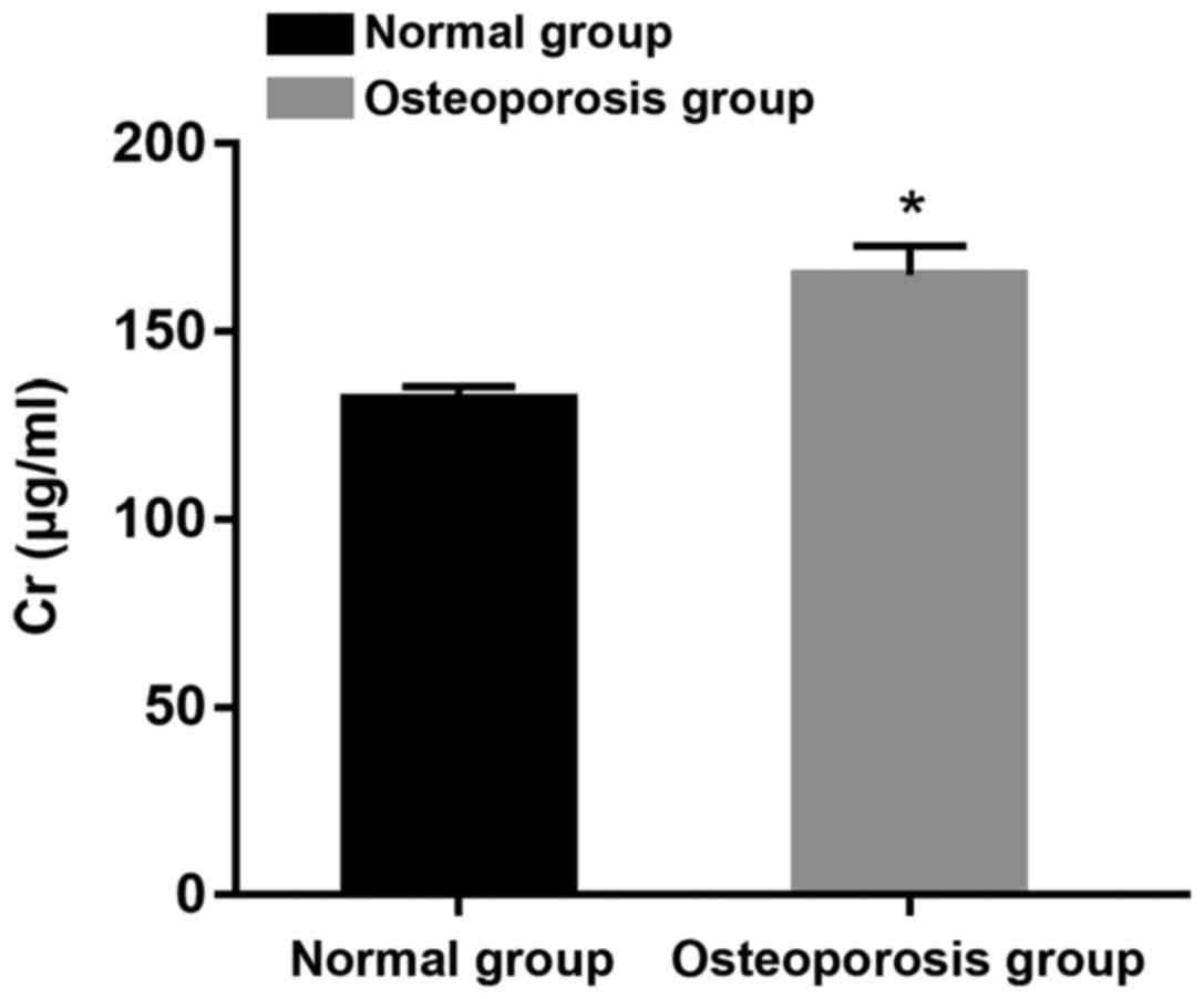
Comparison of Cr between normal and
osteoporosis group
Patients were divided into two groups: normal and
osteoporosis group on the basis of BMD. It was found that Cr
content of patients with osteoporosis was noticeably higher than
that in normal group, which showed a statistically significant
difference (P<0.05) (Fig. 1).
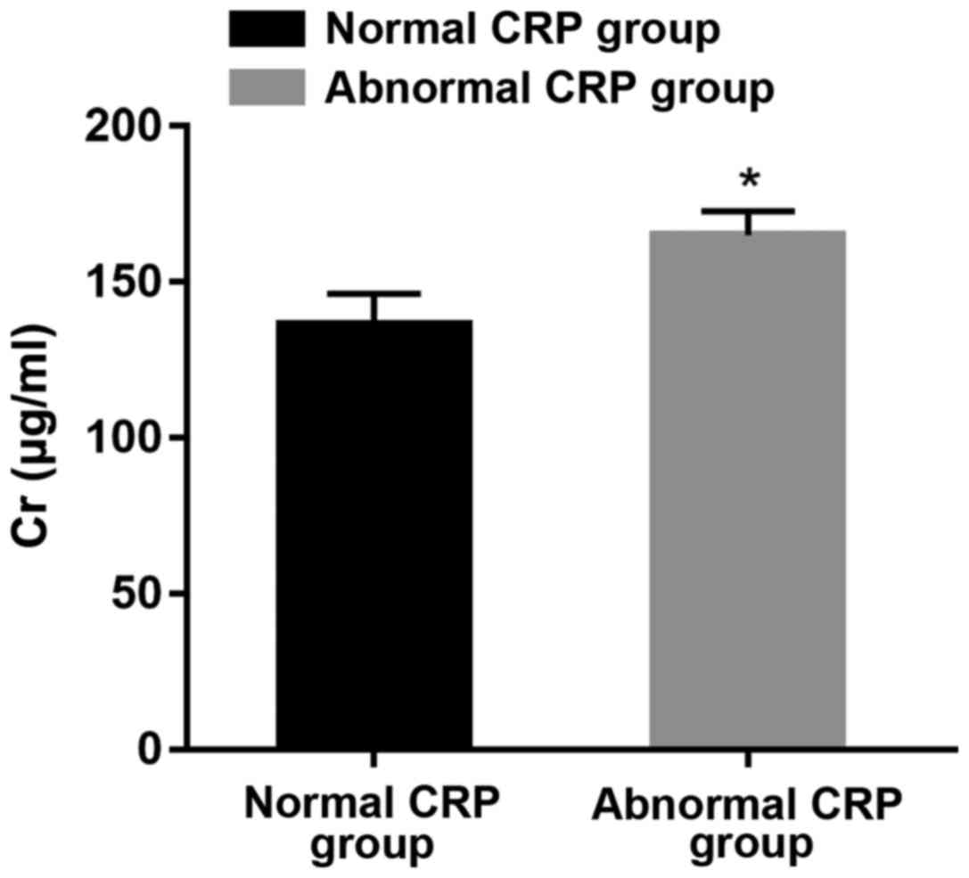
Comparison of Cr content between
groups with normal and abnormal CRP
According to the comparison between the two groups
of patients, it was suggested that the Cr content in the group with
abnormal CRP was significantly higher than that in the normal group
(P<0.05) (Fig. 2).
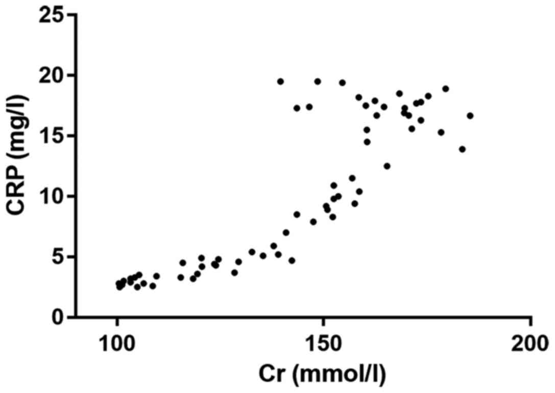
Correlation analysis of Cr and
CRP
Through the correlation analysis, it was found that
there was a positive correlation between Cr and CRP (r=0.6961,
P<0.001). Thus, with the increase of Cr level, the content of
CRP was higher (Fig. 3).
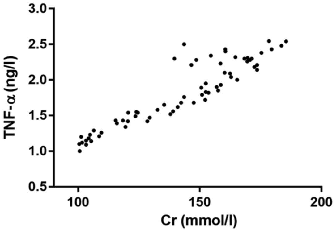
Analysis of the relationship between
Cr and TNF-α
The correlation analysis showed that there was a
positive correlation between Cr and TNF-α (r=0.8969, P<0.001),
which meant that with the increase of Cr level in the body, the
content of TNF-α in vivo was higher (Fig. 4).
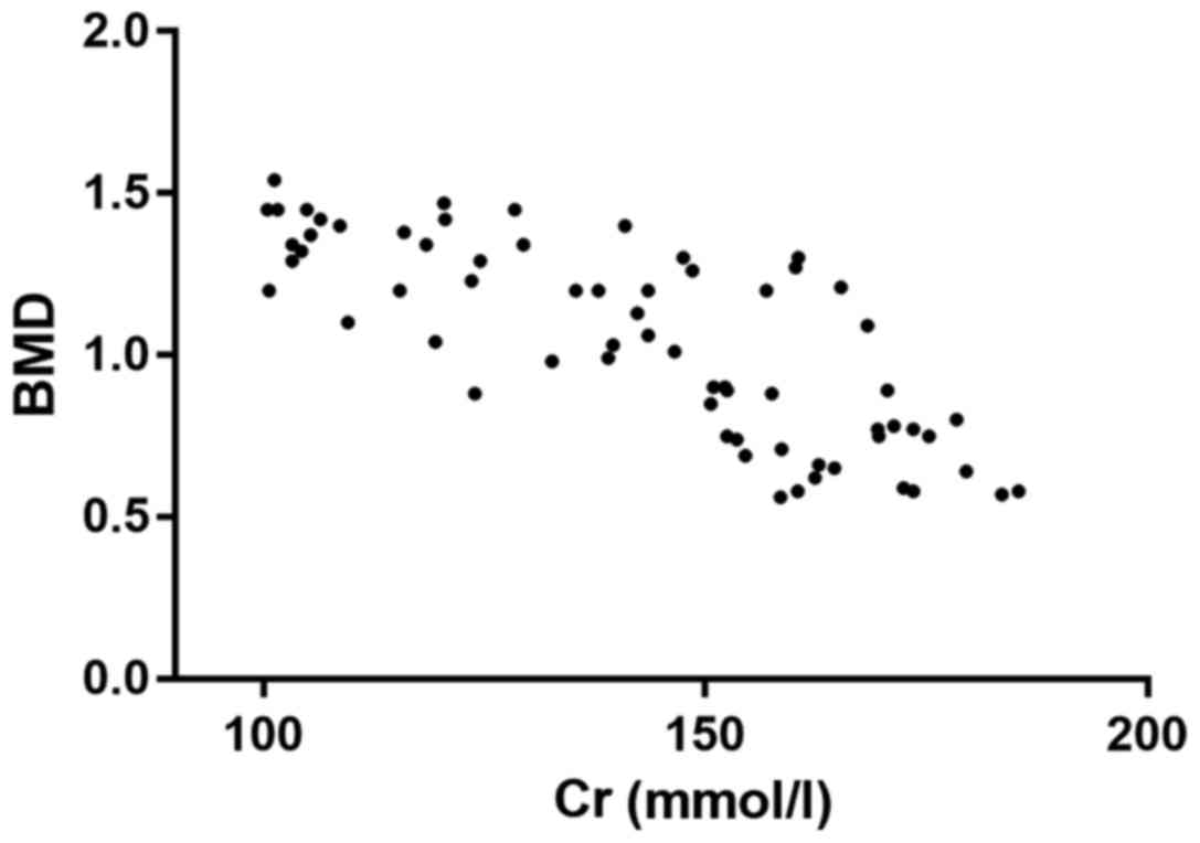
Correlation analysis of Cr and
BMD
The correlation analysis showed that there was a
negative correlation between Cr and BMD (r=0.5472, P<0.001).
This meant that with the increase of Cr level, BMD in vivo
was lower (Fig. 5).
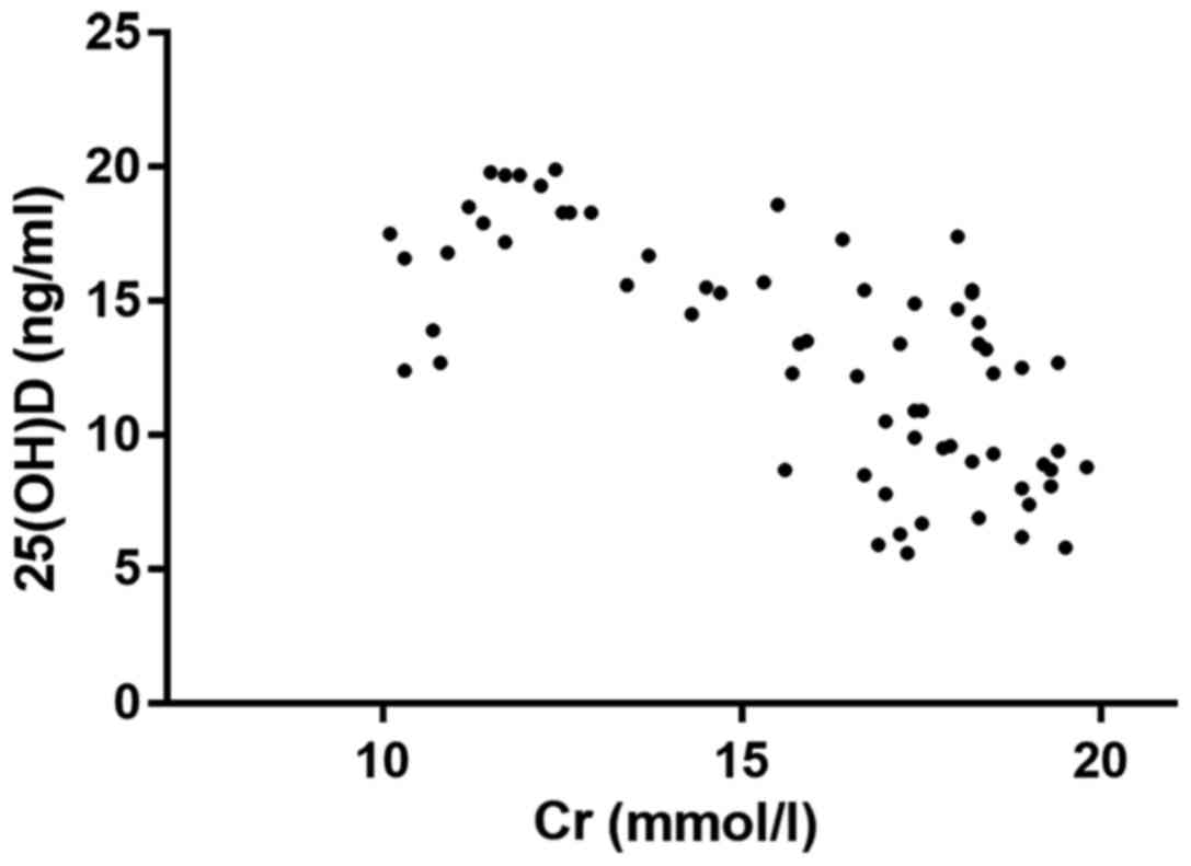
Analysis of the correlation between Cr
and 25(OH)D
Through the correlation analysis, it was found that
there was a negative correlation between Cr and 25(OH)D (r=0.4733,
P<0.001). In other words, with the increase of Cr level, 25(OH)D
was lower (Fig. 6).
Discussion
CKD has a high morbidity rate in clinical practice
and causes great inconvenience to patient life and work (11). It leads to many complications
including inflammatory infection, osteoporosis and vitamin D
deficiency (12,13). To cure and prevent CKD effectively,
in-depth studies of the relation of CKD with inflammatory factors,
osteoporosis and vitamin D deficiency should be conducted,
providing more therapy targets for CKD.
Studies have shown that patients with CKD will
suffer from various inflammations, of which the pathogenic factors
are mainly renal disease and infection immunodeficiency.
Inflammatory infection is not caused by bacteria or viruses, but by
higher concentration of IL-6 released by T lymphocytes and
increased expression levels of CRP and TNF-α after the antigen
stimulation in CKD patients (14,15).
Continuous accumulation of these inflammatory factors produces
physiological damage to kidneys and in turn leads to
microinflammation in the kidneys, which causes the increased
content of inflammation such as IL-6 (16–18).
This study showed that there are correlations between the severity
of renal inflammation and inflammatory factors including IL-6,
TNF-α and CRP. The more severe the disease is, the higher the
content of the above inflammatory factors is, which indicates that
the severity of CKD is positively correlated with inflammation.
Abnormalities of serum phosphate and serum calcium
are detected in patients with CKD, in which the content of serum
phosphate is continuously elevated, and that of serum calcium is
continuously decreased with the aggravation of the disease. This is
considered to be related to metabolic acidosis clinically. Acidosis
will destroy homeostasis of serum phosphate, hinder the active
transport of small intestinal mucosa and cause passive diffusion of
phosphate and abnormal transfer of internal loading of phosphorus.
The significant decrease in content of serum calcium in patients
with CKD is believed clinically to be associated with elevation of
serum phosphate, abnormal arginine intake of calcium and vitamin D
deficiency. As an important indicator of bone metabolism in
patients, the marked reduction of BMD indicates the reduction of
bone mass and high possibility of bone fracture and osteoporosis
(19,20). The present study showed that with the
deterioration of CKD, the value of BMD is decreased significantly,
indicating that patients' osteoporosis is more severe with the
worsening of the disease condition. Related research has shown that
too high content of serum phosphate will lead to over release of
inflammatory factors in patients. In addition, serum phosphate
stimulates monocyte and macrophage to release massive inflammatory
factors and activates the nuclear factor κB (NF-κB) signal pathway,
leading to increased content of TNF-α. It is speculated that there
may be a relation of BMD and IL-6 with the above mechanism.
Vitamin D deficiency is mainly evaluated through the
detection of 25(OH)D that in vivo is combined with the
corresponding receptor so as to form interaction between serum
phosphate and serum calcium. Vitamin D deficiency is very common in
patients with CKD, and clinical studies have found that vitamin D
deficiency has become an important risk factor for CKD. In this
study, the inflammation degree in CKD was found to be negatively
correlated with 25(OH)D, and as the condition became more severe,
the content of this factor was lower, which is similar to findings
of previous clinical studies (21).
Hence, diagnosis, treatment and prevention of CKD can be conducted
by measuring the content of this factor.
In summary, the severity of CKD in patients is
correlated with inflammatory factors, osteoporosis and vitamin D
deficiency. In addition, more serious disease condition causes the
release of more inflammatory factors, higher content of serum
phosphate, lower content of serum calcium and more severe vitamin D
deficiency as well as osteoporosis.
Acknowledgements
Not applicable.
Funding
No funding was received.
Availability of data and materials
The datasets used and/or analyzed during the current
study are available from the corresponding author on reasonable
request.
Authors' contributions
CL was responsible for the data analyses and wrote
the manuscript. CL and HL collected the clinical and laboratory
information. Both authors read and approved the final
manuscript.
Ethics approval and consent to
participate
The study was approved by the Ethics Committee of
the Union Hospital (Wuhan, China) and informed consents were signed
by the patients or the guardians.
Patient consent for publication
Not applicable.
Competing interests
The authors declare that they have no competing
interests.
References
|
1
|
Chen J, Gu D, Chen CS, Wu X, Hamm LL,
Muntner P, Batuman V, Lee CH, Whelton PK and He J: Association
between the metabolic syndrome and chronic kidney disease in
Chinese adults. Nephrol Dial Transplant. 22:1100–1106. 2007.
View Article : Google Scholar : PubMed/NCBI
|
|
2
|
Cruz MC, Andrade C, Urrutia M, Draibe S,
Nogueira-Martins LA and Sesso Rde C: Quality of life in patients
with chronic kidney disease. Clinics (Sao Paulo). 66:991–995. 2011.
View Article : Google Scholar : PubMed/NCBI
|
|
3
|
Jepson RE: Current understanding of the
pathogenesis of progressive chronic kidney disease in cats. Vet
Clin North Am Small Anim Pract. 46:1015–1048. 2016. View Article : Google Scholar : PubMed/NCBI
|
|
4
|
Rabbani N and Thornalley PJ: Advanced
glycation end products in the pathogenesis of chronic kidney
disease. Kidney Int. 93:803–813. 2018. View Article : Google Scholar : PubMed/NCBI
|
|
5
|
Arnold R, Issar T, Krishnan AV and Pussell
BA: Neurological complications in chronic kidney disease. JRSM
Cardiovasc Dis. 5:20480040166776872016.PubMed/NCBI
|
|
6
|
Huan L, Yuezhong L, Chao W and HaiTao T:
The urine albumin-to-creatinine ratio is a reliable indicator for
evaluating complications of chronic kidney disease and progression
in IgA nephropathy in China. Clinics (Sao Paulo). 71:243–250. 2016.
View Article : Google Scholar : PubMed/NCBI
|
|
7
|
Lips P, Goldsmith D and de Jongh R:
Vitamin D and osteoporosis in chronic kidney disease. J Nephrol.
30:671–675. 2017. View Article : Google Scholar : PubMed/NCBI
|
|
8
|
Shigematsu T, Muraoka R, Sugimoto T and
Nishizawa Y: Risedronate therapy in patients with mild-to-moderate
chronic kidney disease with osteoporosis: Post-hoc analysis of data
from the risedronate phase III clinical trials. BMC Nephrol.
18:662017. View Article : Google Scholar : PubMed/NCBI
|
|
9
|
Marciniak C, Gabet J, Lee J, Ma M, Brander
K and Wysocki N: Osteoporosis in adults with cerebral palsy:
Feasibility of DXA screening and risk factors for low bone density.
Osteoporos Int. 27:1477–1484. 2016. View Article : Google Scholar : PubMed/NCBI
|
|
10
|
Ward C, Contino K, Patel A, Mbei EE, Roy
S, Hunter K and Gandhi S: The association of serum
25-Hydroxyvitamin D status in patients with osteoarthritis in the
primary care office. N Am J Med Sci. 8:47–55. 2016. View Article : Google Scholar : PubMed/NCBI
|
|
11
|
Pongpirul W, Pongpirul K, Ananworanich J,
Klinbuayaem V, Avihingsanon A and Prasithsirikul W: Chronic kidney
disease incidence and survival of Thai HIV-infected patients. AIDS.
32:393–398. 2018.PubMed/NCBI
|
|
12
|
Michishita R, Matsuda T, Kawakami S,
Tanaka S, Kiyonaga A, Tanaka H, Morito N and Higaki Y: The
association between changes in lifestyle behaviors and the
incidence of chronic kidney disease (CKD) in middle-aged and older
men. J Epidemiol. 27:389–397. 2017. View Article : Google Scholar : PubMed/NCBI
|
|
13
|
Kochi M, Kohagura K, Shiohira Y, Iseki K
and Ohya Y: Chronic kidney disease, inflammation, and
cardiovascular disease risk in rheumatoid arthritis. J Cardiol.
71:277–283. 2018. View Article : Google Scholar : PubMed/NCBI
|
|
14
|
Li ZY, Zheng Y, Chen Y, Pan M, Zheng SB,
Huang W, Zhou ZH and Ye HY: Brazilin ameliorates diabetic
nephropathy and inflammation in db/db mice. Inflammation.
40:1365–1374. 2017. View Article : Google Scholar : PubMed/NCBI
|
|
15
|
Batchu SN, Hughson A, Gerloff J, Fowell DJ
and Korshunov VA: Role of Axl in early kidney inflammation and
progression of salt-dependent hypertension. Hypertension.
62:302–309. 2013. View Article : Google Scholar : PubMed/NCBI
|
|
16
|
Garcia-Bello JA, Gómez-Díaz RA,
Contreras-Rodríguez A, Talavera JO, Mondragón-González R,
Sanchez-Barbosa L, Diaz-Flores M, Valladares-Salgado A, Gallardo
JM, Aguilar-Kitsu A, et al: Carotid intima media thickness,
oxidative stress, and inflammation in children with chronic kidney
disease. Pediatr Nephrol. 29:273–281. 2014. View Article : Google Scholar : PubMed/NCBI
|
|
17
|
Lim GB: Arrhythmias: IL-6 and risk of
atrial fibrillation in chronic kidney disease. Nat Rev Cardiol.
13:1832016. View Article : Google Scholar : PubMed/NCBI
|
|
18
|
Xun C and Zhao Y: Potential role of
soluble TNF-α receptors in diagnosis of patients with chronic
kidney disease. Ann Clin Lab Sci. 47:310–314. 2017.PubMed/NCBI
|
|
19
|
Peltonen S, Biancari F, Lindgren L,
Mäkisalo H, Honkanen E and Lepäntalo M: Outcome of infrainguinal
bypass surgery for critical leg ischaemia in patients with chronic
renal failure. Eur J Vasc Endovasc Surg. 15:122–127. 1998.
View Article : Google Scholar : PubMed/NCBI
|
|
20
|
Kumar S, Mavuduru RS and Sharma V:
Pan-ureteric transitional cell carcinoma in a patient of autosomal
dominant polycystic kidney disease with chronic renal failure. NDT
Plus. 2:76–77. 2009.PubMed/NCBI
|
|
21
|
Levin A, Le Barbier M, Er L, Andress D,
Sigrist MK and Djurdjev O: Incident isolated 1,25(OH)(2)D(3)
deficiency is more common than 25(OH)D deficiency in CKD. J
Nephrol. 25:204–210. 2012. View Article : Google Scholar : PubMed/NCBI
|