|
1
|
Gihus NE and Verschuuren JJ: Myasthenia
gravis: Subgroup classification and therapeutic strategies. Lancet
Neurol. 14:1023–1036. 2015. View Article : Google Scholar : PubMed/NCBI
|
|
2
|
Song Y, Zhou L, Miao F, Chen G, Zhu Y, Gao
X, Wang Y, Pang L, Zhao C, Sun X and Chen Z: Increased frequency of
thymic T follicular helper cells in myasthenia gravis patients with
thymoma. J Thorac Dis. 8:314–322. 2016. View Article : Google Scholar : PubMed/NCBI
|
|
3
|
Truffault F, de Montreville V, Eymard B,
Sharshar T, Le Panse R and Berrih-Aknin S: Thymic germinal centers
and corticosteroids in myasthenia gravis: An immunopathological
study in 1035 cases and a critical review. Clin Rev Allergy
Immunol. 52:108–124. 2017. View Article : Google Scholar : PubMed/NCBI
|
|
4
|
Domeier PP, Schell SL and Rahman ZS:
Spontaneous germinal centers and autoimmunity. Autoimmunity.
50:4–18. 2017. View Article : Google Scholar : PubMed/NCBI
|
|
5
|
Rückert JC, Ismail M, Badakhshi H, Meisel
A and Swierzy M: Thymectomy in myasthenia and/or thymoma. Zentralbl
Chir. 139:121–134. 2014.(In German). View Article : Google Scholar : PubMed/NCBI
|
|
6
|
Xie Y, Li HF, Sun L, Kusner LL, Wang S,
Meng Y, Zhang X, Hong Y, Gao X, Li Y and Kaminski HJ: The role of
osteopontin and its gene on glucocorticoid response in myasthenia
gravis. Front Neurol. 8:2302017. View Article : Google Scholar : PubMed/NCBI
|
|
7
|
Moser B, Janik S, Schiefer AI, Müllauer L,
Bekos C, Scharrer A, Mildner M, Rényi-Vámos F, Klepetko W and
Ankersmit HJ: Expression of RAGE and HMGB1 in thymic epithelial
tumors, thymic hyperplasia and regular thymic morphology. PLoS One.
9:e941182014. View Article : Google Scholar : PubMed/NCBI
|
|
8
|
Yablonsky P, Pischik V, Tobina MG and
Atiukov M: The results of video-assisted thoracoscopic thymectomies
in Saint Petersburg, Russia: 20-year of experience. J Vis Surg.
3:1132017. View Article : Google Scholar : PubMed/NCBI
|
|
9
|
Ottlakan A, Borda B, Morvay Z, Maraz A and
Furak J: The effect of diagnostic imaging on surgical treatment
planning in diseases of the thymus. Contrast Media Mol Imaging.
2017:93072922017. View Article : Google Scholar : PubMed/NCBI
|
|
10
|
De Roxas RC, Bagnas MA, Baldonado JJ,
Rivera JP and Roxas AA: Clinical profile and outcome of
postthymectomy versus non-thymectomy myasthenia gravis patients in
the Philippine general hospital: A 6-year retrospective study.
Front Neurol. 7:962016. View Article : Google Scholar : PubMed/NCBI
|
|
11
|
Li HF, Hong Y, Xie Y, Hao HJ and Sun RC:
Precision medicine in myasthenia graves: Begin from the data
precision. Ann Transi Med. 4:1062016. View Article : Google Scholar
|
|
12
|
Mao ZF, Mo XA, Qin C, Lai YR and Hackett
ML: Incidence of thymoma in myasthenia gravis: A systematic review.
J Clin Neurol. 8:161–169. 2012. View Article : Google Scholar : PubMed/NCBI
|
|
13
|
Araki T, Nishino M, Gao W, Dupuis J,
Washko GR, Hunninghake GM, Murakami T, O'Connor GT and Hatabu H:
Anterior mediastinal masses in the framingham heart study:
Prevalence and CT image characteristics. Eur J Radiol Open.
2:26–31. 2015. View Article : Google Scholar : PubMed/NCBI
|
|
14
|
Tajima A, Pradhan I, Trucco M and Fan Y:
Restoration of thymus function with bioengineered thymus organoids.
Curr Stem Cell Rep. 2:128–139. 2016. View Article : Google Scholar : PubMed/NCBI
|
|
15
|
Nagakubo D, Krauth B and Boehm T: Genetic
and non-genetic determinants of thymic epithelial cell number and
function. Sci Rep. 7:103142017. View Article : Google Scholar : PubMed/NCBI
|
|
16
|
Kranich J and Krautler NJ: How follicular
dendritic cells shape the B-cell antigenome. Front Immunol.
7:2252016. View Article : Google Scholar : PubMed/NCBI
|
|
17
|
Mlika M, Gattoufi W, Zribi H, Braham E,
Marghli A and El Mezni F: A unilocular thymic cyst associated with
true thymic hyperplasia: A challenging diagnosis especially in a
child. Int Med Case Rep J. 8:215–218. 2015. View Article : Google Scholar : PubMed/NCBI
|
|
18
|
Pereira BI and Akbar AN: Convergence of
innate and adaptive immunity during human aging. Front Immunol.
7:4452016. View Article : Google Scholar : PubMed/NCBI
|
|
19
|
Sidler C, Kovalchuk O and Kovalchuk I:
Epigenetic regulation of cellular senescence and aging. Front
Genet. 8:1382017. View Article : Google Scholar : PubMed/NCBI
|
|
20
|
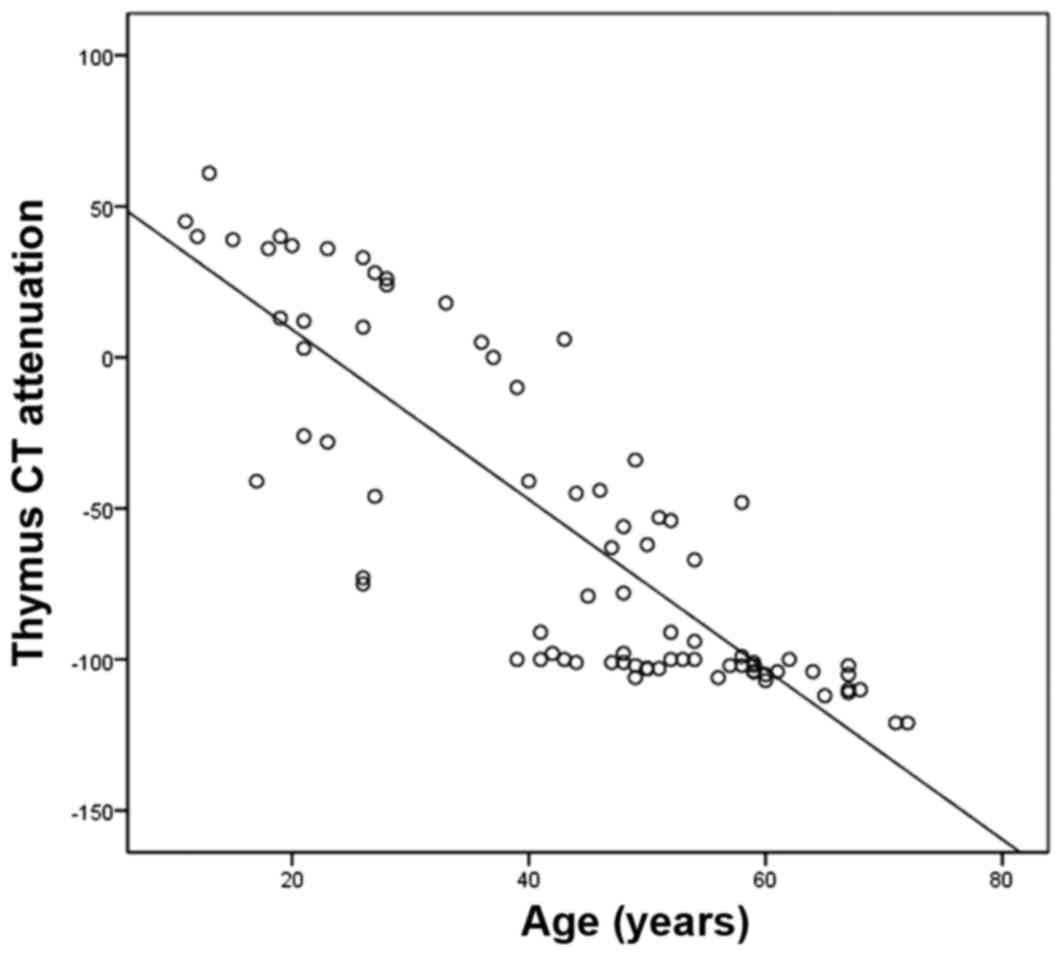
St Amour TE, Siegel MJ, Glazer HS and
Nadel SN: CT appearances of the normal and abnormal thymus in
childhood. J Comput Assist Tomogr. 11:645–650. 1987. View Article : Google Scholar : PubMed/NCBI
|
|
21
|
Francis IR, Glazer GM, Bookstein FL and
Gross BH: The thymus: Reexamination of age-related changes in size
and shape. AJR Am J Roentqenol. 145:249–254. 1985. View Article : Google Scholar
|
|
22
|
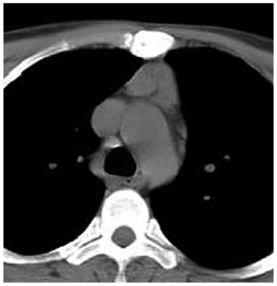
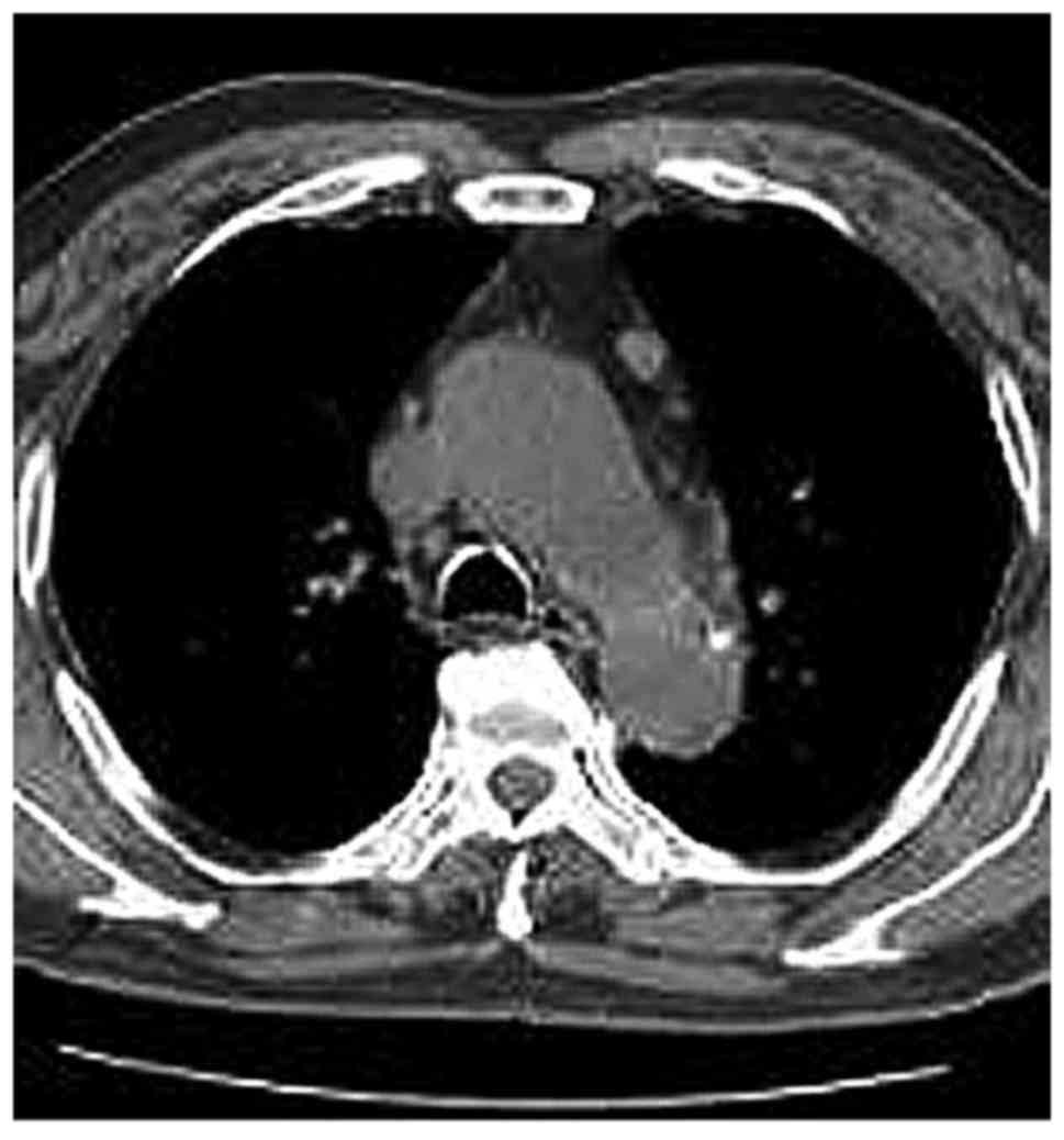
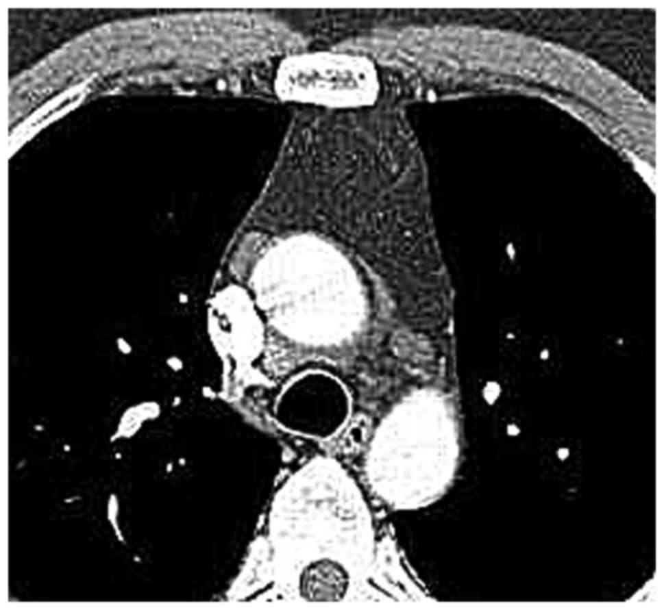
Nishino M, Ashiku SK, Kocher ON, Thurer
RL, Boiselle PM and Hatabu H: The thymus: A comprehensive review.
Radiographics. 26:335–348. 2006. View Article : Google Scholar : PubMed/NCBI
|
|
23
|
Manchanda S, Bhalla AS, Jana M and Gupta
AK: Imaging of the pediatric thymus: Clinicoradiologic approach.
World J Clin Pediatr. 6:10–23. 2017. View Article : Google Scholar : PubMed/NCBI
|
|
24
|
Shimamoto A, Ashizawa K, Kido Y, Hayashi
H, Nagayasu T, Kawakami A, Mukae H, Hayashi T, Ohtsubo M,
Shigematsu K, et al: CT and MRI findings of thymic carcinoid. Br J
Radiol. 90:201503412017. View Article : Google Scholar : PubMed/NCBI
|
|
25
|
Nishikawa N, Nagai M, Tsujii T, Kyaw WT,
Tanabe N, Iwaki H, Yabe H, Ando R and Nomoto M: Treatment of
myasthenia gravis in patients with elderly onset at advanced age.
Jpn Clin Med. 6:9–13. 2015. View Article : Google Scholar : PubMed/NCBI
|