Introduction
Lumbar degenerative disc disease is a common disease
that occurs in older people and develops with age. In the past, the
standard treatment for this disease has been internal fixation
surgery (1–3). However, traditional open surgery
results in considerable trauma in senior patients. It has been
reported that age is an independent factor in the severity of
lumbar degenerative disc disease; the disease becomes more severe
and complex with age (4). At the
same time, due to physiological decline, senior patients are often
affected by a variety of systemic chronic diseases, with
significant increases in surgical risk (5). Li et al (6) demonstrated that the incidence and
mortality rate of patients undergoing traditional open surgery
increased with age; in patients aged over 65 years the incidence
was 11.6% and the mortality rate was 0.15%, leading to difficulties
in the treatment of senior patients.
Currently, a variety of spinal minimally invasive
technologies offer novel strategies that may avoid the
aforementioned problems. Percutaneous transforaminal endoscopic
discectomy (PTED) has quickly gained attention because it has
numerous notable advantages, including small incision, short
hospitalization time, small economic burden and fewer surgical
complications (7–10). However, the limited scope of
decompression and reduced capacity to recover the stability of the
spine have made the indications and contraindications of the
surgery the focus of academic debate (11). Kim et al (12) proposed that the application of PTED
should be carefully considered in patients aged over 57 years as
these patients have a higher reoperation risk.
In the current study, the records of senior patients
aged over 70 years with lumbar degenerative disc disease were
reviewed, and efficacy of PTED in the treatment of lumbar
degenerative disc disease for senior patients was evaluated. The
current study provides support for the clinical application of PTED
in treating elderly lumbar degenerative disc disease.
Materials and methods
Subjects
This retrospective study was approved by the Ethics
Committee of The Third Hospital of Hebei Medical University
(Shijiazhuang, China). All patients provided written informed
consent for their inclusion in the study. A total of 318 patients
aged >70 years with lumbar degenerative disc disease were
selected in the Department of Spine Surgery in The Third Hospital
of Hebei Medical University from June 2012 to June 2015. The
patients were allocated to two groups according to their choice of
treatment: A PTED group and an open surgery group. In the PTED
group, there were 41 patients, including 17 males and 24 females,
aged from 70 to 83 years, with a mean age of 74 years. All patients
in this group underwent PTED. In the open surgery group, there were
277 patients, including 102 males and 175 females, aged from 70 to
79 years with a mean age of 73 years. The patients underwent
traditional open reduction and internal fixation. Among the 277
patients, 75 underwent transforaminal lumbar interbody fusion
(TLIF) and 202 underwent posterior lumbar interbody fusion
(PLIF).
The inclusion criteria were as follows: Lumbar
degenerative disc disease patients aged >70 years with single or
bilateral lower extremity numbness, pain and intermittent
claudication as the clinical manifestations; patients whose imaging
findings were consistent with the symptoms and signs of
degenerative manifestations, such as intervertebral disc protrusion
and spinal stenosis; patients with ineffective conservative
treatment for >6 months; patients undergoing single segment
TLIF, PLIF or PTED.
The exclusion criteria were as follows: Patients
<70 years old; patients with Grade II or above lumbar
spondylolisthesis; patients with elevated infection indicators,
including erythrocyte sedimentation rate and C-reactive protein;
patients with lumbar trauma, cancer, severe osteoporosis or
congenital malformations; patients with rheumatoid arthritis
disease or other serious systemic diseases, or metal allergy;
patients with incomplete data or who were lost to follow-up.
Surgery
In the PTED group, nucleus removal was conducted
using the transforaminal endoscopic surgical system technique
(13). In the open surgery group,
PLIF (14) and TLIF (15) were used for decompression of
laminectomy, nucleus removal, and cage and pedicle screw
implantation. The surgeries of the patients in the two groups were
completed by the same surgical team.
Postoperative management
Patients in the PTED group were given grade II
nursing with the same diet as pre-operation (16), and they did moderate exercise out of
bed following rest for 12 h. Blood glucose was monitored for the
preoperative underlying disease, and the preoperative drugs were
continually used for the treatment of underlying disease.
Patients in the open surgery group were given grade
I nursing with oxygen inhalation. Vital signs were monitored for 24
h and patients were prohibited from eating or drinking for 6 h,
then given liquid food for 1 day. Once defecation was normal, the
preoperative diet was restored. After routine rehydration and
anti-infective treatment, the patients received anticoagulant
therapy with the subcutaneous injection of low molecular weight
heparin 24 h after surgery. Patients rested in bed and were
prohibited from all strenuous exercise. Limbs were passively
exercised, and the dressing was regularly replaced on the wound.
Biochemical indicators were closely monitored. Common postoperative
symptoms, including fever, anemia, electrolyte imbalance and
coagulation dysfunction, were promptly treated with supportive
treatments, including antipyresis, blood transfusion and fluid
infusion. Once the condition had been stable for 7 days following
surgery, without the formation of deep venous thrombosis of the
lower limbs, moderate activity out of bed was allowed. The suture
was removed 12–14 days following surgery, depending on the
condition of wound.
Follow-up and indices
All patients were followed up for 12–48 months (mean
follow-up, 21.6 months). Surgery time was regarded as the time from
skin incision to the incision being completely sutured.
Intraoperative fluoroscopy time was regarded as the time of
intraoperative fluoroscopy exposure using C-arm X-ray. The average
exposure time was 1 sec for each fluoroscopy. Japan Orthopedic
Association (JOA) evaluation and treatment scores (17) were used to evaluate nerve function
prior to surgery, 1 week following surgery and during the 12-month
follow-up. Visual analog scale (VAS) scores (18) were used to evaluate the severity of
pain prior to surgery, 1 week following surgery and during the
12-month follow-up. The Charlson Comorbidity Index (CCI) was used
to evaluate the degree of severity of preoperative underlying
disease (Table I).
 | Table I.Charlson Comorbidity Index. |
Table I.
Charlson Comorbidity Index.
| Disease | Score |
|---|
| Myocardial
infarction | 1 |
| Congestive heart
failure | 1 |
| Peripheral vascular
disease | 1 |
| Cerebrovascular
disease | 1 |
| Dementia | 1 |
| Chronic lung
disease | 1 |
| Connective tissue
disease | 1 |
| Ulcer | 1 |
| Mild liver
disease | 1 |
| Diabetes | 1 |
| Hemiplegia | 2 |
| Moderate/severe
kidney disease | 2 |
| Diabetes combined
with organ damage | 2 |
| Tumor | 2 |
| Leukemia | 2 |
| Lymphoma | 2 |
| Moderate/severe liver
disease | 2 |
| Metastatic
tumors | 2 |
| Acquired
immunodeficiency syndrome | 2 |
Statistical analysis
SPSS 22.0 (IBM Corp., Armonk, NY, USA) was used for
data analysis. Measurement data were expressed as the mean ±
standard deviation. Kolmogorov-Smirnov test was used to determine
the distribution of the data. The paired sample t-test was used to
analyze paired data. Comparisons between groups in terms of age,
surgery time, intraoperative fluoroscopy time, intraoperative blood
loss, VAS and JOA scores prior to surgery, 1 week following surgery
and 12 months following surgery, and preoperative CCI score were
analyzed by independent sample t-test. Comparisons within groups in
terms of VAS and JOA scores prior to surgery and at 12 months
following surgery were analyzed by paired sample t-test. The
incidence of deep venous thrombosis was compared between groups by
Pearson's chi-squared test. The incidence of postoperative
cerebrospinal fluid leakage was compared by Fisher's exact
probability test (test level, α=0.05). P<0.05 was considered to
indicate a statistically significant difference.
Results
Comparison of observation indices
between the PTED group and the open surgery group
As indicated in Table
II, there were significant differences in surgery time,
intraoperative fluoroscopy time, and intraoperative blood loss
between the PTED group and the open surgery group (P<0.001),
whereas there was no significant difference in age. The surgery
time in the PTED group was significantly reduced compared with the
open surgery group (74.5±19.72 vs. 169.8±24.5 min). The
intraoperative fluoroscopy time in the PTED group was significantly
longer compared with that in the open surgery group (23.7±6.08 vs.
4.9±1.8 sec). Intraoperative blood loss in the PTED group was
significantly reduced when compared with the open surgery group
(13.5±4.6 vs. 668.0±260.4 ml).
| Table II.Comparison of observation indices
between the two groups. |
Table II.
Comparison of observation indices
between the two groups.
| Index | PTED group | Open surgery
group | P-value |
|---|
| Age (year) | 74.30±3.13 | 73.40±2.48 | 0.13 |
| Surgery time
(min) |
74.50±19.72 | 169.80±24.50 | <0.001 |
| Intraoperative
fluoroscopy time (sec) | 23.70±6.08 |
4.90±1.80 | <0.001 |
| Intraoperative
blood loss (ml) | 13.50±4.60 |
668.00±260.40 | <0.001 |
| Preoperative VAS
score |
6.20±1.24 |
6.10±1.40 | 0.67 |
| Preoperative JOA
score | 11.90±4.09 | 11.50±3.88 | 0.32 |
| VAS score 1 week
after surgery |
2.60±0.85 |
3.10±0.79 | <0.001 |
| JOA score 1 week
after surgery | 23.60±2.28 | 19.40±1.79 | <0.001 |
| VAS score 12 months
after surgery |
2.00±0.97 |
2.09±0.90 | 0.56 |
| JOA score 12 months
after surgery | 23.40±2.14 | 23.60±2.29 | 0.64 |
| Preoperative CCI
score |
4.44±1.62 |
2.78±0.92 | <0.001 |
| Incidence rate of
postoperative deep venous thrombosis of lower limbs (%) | 12.10 | 28.20 | 0.03 |
| Incidence rate of
postoperative cerebrospinal fluid leakage (%) | 4.90 | 7.60 | 0.75 |
Comparison of pain and neurological
function between the PTED group and the open surgery group
There were significant differences in VAS score and
JOA score at 1 week after surgery between the PTED group and the
open surgery group (P<0.001), whereas no significant differences
were identified in preoperative VAS score, preoperative JOA score,
VAS score at 12 months after surgery or JOA score at 12 months
after surgery between the two groups (Table II). Furthermore, there were
significant differences in the VAS and JOA scores prior to surgery
and at 12 months after surgery within each group (P<0.001;
Table III). Preoperative CCI in
the PTED group was significantly higher compared with the open
surgery group (4.44±1.62 vs. 2.78±0.92; P<0.001; Table II). In summary, these results
indicated that lumbar and leg pain was significantly alleviated,
and neurological function was significantly improved in the two
groups at 12 months after surgery.
| Table III.Comparison of pain and neurological
function between the two groups before and after surgery. |
Table III.
Comparison of pain and neurological
function between the two groups before and after surgery.
|
| PTED group | Open surgery
group |
|---|
|
|
|
|
|---|
| Time | VAS | JOA | VAS | JOA |
|---|
| Preoperative | 6.20±1.24 | 11.90±4.09 | 6.10±1.40 | 11.50±3.88 |
| 12 months after
surgery | 2.00±0.97 | 23.40±2.14 | 2.09±0.90 | 23.60±2.29 |
| P-value | <0.001 | <0.001 | <0.001 | <0.001 |
Benefits of PTED surgery
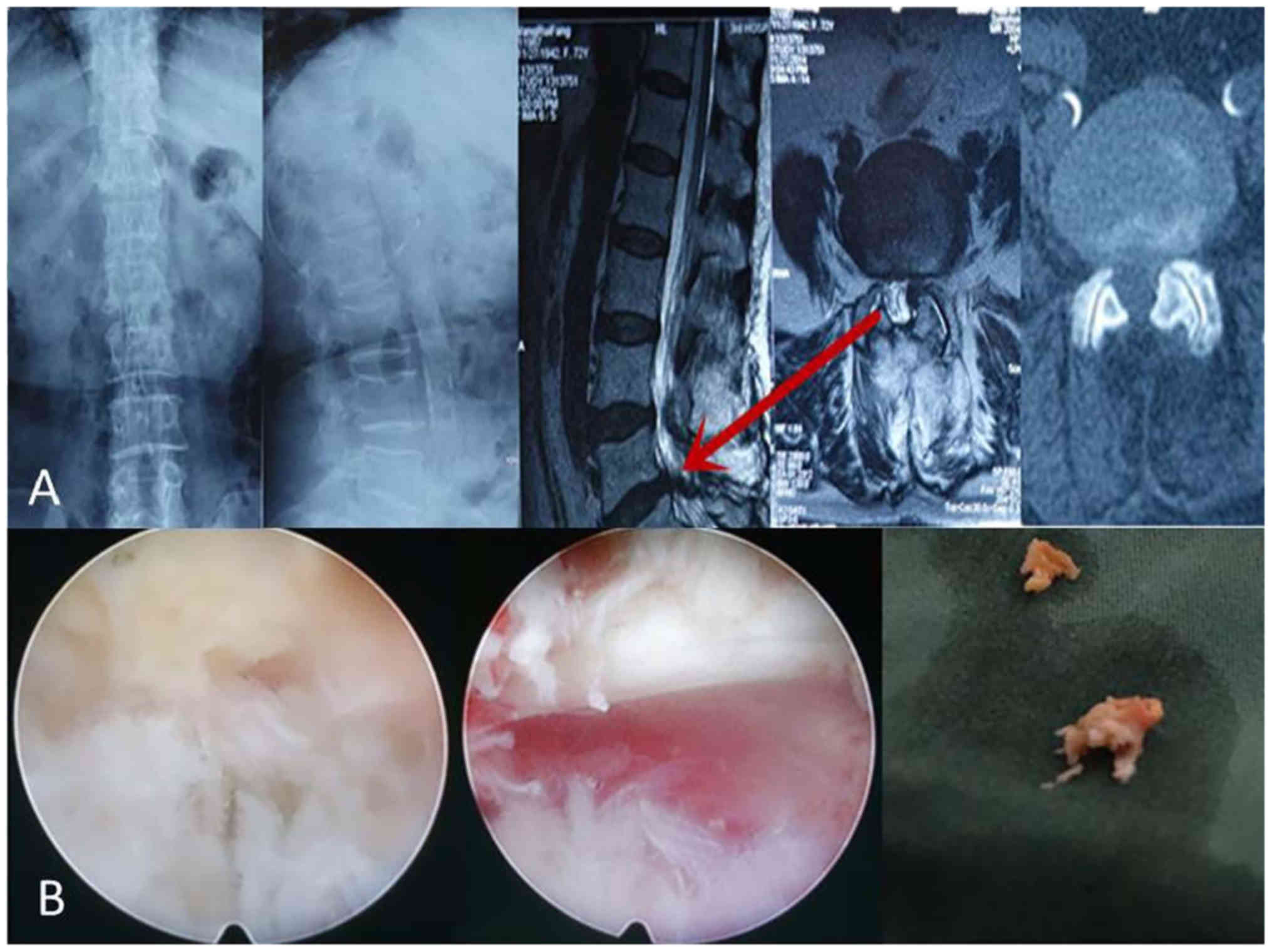
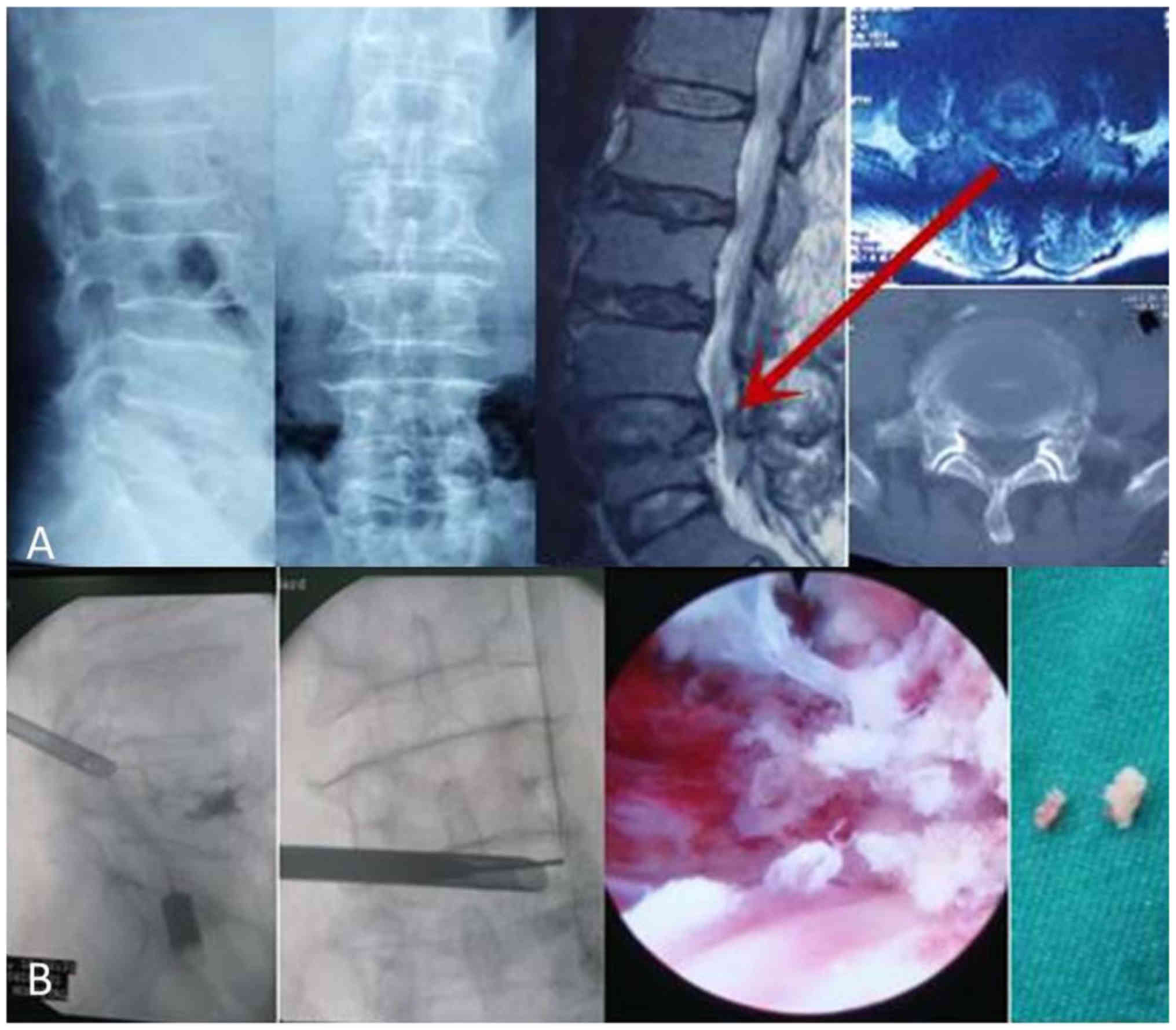
Two senior patients had underlying diseases,
including L2-3 disc herniation and left nerve root compression
(Figs. 1 and 2). The intervertebral disc decompression
following a PTED on senior patients with underlying disease was
good as the VAS score was reduced and JOA score improved (data not
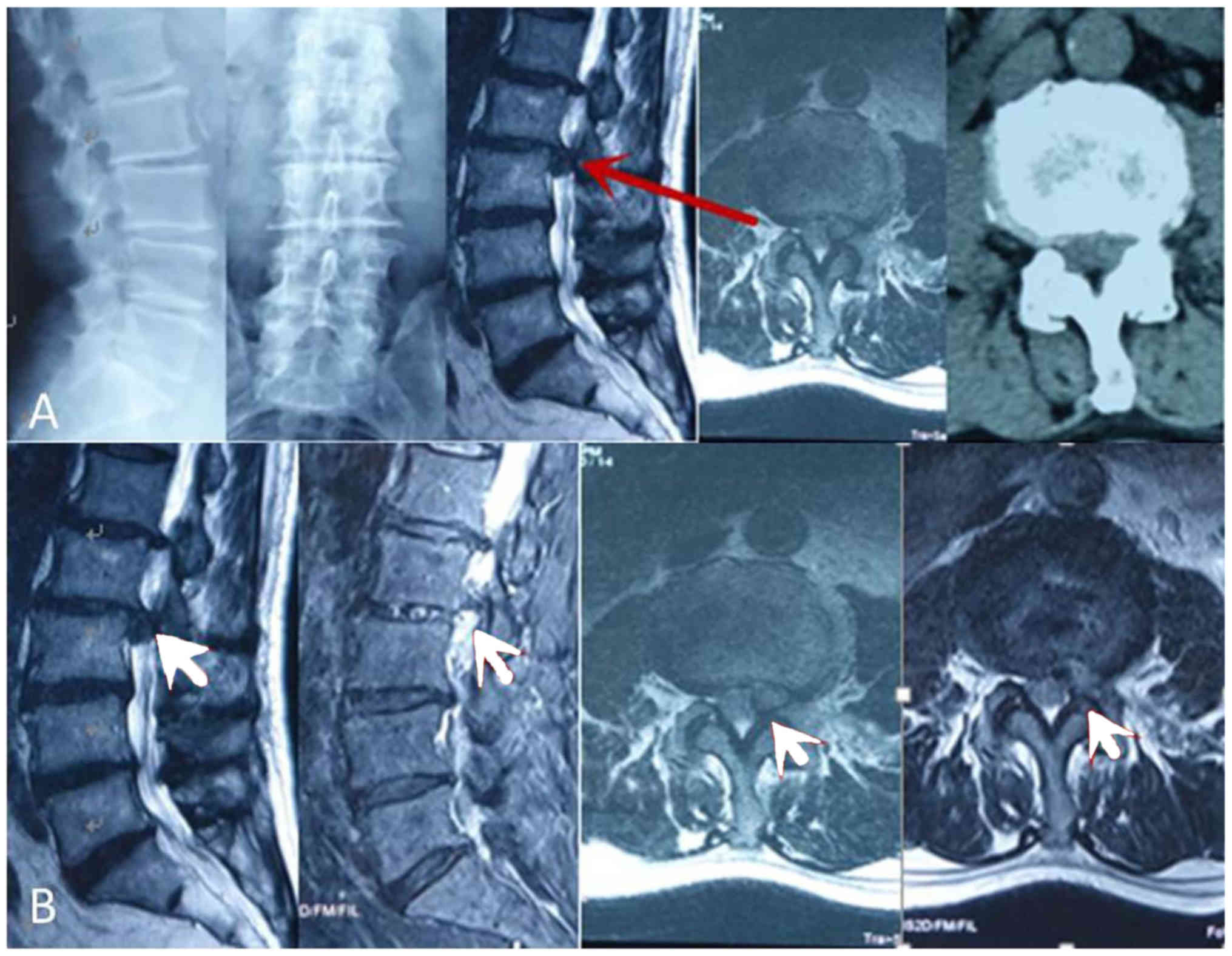
shown). Another senior patient had a L3-4 disc herniation, stenosis
of left nerve root canal and an intervertebral foramen (Fig. 3). Following the PTED surgery under
local anesthesia, the compression of the nerve root and the pain
experienced by the senior patient were largely relieved (data not
shown). After removal of nucleus pulposus following a PTED
(Fig. 4), the VAS score was reduced
and the JOA score was improved, suggesting the recovery of nerve
function (Table III). The last
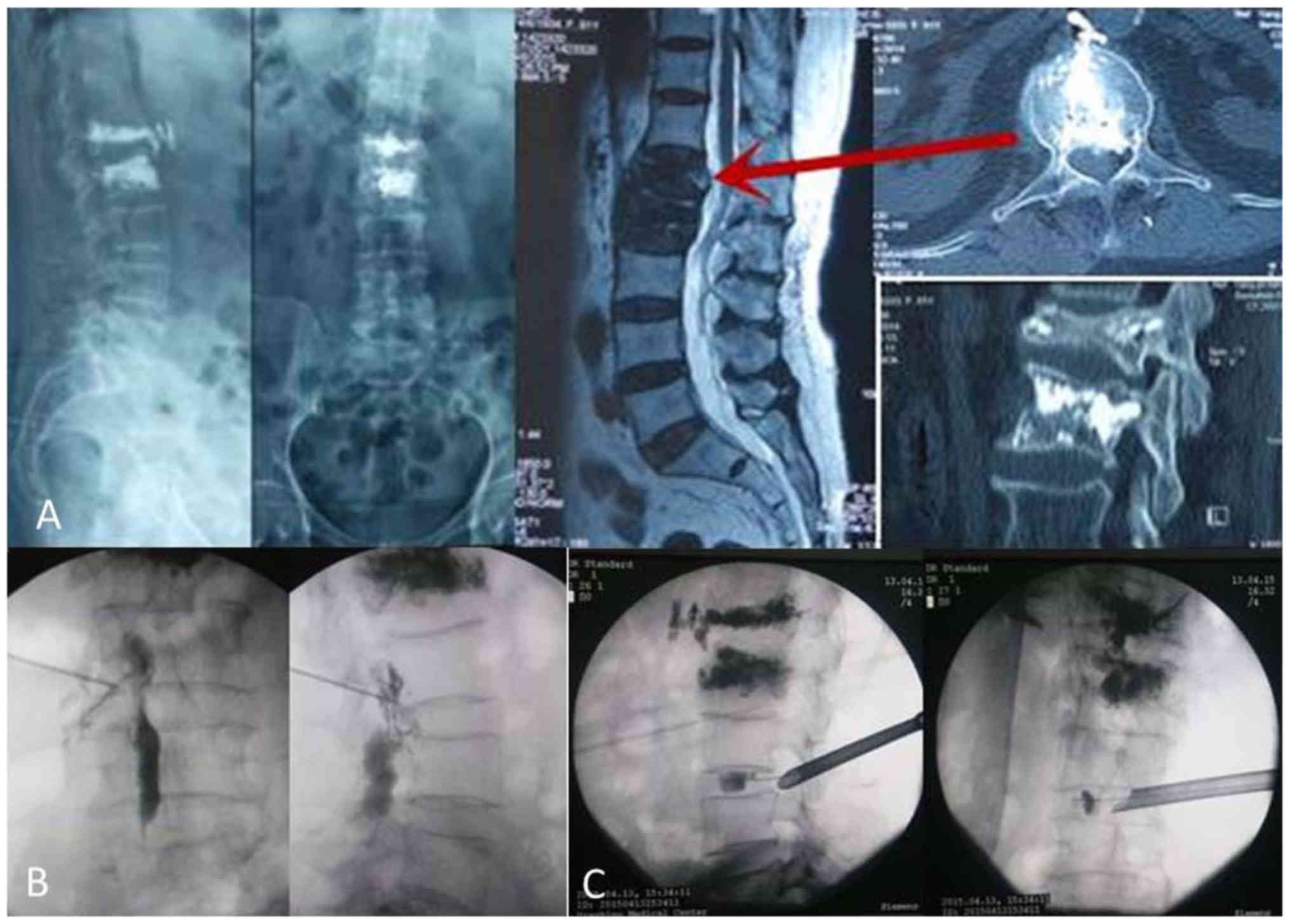
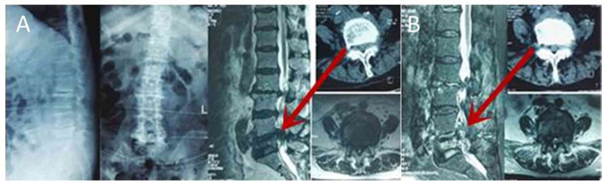
senior patient had multi-segment lumbar disc herniation and spinal
stenosis with degenerative scoliosis prior to surgery (Fig. 5A); there was a remarkable
decompression effect of spinal canal following PTED surgery
(Fig. 5B).
Postoperative complications
The incidence of postoperative deep venous
thrombosis of lower limbs in the PTED group was significantly lower
compared with the open surgery group (12.1 vs. 28.2%; P<0.05),
whereas no significant difference in the incidence of postoperative
cerebrospinal fluid leakage was identified between the two groups
(Table II).
In the open surgery group, the incidence of
constipation, urinary system infection and wound infection
following surgery was 56.7, 19.5 and 9.4%, respectively. Patients
undergoing intraoperative and postoperative transfusion therapy
accounted for 44.8%. There were 12 cases of gastrointestinal
hemorrhagic stress ulcer, 3 of pneumonia, 2 of pulmonary embolism,
1 of mortality following myocardial infarction, 1 of mortality
following cerebral infarction and 1 of hemiplegia following
cerebral hemorrhage. Patients in the PTED group did not experience
any complications.
Discussion
Lumbar degenerative disc disease is a
pathophysiological progress that develops with lumbar tissue aging,
including lumbar disc herniation, lumbar spinal stenosis, lumbar
spondylolisthesis and lumbar instability (15). Therefore, instability of the spine is
an important part of lumbar degeneration. Studies have hypothesized
that the first cause of the instability of the spine is loss of
intervertebral height, followed by dehydration of intervertebral
disc or nucleus pulposus (16,19,20). In
addition, instability of the spine contributes to lumbar segments
exceeding the normal range and exhibiting abnormal activities, thus
causing a range of clinical symptoms, including continuous low back
pain with or without radiation pain in the buttocks and the
posterolateral lower extremities (17,18). For
patients in good physical condition and with strong surgical
tolerance, traditional open surgeries, including lumbar
decompression, intervertebral discectomy and interbody fusion, are
commonly used. These methods have numerous advantages, including
complete decompression, instantly restored spinal stability and
stable surgical efficacy. Currently, PLIF and TLIF are the standard
procedures in the treatment of lumbar degenerative disc disease
(21–24). PTED is a technically demanding
procedure with a steep learning curve, and therefore requires an
experienced surgeon (25).
Therefore, it is difficult to apply in primary hospitals. For
patients with severe lumbar degenerative disc disease, intraspinal
vascular hyperplasia often occurs, resulting in a large volume of
bleeding during PTED surgery, which will cover the surgical field.
Currently, PTED cannot achieve fixation of instability segments of
the spine; therefore, degenerative clinical symptoms, mainly caused
by instability of the lumbar spine, are not yet suitable for PTED
treatment.
However, traditional open surgery also has many
drawbacks, including a large surgical incision, considerable
intraoperative blood loss, slow recovery after surgery, and a wide
range of complications (26). It has
been demonstrated that lumbar degenerative disc disease is likely
to become more severe and complex with age (4). At the same time, the incidence and
severity of underlying diseases in other systems also increases
with age. Therefore, during surgery on senior patients with lumbar
degenerative disc disease, surgeons are likely to face many
problems, including long surgery times and large surgical
trauma.
In recent decades, the concept of minimally invasive
surgery has been widely accepted, and a variety of spinal minimally
invasive techniques have emerged (7,27–32). The
technique of intervertebral foramen nucleus removal can achieve
decompression of the spinal canal and can be performed with local
anesthesia and a small incision, which has been widely studied and
developed (7–10). PTED addressed many of the
aforementioned problems in senior patients and some studies
indicated it was an ideal surgery option for senior patients
(33). However, PTED is not able to
achieve intervertebral fusion, internal fixation or reconstruction
of spine stability. It has been indicated that the recurrence rate
is higher in patients older than 57 years treated with PTED when
compared with patients treated with open surgery at 3–4 years after
surgery (12). Therefore, the use of
PTED in senior patients requires further investigation.
Compared with traditional open surgery, PTED is a
minimally invasive surgery that offers direct access to the lesion.
It avoids the destruction of the paravertebral muscles, vertebral
lamina, spinous process and posterior spinal muscular ligamentous
complex, so it has a minimal effect on the stability of the spine
and involves minimal intraoperative bleeding. Patients are able to
regain function quickly following surgery, and the time of
hospitalization is greatly shortened, which reduces the economic
burden for patients. One of the major advantages of PTED is the
protection of the stability of the spine. In the current study,
postoperative recurrence or instability were not observed during
the follow-up period, which may be associated with the relatively
stable state of spine in the elderly patients and the absorption of
degenerated nucleus pulposus. These results were consistent with
previous reports (18,24).
In the current study, perioperative indices and
short-term results of senior patients with lumbar degenerative disc
disease were compared following treatment with PTED and traditional
open surgery. There was no significant difference in age between
the two groups, but the preoperative CCI in the PTED group was
significantly higher compared with the open surgery group,
indicating that the severity of preoperative underlying disease was
greater in the PTED group. Open internal fixation surgery was a
more established treatment option and was still the preferred
surgery for patients. For patients in poor physical condition that
could not undergo open internal fixation surgery, PTED was
preferred, possibly resulting in the higher levels of preoperative
CCI in the PTED group. Compared with traditional open fixation
surgery, PTED surgery has advantages in terms of surgery time and
intraoperative blood loss, but the intraoperative radiation
exposure time was significantly higher when compared with open
surgery, which was inconsistent with previous reports (8,34). The
comparison of pain and neurological scores of patients in the two
groups prior to and 12 months after surgery indicated that both
surgeries could significantly alleviate pain and improve nerve
function of the lower limbs, and there was no significant
difference in short-term effects between the two surgeries.
However, the pain scores were lower and neurological scores were
higher among patients in the PTED group compared with the open
surgery group at 1 week after surgery. These findings may be
associated with the small incision and minimal injury to
surrounding tissues during PTED surgery. Patients undergoing PTED
surgery recovered faster, got out of bed earlier and suffered less
perioperative pain, which was beneficial for the fast recovery of
postoperative neurological function.
Patients in the open surgery group suffered from
multiple postoperative complications, while patients in PTED group
did not experience these complications. Patients with moderate
levels of activity 12 h after PTED surgery protect the circulation
of lower extremities (35), which is
likely to lead to non-occurrence of postoperative complications. In
addition, the use of low molecular weight heparin in the open
surgery group could cause complications, particularly for the
senior patients with distinct deterioration of cardiovascular
function and poor stability of coagulation.
Four advantages of PTED were indicated in the
treatment of lumbar degenerative disc disease for senior patients.
First, in minimally invasive surgery under local anesthesia, there
is a reduced requirement for high surgical tolerance. The surgery
was suitable for patients with underlying diseases. Second, there
was no constraint with regards to general anesthesia
contraindications. After PTED surgery under local anesthesia, the
compression of the nerve root and pain in patients was relieved,
and neurological function was well recovered. Third, PTED surgery
was predicted to be effective in repairing lumbar disc herniation
and multi-segment disc degeneration in patients with degenerative
scoliosis. Lastly, the surgery was associated with fast recovery
and fewer complications caused by long-term bed rest. Therefore,
PTED surgery can achieve bilateral decompression, markedly
shortened surgery time and improved prognosis for patients with
neurological symptoms of both lower extremities.
Several points regarding PTED surgery are worth
noting. First, due to the deformity of local anatomical structure
of the spine, favorable images were of vital importance for
patients with degenerative scoliosis. Surgery should be performed
after adjusting the standard post-anterior position images of the
responsible segments to avoid neurovascular injury. Second, for
patients with distinct spinal stenosis, surgeons should pay
attention to the decompression of lateral recess and removal of
hypertrophic yellow ligament when removing the nucleus pulposus.
Lastly, patients with severe degeneration were often accompanied by
intravascular vascular hyperplasia with bleeding in PTED surgery,
which obscured the surgical field. For older patients, their
ability to coagulate was dysfunctional and the hemostatic drugs
were not effective, to combat this complication surgeons can rotate
the angle of channel or close the outlet using water pressure to
stop bleeding. If the bleeding still cannot be effectively stopped,
surgeons can fill the channel with hemostatic material.
Treatment selection should not be guided only by
patient age. Surgeons should note the underlying diseases and
physical tolerance of patients, and clinical treatment should be
guided by functional examination of main organs. There was no
recurrence in the PTED group during the follow-up period, which may
be associated with relatively stable lumbar vertebrae. However, in
the current study, the sample size of the PTED group was limited
with a short follow-up time, leading to the ineffective evaluation
of the postoperative recurrence of lumbar degenerative disc
disease, which requires further study.
In conclusion, PTED resulted in reduced trauma and
lower incidence of severe complications in the treatment of senior
patients with lumbar degenerative disc disease compared with open
surgery. Therefore, PTED is a safe and effective minimally invasive
method for senior patients with lumbar degenerative disc disease,
particularly those with underlying diseases and high anesthesia
risk.
Acknowledgements
Not applicable.
Funding
No funding was received.
Availability of data and materials
The datasets used and/or analyzed during the current
study are available from the corresponding author on reasonable
request.
Authors' contributions
JYB and WZ designed the study, recruited the
patients, analyzed the data and drafted the manuscript. XZL, JHC
and XZW analyzed the data and revised the manuscript, WYD and YS
collected and analyzed the pre-, intra- and postoperative data. All
authors reviewed and approved the final manuscript.
Ethics approval and consent to
participate
All experiments were approved by the Ethics
Committee of The Third Hospital of Hebei Medical University
(Shijiazhuang, China).
Patient consent for publication
Not applicable.
Competing interests
All authors declare that they have no competing
interests.
References
|
1
|
Siebert E, Prüss H, Klingebiel R, Failli
V, Einhäupl KM and Schwab JM: Lumbar spinal stenosis: Syndrome,
diagnostics and treatment. Nat Rev Neurol. 5:392–403. 2009.
View Article : Google Scholar : PubMed/NCBI
|
|
2
|
Aliabadi H and Isaacs R: Lumbar spinal
stenosis: A brief review. Neurosurg Quart. 19:200–206. 2009.
View Article : Google Scholar
|
|
3
|
Bresnahan L, Ogden AT, Natarajan RN and
Fessler RG: A biomechanical evaluation of graded posterior element
removal for treatment of lumbar stenosis: Comparison of a minimally
invasive approach with two standard laminectomy techniques. Spine
(Phila Pa 1976). 34:17–23. 2009. View Article : Google Scholar : PubMed/NCBI
|
|
4
|
Kambin P: Arthroscopic microdiskectomy. Mt
Sinai J Med. 58:159–164. 1991.PubMed/NCBI
|
|
5
|
Piñera AR, Duran C, Lopez B, Saez I,
Correia E and Alvarez L: Instrumented lumbar arthrodesis in elderly
patients: Prospective study using cannulated cemented pedicle screw
instrumentation. Eur Spine J. 20 Suppl 3:S408–S414. 2011.
View Article : Google Scholar
|
|
6
|
Li G, Patil CG, Lad SP, Ho C, Tian W and
Boakye M: Effects of age and comorbidities on complication rates
and adverse outcomes after lumbar laminectomy in elderly patients.
Spine (Phila Pa 1976). 33:1250–1255. 2008. View Article : Google Scholar : PubMed/NCBI
|
|
7
|
Yeung AT: The evolution of percutaneous
spinal endoscopy and discectomy: State of the art. Mt Sinai J Med.
67:327–332. 2000.PubMed/NCBI
|
|
8
|
Yeung AT and Tsou PM: Posterolateral
endoscopic excision for lumbar disc herniation: Surgical technique,
outcome, and complications in 307 consecutive cases. Spine (Phila
Pa 1976). 27:722–731. 2002. View Article : Google Scholar : PubMed/NCBI
|
|
9
|
Yeung AT and Yeung CA: Minimally invasive
techniques for the management of lumbar disc herniation. Orthop
Clin North Am. 38:363–372, abstract vi. 2007. View Article : Google Scholar : PubMed/NCBI
|
|
10
|
Liao Z, Chen W and Wang CH: Transforaminal
percutaneous endoscopic surgery for far lateral lumbar
intervertebral disk herniation. Orthopedics. 37:e717–e727. 2014.
View Article : Google Scholar : PubMed/NCBI
|
|
11
|
Lee SH, Kang BU, Ahn Y, Choi G, Choi YG,
Ahn KU, Shin SW and Kang HY: Operative failure of percutaneous
endoscopic lumbar discectomy: A radiologic analysis of 55 cases.
Spine (Phila Pa 1976). 31:E285–E290. 2006. View Article : Google Scholar : PubMed/NCBI
|
|
12
|
Kim CH, Chung CK, Choi Y, Shin S, Kim MJ,
Lee J and Park BJ: The selection of open or percutaneous endoscopic
lumbar discectomy according to an age cut-off point: Nationwide
cohort study. Spine (Phila Pa 1976). 40:E1063–E1070. 2015.
View Article : Google Scholar : PubMed/NCBI
|
|
13
|
Hoogland T, Schubert M, Miklitz B and
Ramirez A: Transforaminal posterolateral endoscopic discectomy with
or without the combination of a low-dose chymopapain: A prospective
randomized study in 280 consecutive cases. Spine (Phila Pa 1976).
31:E890–E897. 2006. View Article : Google Scholar : PubMed/NCBI
|
|
14
|
Schlegel KF and Pon A: The biomechanics of
posterior lumbar interbody fusion (PLIF) in spondylolisthesis. Clin
Orthop Relat Res. 193:115–119. 1985.
|
|
15
|
Suri P, Miyakoshi A, Hunter DJ, Jarvik JG,
Rainville J, Guermazi A, Li L and Katz JN: Does lumbar spinal
degeneration begin with the anterior structures? A study of the
observed epidemiology in a community-based population. BMC
Musculoskelet Disord. 12:2022011. View Article : Google Scholar : PubMed/NCBI
|
|
16
|
Wang X, Tao L, Cui Z, Du Y and Yin H:
Application of rehabilitation nursing on spinal surgery. J
Changchun Univ Trad Chin Med. 23:130–132. 2017.(In Chinese).
|
|
17
|
Kato S, Oshima Y, Oka H, Chikuda H,
Takeshita Y, Miyoshi K, Kawamura N, Masuda K, Kunogi J, Okazaki R,
et al: Comparison of the Japanese Orthopaedic Association (JOA)
Score and Modified JOA (mJOA) score for the assessment of cervical
myelopathy: A multicenter observational study. PLoS One.
10:e01230222015. View Article : Google Scholar : PubMed/NCBI
|
|
18
|
Zanoli G, Strömqvist B and Jönsson B:
Visual analog scales for interpretation of back and leg pain
intensity in patients operated for degenerative lumbar spine
disorders. Spine. 26:(Phila Pa 1976). 2375–2380. 2001. View Article : Google Scholar : PubMed/NCBI
|
|
19
|
Galbusera F, van Rijsbergen M, Ito K,
Huyghe JM, Brayda-Bruno M and Wilke HJ: Ageing and degenerative
changes of the intervertebral disc and their impact on spinal
flexibility. Eur Spine J. 23 Suppl 3:S324–S332. 2014.PubMed/NCBI
|
|
20
|
Zhao F, Pollintine P, Hole BD, Dolan P and
Adams MA: Discogenic origins of spinal instability. Spine (Phila Pa
1976). 30:2621–2630. 2005. View Article : Google Scholar : PubMed/NCBI
|
|
21
|
Jeong SH, Kim HS and Kim SW: Mini-open
PLIF for moderate to high grade spondylolisthesis: Technique to
achieve spontaneous reduction. Korean J Spine. 12:251–255. 2015.
View Article : Google Scholar : PubMed/NCBI
|
|
22
|
Song D, Chen Z, Song D and Li Z:
Comparison of posterior lumbar interbody fusion (PLIF) with
autogenous bone chips and PLIF with cage for treatment of
double-level isthmic spondylolisthesis. Clin Neurol Neurosurg.
138:111–116. 2015. View Article : Google Scholar : PubMed/NCBI
|
|
23
|
Bai J, Zhang W, Zhang X, Sun Y, Ding W and
Shen Y: A clinical investigation of contralateral neurological
symptom after transforaminal lumbar interbody fusion (TLIF). Med
Sci Monit. 21:1831–1838. 2015. View Article : Google Scholar : PubMed/NCBI
|
|
24
|
Tian Y and Liu X: Clinical outcomes of two
minimally invasive transforaminal lumbar interbody fusion (TLIF)
for lumbar degenerative diseases. Eur J Orthop Surg Traumatol.
26:745–751. 2016. View Article : Google Scholar : PubMed/NCBI
|
|
25
|
Lee DY and Lee SH: Learning curve for
percutaneous endoscopic lumbar discectomy. Neurol Med Chir (Tokyo).
48:383–389. 2008. View Article : Google Scholar : PubMed/NCBI
|
|
26
|
Tanaka N, An HS, Lim TH, Fujiwara A, Jeon
CH and Haughton VM: The relationship between disc degeneration and
flexibility of the lumbar spine. Spine J. 1:47–56. 2001. View Article : Google Scholar : PubMed/NCBI
|
|
27
|
Schaffer JL and Kambin P: Percutaneous
posterolateral lumbar discectomy and decompression with a
6.9-millimeter cannula. Analysis of operative failures and
complications. J Bone Joint Surg Am. 73:822–831. 1991. View Article : Google Scholar : PubMed/NCBI
|
|
28
|
Schreiber A, Suezawa Y and Leu H: Does
percutaneous nucleotomy with discoscopy replace conventional
discectomy? Eight years of experience and results in treatment of
herniated lumbar disc. Clin Orthop Relat Res. 35–42. 1989.
View Article : Google Scholar : PubMed/NCBI
|
|
29
|
Choy DS: Percutaneous laser disc
decompression (PLDD): A first line treatment for herniated discs. J
Clin Laser Med Surg. 19:1–2. 2001. View Article : Google Scholar : PubMed/NCBI
|
|
30
|
Ozgur BM, Aryan HE, Pimenta L and Taylor
WR: Extreme Lateral Interbody Fusion (XLIF): A novel surgical
technique for anterior lumbar interbody fusion. Spine J. 6:435–443.
2006. View Article : Google Scholar : PubMed/NCBI
|
|
31
|
Mummaneni PV and Rodts GE Jr: The
mini-open transforaminal lumbar interbody fusion. Neurosurgery. 57
Suppl 4:S256–S261. 2005.
|
|
32
|
Ozgur BM, Hughes SA, Baird LC and Taylor
WR: Minimally disruptive decompression and transforaminal lumbar
interbody fusion. Spine J. 6:27–33. 2006. View Article : Google Scholar : PubMed/NCBI
|
|
33
|
Jasper GP, Francisco GM and Telfeian AE: A
retrospective evaluation of the clinical success of transforaminal
endoscopic discectomy with foraminotomy in geriatric patients. Pain
Physician. 16:225–229. 2013.PubMed/NCBI
|
|
34
|
Hoogland T, van den Brekel-Dijkstra K,
Schubert M and Miklitz B: Endoscopic transforaminal discectomy for
recurrent lumbar disc herniation: A prospective, cohort evaluation
of 262 consecutive cases. Spine (Phila Pa 1976). 33:973–978. 2008.
View Article : Google Scholar : PubMed/NCBI
|
|
35
|
Nicol M, Sun Y, Craig N and Wardlaw D:
Incidence of thromboembolic complications in lumbar spinal surgery
in 1,111 patients. Eur Spine J. 18:1548–1552. 2009. View Article : Google Scholar : PubMed/NCBI
|