Case report
A 27-year old Caucasian woman presented herself at
the Department of Dermatology for a recurrent pruritic skin
eruption during the last 3 months. The patient was breastfeeding
for the last 10 months a healthy baby that was born in normal
vaginal delivery. The patient recalls having used oral
contraceptives as a preventive birth control method for 2 to 5
years prior to the conception that occurred naturally, without need
of hormonal treatment or in vitro fertilization.
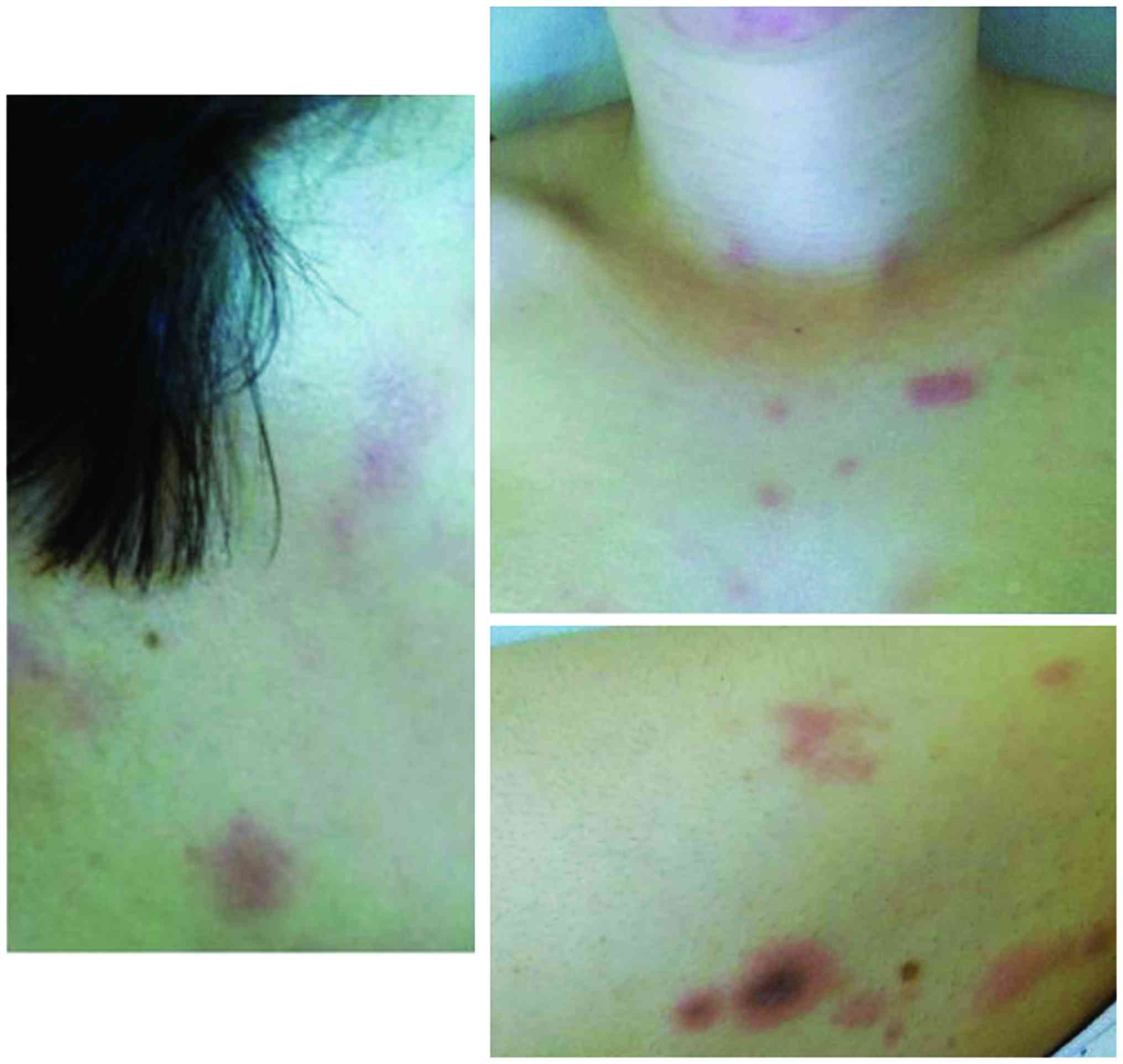
The cutaneous examination revealed well demarcated,
round and oval shaped erythemato-edematous papules and plaques on
the cheeks, chin, neck, upper and lower limbs, as well as some
target-like lesions, ranging from 5 to 20 mm (Fig. 1). The patient reported a sudden onset
of the skin eruption 3 months prior to the medical visit, starting
a few days before the menses and lasting until after the completion
of menstruation, with clinical worsening at every episode.
A 4-mm punch biopsy was performed and revealed on
the histopathological examination spongiosis, a perivascular and
diffuse lymphocytic infiltrate in the dermis, a few eosinophils and
apoptosis of the basal cells.
Intradermal testing with 0.01 ml progesterone (50
mg/ml progesterone at a dilution of 1:10 in aqueous solution) read
at 20 min was positive, showing a 5 mm wheal, compared to no skin
reaction to the saline solution control.
The patient was advised to take oral contraceptives
and mid-potency topical corticosteroids but chose to apply only
topical treatment. Two months later, the patient became pregnant
and delivered a healthy baby on term. The skin eruptions ceased
during the second pregnancy and did not reappear at a 1 year
follow-up postpartum (1,2).
Discussion
Progesterone hypersensitivity or autoimmune
progesterone dermatitis is a rare disorder that has been scarcely,
but worldwide reported. Initially, most cases were associated with
exogenous progesterone intake (3);
on an extensive review of published data, only 44.95% of patients
recalled exogenous progesterone exposure, however, there was a lack
of recorded history for 35.96% of patients (4). Onset of the disease was also noted in
relation to pregnancy, intrapartum, postpartum or after medical
abortion (5).
Diagnosis is based on cutaneous eruptions related to
increase in progesterone levels and may be confirmed by
intracutaneous progesterone testing or progesterone-specific IgE
serum. Other available but less useful diagnostic methods include:
intramuscular progesterone injection, oral progesterone intake,
patch testing (with a low negative predictive value), in
vitro leukocyte histamine release assay and
interferon-gamma-release assay (6).
Clinical presentation is highly variable and includes all urticaria
manifestations with or without angioedema, vesiculobullous,
eczematous or purpuric lesions, as well as cutaneous eruptions
resembling Stevens-Johnson syndrome, erythema multiforme or fixed
drug eruptions (4); asthma and
anaphylaxis may rarely accompany the symptoms (7).
A review of clinical treatment outcomes was
published and reviewed in 2015, summarizing the published data at
that time. It found that out of 86 patients with recorded clinical
progression, one patient had achieved disease control without
treatment and only 4 patients (4.65%) achieved complete remission
without therapy. Of those who received treatment, complete
remission was achieved primarily through total abdominal
hysterectomy/bilateral salpingo-oophorectomy (14 cases,
representing 16.27% of all patients and 51.85% of all patients with
complete remission). Medical treatments such as antihistamines,
systemic corticosteroids, conjugated estrogen/ethinyl estradiol,
gonadotropin releasing hormone, oral contraceptive were mostly
associated with disease control or remission (4). Progesterone desensitization was
employed in 6 patients, resulting in complete remission in 4,
clinical recurrence in one case and disease control in another
patient. Oral and intramuscular desensitization protocols were
further reported in a retrospective case-series of 11 patients,
showing dermatologic symptom improvement in 88% of cases and
cessation of asthma/anaphylactic symptoms (7). The remaining 13 patients, underwent
hormone therapy; 62% of this group also used additional treatments
such as antihistamines, topical steroids, montelukast for symptom
management. Efficacy of hormonal therapy should mostly be dose and
product dependent, however, it is not always true. A 39 year-old
multiparous woman diagnosed with autoimmune progesterone dermatitis
underwent treatment with vaginal hormonal contraceptives which
exacerbated the skin lesions, while oral contraceptive induced a
complete symptom resolution (8).
In the present case, we were pleasantly surprised by
the evolution of the patient: after onset, the cyclical clinical
lesions gradually worsened (showing apoptosis of basal cells on the
histopathological examination) but were kept under control with
topical corticosteroids. During pregnancy, there is a natural
inhibition of ovulation which explains disease remission, however,
symptoms may later recur. Fortunately, in this case, the patient
presented no disease relapse at a 2 year follow-up from the first
presentation. Symptom resolution without treatment has previously
been recorded (9), as has minimal
symptomatology of no concern to the patient (10). Pregnancy was reported to be
associated with symptom release in women diagnosed with autoimmune
progesterone dermatitis, possibly due to a gradual increase in
progesterone levels (11) but also
with clinical exacerbation, even anaphylactic shock during delivery
(12).
Patients already diagnosed with progesterone
hypersensitivity benefit from more information and better
surveillance during pregnancy while asymptomatic women might
manifest atypical symptoms. For example, unrevealing workup for
habitual idiopathic pregnancy losses showed immediate
hypersensitivity towards estrogen and progesterone in 50% of
patients and delayed hypersensitivity in 70% of patients (13). Even though most women demonstrated
combined hypersensitivity, some had only estrogen or progesterone
hypersensitivity. A follow-up of women with recurrent pregnancy
loss and sex-hormone hypersensitivity that underwent
desensitization protocols demonstrated 5 conceptions before
desensitization and 16 (out of 26–61%) conceptions and live births
(14).
The above-mentioned studies shift our focus from the
typical patient who has cyclical skin eruptions during the luteal
phase to women who do not have clinical onset of disease but other
symptoms, for example habitual pregnancy losses. From this point of
view, a recent report illustrates the case of a 31 year old woman
with recurrent pregnancy loss who also developed self limited
urticarial or eczematous eruptions in the time-frame corresponding
to higher progesterone levels. Following a positive intradermal
test to progesterone she underwent desensitization and successfully
sustained pregnancy with progesterone supplementation (15).
Unfortunately, not all patients had such a favorable
outcome. A recent article describes the case of a 22-year old woman
with progesterone hypersensitivity who had to undergo bilateral
salpingo-oophorectomy due to unsatisfactory response to various
medical treatments including antihistamines, oral contraceptives,
corticosteroids, leuprolide acetate and estradiol, tamoxifen and
danazol, desensitization protocol, immunomodulators for a period of
48 months (16).
In conclusion, our report addresses the various
treatment outcomes of women with progesterone hypersensitivity and
highlights the possibility of resolution in the absence of a
medical or surgical treatment.
Acknowledgements
Not applicable.
Funding
No funding was received.
Availability of data and materials
The datasets used and/or analyzed during the current
study are available from the corresponding author on reasonable
request.
Authors' contributions
SCS, LU, EC were responsible for the data analysis
and interpretation and contributed to writing the manuscript. SAD,
AV, VD were responsible for the data selection and clinical
interpretation of the data. RC designed the study and interpreted
the data. All authors read and approved the final manuscript. All
authors read and approved the final manuscript.
Ethics approval and consent to
participate
The study was approved by the Ethics Committee of
the Medical Clinic (Cluj-Napoca, Romania), and written consent was
given by the patient.
Patient consent for publication
The patient gave written consent, however, the
authors made efforts to remove identifying information to protect
the privacy of the patient.
Competing interests
The authors declare that they have no competing
interests.
References
|
1
|
Gheorghe I, Tatu AL, Lupu I, Thamer O,
Cotar AI, Pircalabioru GG, Popa M, Cristea VC, Lazar V and
Chifiriuc MC: Molecular characterization of virulence and
resistance features in Staphylococcus aureus clinical strains
isolated from cutaneous lesions in patients with drug adverse
reactions. Rom Biotechnol Lett. 22:12321–12327. 2017.
|
|
2
|
Raţiu MP, Purcărea I, Popa F, Purcărea VL,
Purcărea TV, Lupuleasa D and Boda D: Escaping the economic turn
down through performing employees, creative leaders and growth
driver capabilities in the Romanian pharmaceutical industry.
Farmacia. 59:119–130. 2011.
|
|
3
|
Hart R: Autoimmune progesterone
dermatitis. Arch Dermatol. 113:426–430. 1977. View Article : Google Scholar : PubMed/NCBI
|
|
4
|
Nguyen T and Razzaque Ahmed A: Autoimmune
progesterone dermatitis: Update and insights. Autoimmun Rev.
15:191–197. 2016. View Article : Google Scholar : PubMed/NCBI
|
|
5
|
Mbonile L: Autoimmune progesterone
dermatitis: Case report with history of urticaria, petechiae and
palpable pinpoint purpura triggered by medical abortion. S Afr Med
J. 106:48–50. 2016. View Article : Google Scholar : PubMed/NCBI
|
|
6
|
Li RC, Buchheit KM and Bernstein JA:
Progestogen hypersensitivity. Curr Allergy Asthma Rep. 18:12018.
View Article : Google Scholar : PubMed/NCBI
|
|
7
|
Foer D, Buchheit KM, Gargiulo AR, Lynch
DM, Castells M and Wickner PG: Progestogen hypersensitivity in 24
Cases: Diagnosis, management, and proposed renaming and
classification. J Allergy Clin Immunol Pract. 4:723–729. 2016.
View Article : Google Scholar : PubMed/NCBI
|
|
8
|
Camões S, Sampaio J, Rocha J, Tiago P and
Lopes C: Autoimmune progesterone dermatitis: Case report of an
unexpected treatment reaction. Australas J Dermatol. 58:e132–e134.
2017. View Article : Google Scholar : PubMed/NCBI
|
|
9
|
Herzberg AJ, Strohmeyer CR and
Cirillo-Hyland VA: Autoimmune progesterone dermatitis. J Am Acad
Dermatol. 32:333–338. 1995. View Article : Google Scholar : PubMed/NCBI
|
|
10
|
Özmen İ and Aktürk E: Autoimmune
progesterone dermatitis presenting with purpura. Cutis. 98:E12–E13.
2016.
|
|
11
|
García-Ortega P and Scorza E: Progesterone
autoimmune dermatitis with positive autologous serum skin test
result. Obstet Gynecol. 117:495–498. 2011. View Article : Google Scholar : PubMed/NCBI
|
|
12
|
O'Rourke J, Khawaja N, Loughrey J and
McKenna P: Autoimmune progesterone dermatitis in a parturient for
emergency caesarean section. Int J Obstet Anesth. 13:275–278. 2004.
View Article : Google Scholar : PubMed/NCBI
|
|
13
|
Untersmayr E, Jensen AN and Walch K: Sex
hormone allergy: Clinical aspects, causes and therapeutic
strategies - Update and secondary publication. World Allergy Organ
J. 10:452017. View Article : Google Scholar : PubMed/NCBI
|
|
14
|
Itsekson AM, Soriano D, Zolti M, Seidman
DS and Carp HJ: Intradermal sex hormone desensitization for relief
of premenstrual symptoms may improve the obstetric outcome of women
with recurrent pregnancy loss. Gynecol Endocrinol. 29:169–172.
2013. View Article : Google Scholar : PubMed/NCBI
|
|
15
|
Kuruvilla M, Vanijcharoenkarn K, Wan J,
Pereira N and Chung P: Exogenous progesterone hypersensitivity
associated with recurrent pregnancy loss J Allergy Clin Immunol
Pract. 6:1412–1413. 2018.PubMed/NCBI
|
|
16
|
Drayer SM, Laufer LR and Farrell ME:
Autoimmune progesterone dermatitis presenting as Stevens-Johnson
Syndrome. Obstet Gynecol. 130:881–884. 2017. View Article : Google Scholar : PubMed/NCBI
|