Introduction
Diabetes mellitus is one of the most common types of
chronic disease with extensive morbidity and mortality worldwide
(1). Diabetic retinopathy (DR) is
the most common microvascular complication of diabetes and is a
frequent cause of preventable blindness worldwide (2). In 2010, 3.7 million people were
visually impaired and 0.8 million were blind due to DR (3). Various metabolic disorders have been
associated with the onset of DR (4);
however, the connection between metabolic abnormalities and the
development of DR requires further investigation. At present, the
molecular mechanisms underlying the pathogenesis of DR remain
unknown and there are no effective treatments or preventative
approaches for DR. The present study aimed to investigate risk
factors for developing DR and determine a novel target for the
treatment of this particular complication.
Autophagy is a conserved metabolic process that is
characterized by the degradation and recycling of dysfunctional
proteins or organelles (5). In the
process of autophagy, the autophagosome is formed for the isolation
of targeted or non-specific materials (6); microtubule-associated protein 1 light
chain 3 (LC3B), comprising two forms (LC3B-I and LC3B-II), is
essential for the formation of the autophagosome (7). Upon the induction of autophagy, LC3B-I
is converted to LC3B-II, which integrates into the membrane of the
autophagosome (8). Studies have
demonstrated that autophagy is one of the major causative factors
involved in the pathogenesis of DR (9,10).
LC3B-II has been associated with the extent of autophagosome
formation (11) and serves as a
valuable molecular biomarker for the detection of autophagic
activity (12); thus, LC3B-II may be
an effective therapeutic target for the treatment of DR.
MicroRNAs (miRNAs/miRs) are small non-coding RNAs,
which modulate the expression of target mRNAs via the
post-transcriptional inhibition of translation (13). It has been revealed that miRNAs may
be directly or indirectly involved in several diseases by
regulating the expression of numerous genes (14). miR-125b-5p has been associated with
the progression of DR by regulating the expression of specificity
protein 1 (15). Furthermore,
miRNA-21 was reported to negatively regulate the expression of
peroxisome proliferator-activated receptor-α in the retina of mice
with diabetes (16) and modulate the
expression of prorenin receptor-induced vascular endothelial growth
factor (VEGF) under hyperglycemic conditions (17). miR-200b was reported to be involved
in the pathogenesis of DR in an VEGF-independent manner (18). These findings indicate the
considerable potential of miRNAs for therapeutic application in the
treatment of DR. Previous studies reported several differentially
expressed miRNAs in the development of early stage DR by using a
miRNA microarray analysis (15,19),
including miR-135b-5p, miR-145-5p, miR-146a-5p, miR-199a-5p and
miR-204. It has also been reported that miR-204-5p was involved in
diabetic keratopathy (20). However,
the role of miR-204-5p in DR remains elusive. Reverse
transcription-quantitative polymerase chain reaction (RT-qPCR)
revealed that miR-204-5p and VEGF were upregulated in the retina of
rats with streptozotocin (STZ)-induced diabetes, whereas the
protein expression levels of LC3B-II and the ratio of
LC3B-II/LC3B-I were significantly decreased. Anti-miR-204-5p
treatment promoted the expression of LC3B-II and the ratio of
LC3B-II/LC3B-I; however, these levels were suppressed in response
to exposure to miR-204-5p mimic. The results of the present study
suggested that miR-204-5p may be involved in the progression of DR
by negatively modulating the expression of LC3B-II. These findings
also indicated that modulation of retinal miR-204-5p expression may
be considered as potential therapeutic strategy for the treatment
of DR.
Materials and methods
Animals and grouping
A total of 60 male Sprague Dawley rats (aged 6–8
weeks; weighing, 180–220 g) were purchased from Laboratory Animal
Services Centre of Hunan Slack Jingda Experimental Animal Co., Ltd.
(Changsha, China). Rats were housed at 18–22°C with 12-h light/dark
cycles and access to standard food and water ad libitum. All
procedures were approved by the Animal Ethics Committee of Second
Affiliated Hospital of Nanchang University (Nanchang, China). Rats
were fed with a high glucose, high fat diet (60% standard chow, 10%
lard, 10% egg yolk powder and 20% sucrose). Following 8 weeks, rats
were divided into two groups: The diabetic group (n=50) and the
control group (n=10). Rats in the diabetes group were injected with
STZ (Sigma-Aldrich; Merck KGaA, Darmstadt, Germany) at 40 mg/kg. At
72 h following STZ treatment, rats were considered diabetic
providing their fasting blood glucose levels were >16.7 mM
(21). The control group received 2
ml of 0.1 M sodium citrate buffer.
Anti-miR-204-5p [adenovirus
AAV-U6-rno-miR-204-5p-CAG-enhanced green fluorescent protein
(EGFP); cat. no. MICA2014002038], miR-204-5p mimic (adenovirus
AAV-U6-mimic-rno-miR-204-5p-CAG-EGFP, 5′-UUCCCUUUGUCAUCCUAUGCCU-3′)
and negative control miRNA (neg-miR, adenovirus
AAV-U6-mimic-rno-negative-miR-CAG-EGFP,
5′-UUCUCCGAACGUGUCACGUTT-3′) were obtained from Shanghai GeneChem
Co., Ltd. (Shanghai, China). Diabetic rats were randomly divided
into three groups: Neg-miR, anti-miR-204-5p and mimic groups.
Diabetic rats in the anti-miR-204-5p group (n=15) were injected
with anti-miR-204-5p adenovirus (4.1×10¹² viral
genome/ml) via the caudal vein. Diabetic rats in the mimic group
(n=15) received miR-204-5p mimic adenovirus
(4.1×10¹²viral genome/ml). The neg-miR group (n=15) were
injected with the same quantity of neg-miR adenovirus. At 8 weeks,
retinal tissues were extracted for further investigation as
detailed below.
Rats retinal epithelial cells (rRECs)
culture
Five diabetic rats were anesthetized by
intraperitoneal injection of pentobarbital sodium (30 mg/kg). Under
anesthesia, the eyeball of diabetic rats was isolated from diabetic
rats and following rats were sacrificed using a lethal dose of
pentobarbital (100 mg/kg body weight). The retina was separated
from the eyeball and washed with Hanks balanced salt solution
(H1020; Beijing Solarbio Science & Technology Co., Ltd.,
Beijing, China) and minced into small pieces (1×1 mm) with surgical
scissors. Following filtration through a 30-µm nylon sieve, the
retina was digested with 2.5% trypsin for 30 min at 37°C. Following
centrifugation (1,500 × g for 5 min) at room temperature, rRECs
were isolated and cultured in Dulbecco's modified Eagle's medium
(DMEM; Gibco; Thermo Fisher Scientific, Inc., Waltham, MA, USA)
containing 20% fetal bovine serum (FBS; Sigma-Aldrich; Merck KGaA)
at 37°C with 5% CO2. Medium was replaced every three
days. rRECs were identified for endothelial homogeneity by testing
for immunoreactivity to factor VII antigen. rRECs were fixed with
4% paraformaldehyde, blocked with 2% BSA for 1 h at room
temperature and incubated with mouse anti-factor VII antibody (cat.
no. ab97614; Abcam, Cambridge, UK) at 4°C overnight. The cells were
washed and incubated with Alexa Fluor® 647-conjugated
donkey anti-rabbit immunoglobulin G secondary antibodies (cat. no.
ab150075; Abcam) at 37°C for 2 h. Then nuclear was stained with
DAPI (Sigma-Aldrich; Merck KGaA) for 10 min at room temperature.
Signals were detected with a fluorescent inverted microscope at a
magnification of ×200. The third passage was prepared for
subsequent experiments.
rRECs were seeded in 6-well plates at
1×10⁶ cells/well and cultured in DMEM supplemented with
10% FBS. At 70–80% density, cells were transfected with 100 nM
miR-204-5p mimic (the mimic group), miR-204-5p inhibitor (the
anti-miR-204-5p group) or neg-miR (the neg-miR group) with
Lipofectamine 3000 (Roche Diagnostics, Basel, Switzerland). The
transfection efficiency was determined by RT-qPCR. At 48 h, cells
were collected for follow-up experiments.
Transmission electron microscopy
rRECs at a density of 1×10⁵ cells/well
were fixed with 2.5% glutaraldehyde in 0.1 M phosphate buffer pH
7.4 for 2 h at room temperature. Following washing with 0.1 M
phosphate buffer, samples were stained with 1% osmium tetroxide for
1 h at room temperature. Following dehydration with an ethanol
gradient, samples were embedded in LX-112 resin and heating at 70°C
overnight. Sections were sliced to ultrathin sections (70 nm) and
imaged with an FEI Tecnai Spirit transmission electron microscope
(FEI; Thermo Fisher Scientific, Inc.) at magnifications of ×1,700
and ×5,000
RT-qPCR analysis
Total RNA was isolated from retinal tissues and
rRECs using TRIzol reagent (cat. no. CW0580S; Beijing CWBio,
Beijing, China) according to the manufacturer's protocol. RNA
quality was evaluated by agarose electrophoresis and the quantity
was determined via spectrophotometry at 260 and 280 nm. A total of
500 ng RNA was reverse transcribed into cDNA using the miRScript
system (GenScript Co., Ltd., Nanjing, China) according to the
manufacturer's protocols. qPCR was performed using SYBR Green qPCR
Super mix (cat. no. CW0957M; Beijing CWBio) on a CFX96 Touch PCR
Detection system (Bio-Rad Laboratories, Inc., Hercules, CA, USA).
The thermocycling conditions were as follows: 95°C for 5 min
followed by 40 cycles of 95°C for 30 sec, 60°C for 30 sec. All
experiments were performed with triplicate. The relative expression
of VEGF was normalized to β-actin using the 2−ΔΔCq
method (22). U6 was used as an
endogenous control to analyze the expression of miR-204-5p. The
primers for RT-qPCR were as follows: VEGF, forward,
5′-GAGTTAAACGAACGTACTTGCAGA-3′ and reverse,
5′-TCTAGTTCCCGAAACCCTGA-3′; β-actin, forward,
5′-TGGCTGGGGTGTTGAAGGTC-3′ and reverse,
5′-ATGGTGGGTATGGGTCAGAAGG-3′; miR-204-5p, forward,
5′-ACACTCCAGCTGGGTTCCCTTTGTCATCCTAT-3′ and reverse,
5′-CTCAACTGGTGTCGTGGA-3′; and U6, forward, 5′-CTCGCTTCGGCAGCACA-3′
and reverse, 5′-AACGCTTCACGAATTTGCGT-3′.
Western blotting
Proteins were isolated from retinal tissues and
rRECs using radioimmunoprecipitation assay buffer (Sigma-Aldrich;
Thermo Fisher Scientific, Inc.). Protein concentration was
determined by bicinchoninic acid kit (Beyotime Institute of
Biotechnology, Shanghai, China) according to the manufacturer's
protocol. Equal amounts of protein (60 µg) were separated using
SDS-PAGE on 12% gels and transferred to polyvinylidene difluoride
membranes (Millipore; Merck KGaA). Following blocking with 5%
non-fat milk for 1 h at room temperature, membranes were probed
with rabbit anti-LC3B antibody (cat. no. ab51520; 1:3,000; Abcam)
or anti-β-actin (cat. no. ab8226; 1:1,000; Abcam) overnight at 4°C.
Then, membranes were washed with TBS plus Tween-20 (0.1%) and
incubated with horseradish peroxidase-conjugated goat anti-rabbit
secondary antibodies (cat. no. sc-516078; 1:1,000; Santa Cruz
Biotechnology, Inc., Dallas, TX, USA) for 2 h at 37°C. Target bands
were developed with an enhanced chemiluminescence detection kit
(Amersham; GE Healthcare, Chicago, IL, USA). Band densities were
calculated using Quantity One software version 4.5.1 (Bio-Rad
Laboratories, Inc.) and normalized to β-actin.
Immunohistochemistry
Retinal tissues were isolated from the different
groups at 8 weeks following STZ injection, fixed with 10% freshly
prepared ice-cold paraformaldehyde at 4°C overnight and cut into
5-µm sections. Sections were incubated with 5% bovine serum albumin
(BSA; Beijing Solarbio Science & Technology Co., Ltd.) at 37°C
for 1 h to block the nonspecific sites. Then, sections were
incubated with rabbit polyclonal anti-LC3B antibody (1:1,000) for 2
h at room temperature. Following washing with PBS (3×), slides were
probed with horseradish peroxidase-conjugated goat anti-rabbit
antibodies for 2 h at room temperature. Immunoperoxidase binding
was visualized by reaction with
diaminobenzidine-H2O2 solution. Five
non-overlapping fields were randomly captured by an inverted
microscope at a magnification of ×400. LC3B-positive cells were
stained in brown or dark yellow.
Statistical analysis
Data are presented as the mean ± standard deviation.
All statistical analysis was performed using SPSS 18.0 software
(SPSS, Inc., Chicago, IL, USA). Statistical analyses were performed
using one-way analysis of variance followed by Dunnett's post hoc
tests. P<0.05 was considered to indicate a statistically
significant difference.
Results
miR-204-5p is overexpressed in
diabetic rats
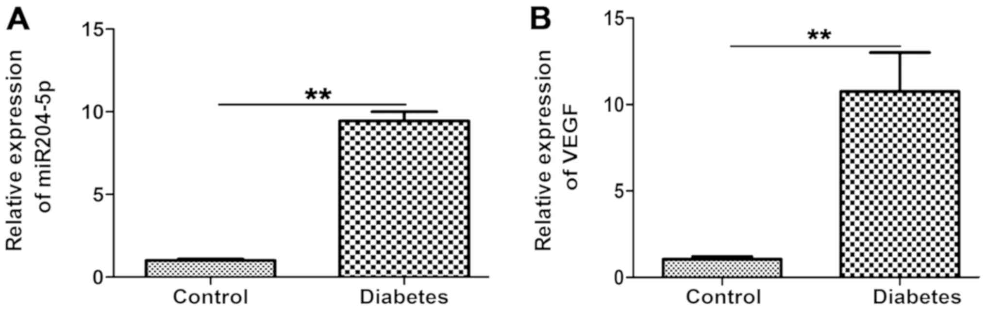
In the present study, a diabetic rat model was
generated by intraperitoneal injection of STZ; the expression of
miR-204-5p in the diabetic rat model was then investigated. To
determine whether the diabetic rat model was successfully
constructed, glucose levels were analyzed at day 3 following STZ
administration. Glucose levels in the STZ-treated group (23.01±3.71
mM) were significantly increased compared with the control group
(4.62±1.51 mM; P<0.01; data not shown). This suggested that a
diabetic rat model was successfully constructed. As presented in
Fig. 1A, compared with the control
group, relative miR-204-5p expression levels in retina tissues of
the diabetes group were significantly upregulated (P<0.01). In
addition, expression VEGF mRNA levels in the diabetes group were
significantly increased compared with the control group (P<0.01;
Fig. 1B). The results indicated that
miR-204-5p and VEGF may be associated with the diabetes and
contribute to the progression of DR.
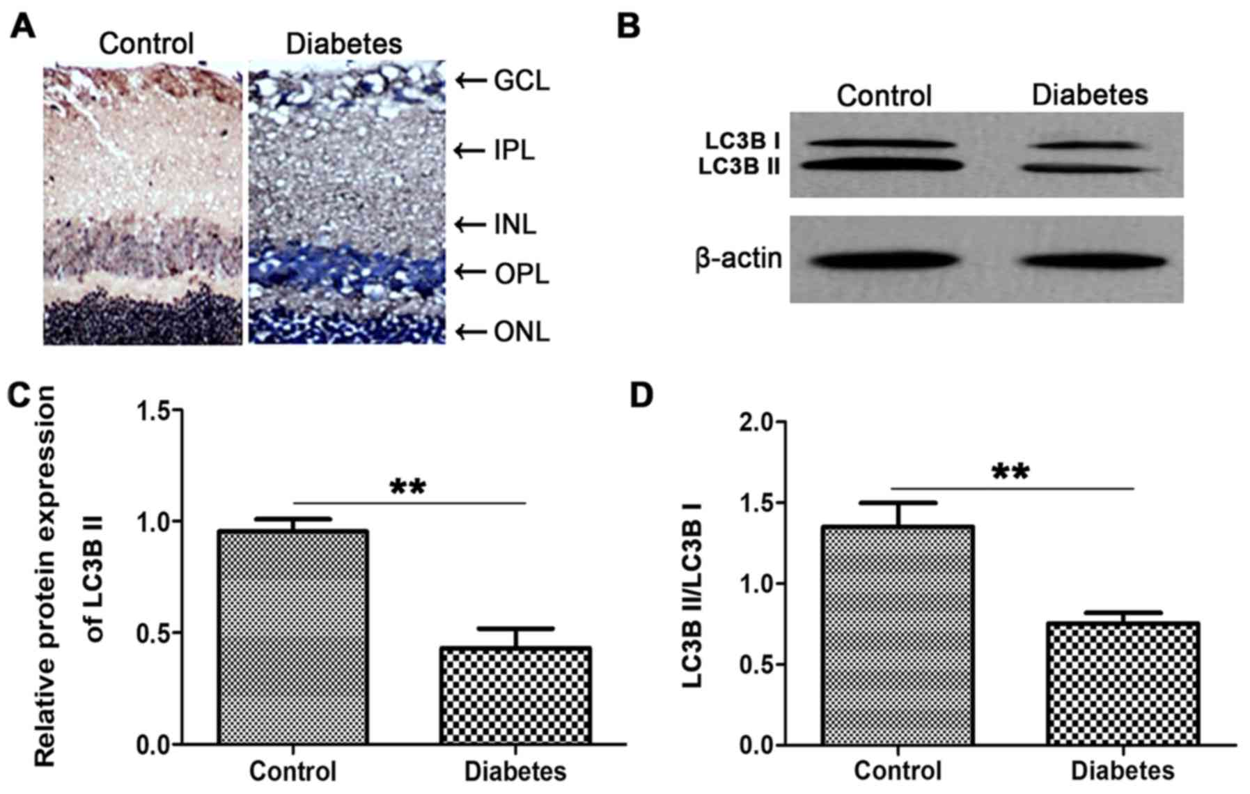
LC3B-II expression and LC3B-II/LC3B-I
are significantly suppressed in diabetic rats
LC3B-II levels are proportional to the number of
autophagic vacuoles (7). To
investigate the role of LC3B in the retina of diabetic rats, LC3B
protein expression was determined via immunohistochemistry. As
presented in Fig. 2A, compared with
the control group, LC3B expression in the diabetic group was
markedly reduced. These results revealed that the expression of
LC3B was inhibited in retina tissues of diabetic rats.
Additionally, protein expression levels of LC3B were
determined by western blotting. Compared with the control group,
expression levels of LC3B-II in the diabetic group were
significantly reduced (P<0.01; Fig.
2B and C). Furthermore, the ratio of LC3B-II/LC3B-I was
significantly decreased in the diabetic group compared with the
control (P<0.01; Fig. 2D). These
data suggested that the expression of LC3B-II and the ratio of
LC3B-II/LC3B-I may be suppressed during the development of DR.
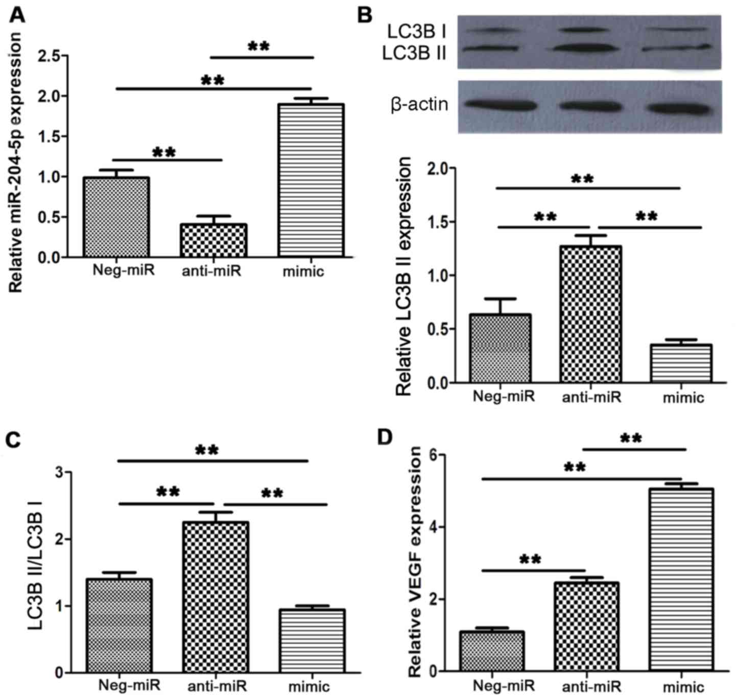
miR-204-5p negatively regulates the
expression of LC3B-II
To further investigate the mechanism by which
miR-204-5p affects the progression of DR, anti-miR-204-5p and
miR-204-5p mimic were administered to rats of the diabetes group.
As presented in Fig. 3A, compared
with the neg-miR group, anti-miR-204-5p treatment significantly
suppressed miR-204-5p expression and miR-204-5p mimic treatment
significantly upregulated miR-204-5p expression (P<0.01). The
results suggested that anti-miR-204-5p and miR-204-5p mimic were
functional in diabetic rats. In addition, compared with the neg-miR
group, injection of anti-miR-204-5p into diabetic rats
significantly enhanced LC3B-II expression and the ratio of
LC3B-II/LC3B-I (P<0.01; Fig. 3B and
C). In the presence of the miR-204-5p mimic these levels
decreased compared with the neg-miR control (P<0.01). mRNA
expression levels of VEGF were significantly upregulated in the
anti-miR-204-5p and the mimic groups compared with the neg-miR
control (P<0.01; Fig. 3D).
Interestingly, miR-204-5p mimic treatment induced a larger increase
in VEGF mRNA expression levels compared with the anti-miR-204-5p
group.
In order to confirm the results in vitro,
rRECs were isolated from diabetic rats and transfected with
anti-miR-204-5p, miR-204-5p mimic or neg-miR. The purity of rRECs
following isolation was determined via factor VII staining. The
percentage of factor VII-positive cells was >97%, indicating
that rRECs were successfully isolated from retina tissues of
diabetic rats (Fig. 4A). As
presented in Fig. 4B, compared with
the neg-miR group, anti-miR-204-5p markedly increased the number of
autophagic vacuoles and a reduction was observed in response to
treatment with miR-204-5p mimic. The results of the transfection
efficiency analysis are presented in Fig. 4C; it was determined that
anti-miR-204-5p successfully inhibited and miR-204-5p mimic
successfully enhanced miR-204-5p expression compared with the
neg-miR control (P<0.01). Anti-miR-204-5p further significantly
promoted LC3B-II expression and significantly increased the
LC3B-II/LC3B-I ratio; miR-204-5p mimic exhibited opposing effects
compared with the neg-miR control (all P<0.01; Fig. 4D and E). Additionally,
anti-miR-204-5p and miR-204-5p mimic significantly upregulated the
expression of VEGF compared with the neg-miR control (P<0.01;
Fig. 4F). These results revealed
that miR-204-5p may be involved in the development of DR by
negatively regulating the expression of LC3B-II.
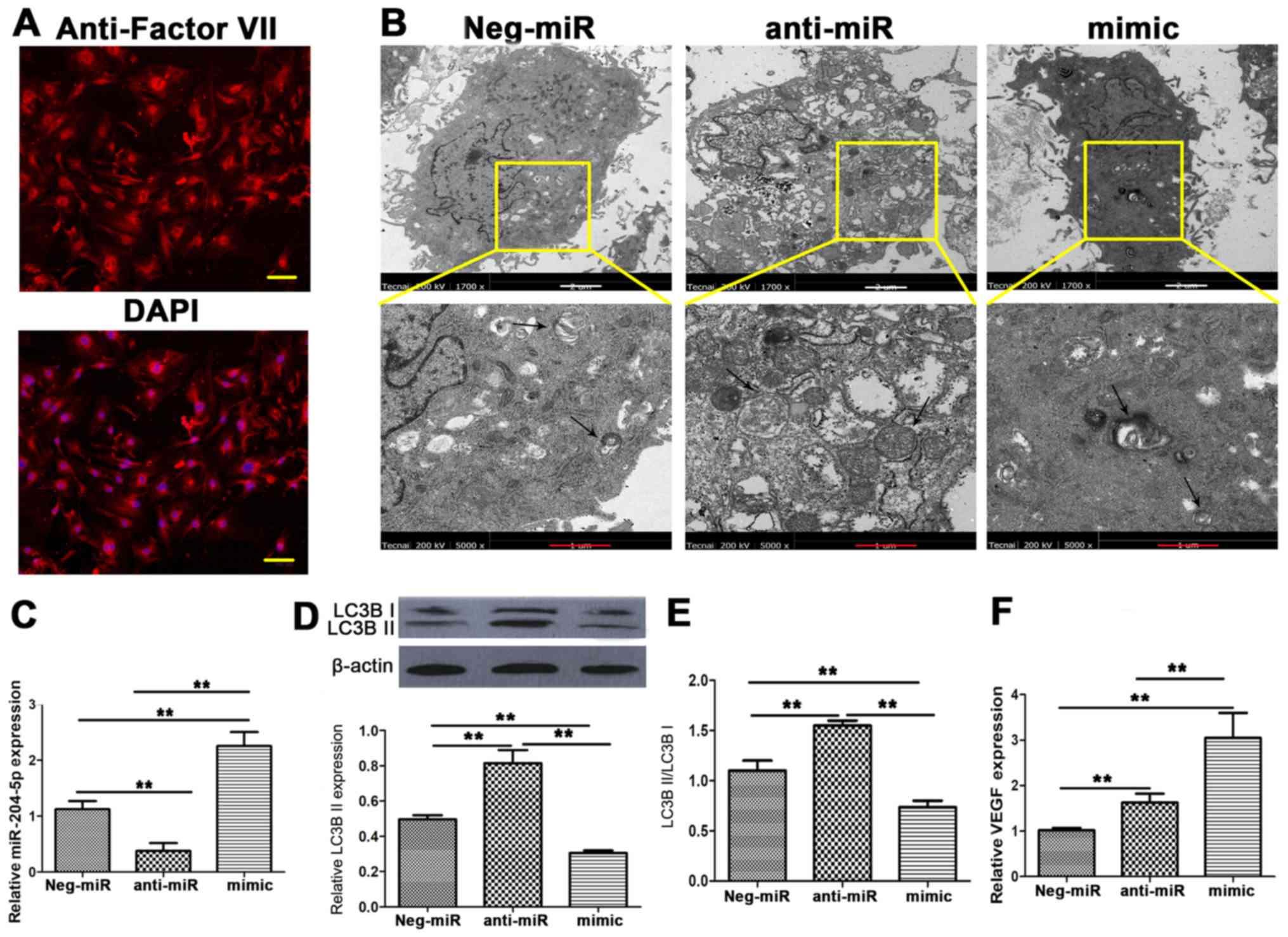 | Figure 4.miR-204-5p downregulates the
expression of LC3B-II and the ratio of LC3B-II/LC3B-I in
vitro. rRECs were isolated from diabetic rats and transfected
with anti-miR, mimic ant neg-miR. (A) Purity of rRECs was
determined via factor VII staining; factor VII-positive cells are
presented in red and nuclei in blue. Scale bar, 100 µm. (B) The
amount of autophagic vacuoles was determined by transmission
electron microscopy (magnification, ×1,700 and ×5,000). Scale bar
in white, 2 µm; scale bar in red, 1 µm. (C) Expression levels of
miR-204-5p in transfected cells. (D) Western blot images and
quantitative analysis of LC3B-II protein expression and (E) the
LC3B-II/LC3B-I ratio. (F) mRNA expression levels of VEGF. The
relative expression of miR-204-5p was normalized to U6 and VEGF was
normalized to β-actin. **P<0.01. rRECs, rat retinal endothelial
cells; miR, microRNA; LC3B, microtubule-associated protein 1 light
chain 3; VEGF, vascular endothelial growth factor; neg-miR, control
miR; anti-miR, anti-miR-204-5p; mimic, miR-204-5p mimic. |
Discussion
DR is a cause of blindness in individuals and
affects their quality of life (23).
It is critical to investigate the pathogenesis of DR and to explore
novel treatment targets. In the present study, it was reported that
miR-204-5p and VEGF mRNA were upregulated in the STZ-induced
diabetes group and expression of LC3B-II and the LC3B-II/LC3B-I
ratio were decreased in diabetic rats compared with the control.
Furthermore, anti-miR-204-5p treatment promoted protein expression
of LC3B-II and the LC3B-II/LC3B-I ratio compared with the neg-miR.
Treatment with miR-204-5p mimic exhibited reverse effects compared
with the control. These findings indicated that miR-204-5p may be
involved in the progression of DR in retina tissues of diabetic
rats by negatively regulating the expression of LC3B-II.
miRNAs are associated with various biological
events, including cell proliferation, differentiation and apoptosis
(24). Previous studies revealed
that miR-204 suppresses the development of numerous types of
cancer, including renal cell carcinoma (25), breast cancer (26) and gastric cancer (27). A recent study demonstrated that
miR-204-5p exerts antitumor effects on melanoma (28) and inhibits the invasion and
metastasis of laryngeal squamous cell carcinoma by suppressing
forkhead box C1 (29). Furthermore,
it was reported that miR-204-5p suppresses inflammation in renal
tubular epithelial cells by directly targeting the interleukin-6
receptor (30). Few studies have
investigated the function of miR-204-5p in the progression of DR;
however, the upregulation of miR-204 in DR has been reported
previously (19). In the STZ-induced
diabetes model of the present study, the expression of miR-204-5p
was upregulated in retina tissues compared with the control, which
suggested a potential association between miR-204-5p and the
development of DR. These findings indicated that miR-204-5p may be
considered as a potential therapeutic target for DR treatment.
Autophagy is a conserved lysosomal degradation
pathway that maintains cell homeostasis (31) and serves an adaptive role to protect
organisms against infections and diabetic complications (32). Autophagy has been proposed as an
important pathway associated with DR; thus, this process may serve
as a potential therapeutic target for the treatment of DR (33). Expression of LC3B is a reported
hallmark of the onset of autophagy (34) and the conversion of LC3B-I to LC3B-II
is a common indicator of autophagy (35). LC3B-II is a specific marker of the
autophagic process as its expression directly correlates with the
number of autophagosomes (11);
however, the association between autophagy and DR is complex.
Studies have demonstrated that STZ-induced diabetic rats exhibit
increased levels of autophagy markers (35,36).
Conversely, it was reported that the number of autophagosomes was
significantly decreased in diabetic rats and that high levels of
glucose suppressed the ratio of LC3B-II/LC3B-I compared with that
in the healthy control group (37).
In the present study, it was observed that expression of LC3B-II
and the LC3B-II/LC3B-I ratio were reduced in retina tissues of
diabetic rats compared with the control group, suggesting a
potential association between DR and autophagic activity.
Additionally, anti-miR-204-5p promoted the expression of LC3B-II
and the LC3B-II/LC3B-I ratio, which were suppressed in response to
miR-204-5p mimic treatment compared with the neg-miR control. The
data of the present study indicated that miR-204-5p negatively
regulated LC3B-II expression in DR. Defective autophagy has been
reported as an early event occurring in the pathogenesis of DR
(10). These findings indicated a
potential novel association between miR-204-5p, DR and
autophagy.
VEGF induces the formation of new blood vessels and
increases the permeability of existing vessels (38). VEGF is mainly expressed in ganglion
cells and retinal pigment epithelial cells (39). Studies have revealed that VEGF
expression is increased (40) and is
a key factor in DR (41). In the
present study, it was reported that the mRNA expression levels of
VEGF in the diabetes group were significantly upregulated compared
with the control group, which was in accordance with the findings
of previous studies (38,41). Anti-miR-204-5p and miR-204-5p mimic
treatment promoted the expression of VEGF compared with the neg-miR
control. These results revealed that miR-205-5p regulated the
expression of VEGF in an unknown way.
The current study has several limitations. It was
suggested that miR-204-5p may be associated with DR and autophagy
by regulating the expression of the autophagy-specific marker
LC3B-II; however, whether miR-204-5p directly or indirectly targets
the expression of LC3B-II remains unknown. Thus, further research
into the targets of miR-204-5p is required.
In conclusion, miR-204-5p and VEGF were reported to
be upregulated in the retina tissues of diabetic rats and may be
associated with the progression of DR. However, the mechanism by
which miR-204-5p affected the VEGF levels remains unknown. Protein
expression levels of LC3B-II and the LC3B-II/LC3B-I ratio were
reduced in retina tissues of diabetic rats compared with the
control. In diabetic rats and isolated rRECs, miR-204-5p-mimic
treatment downregulated LC3B-II expression compared with the
neg-miR control. These results revealed that miR-204-5p promoted
the development of DR via downregulating the expression of LC3B-II.
These results may further provide novel insight into the
association between DR and autophagy.
Acknowledgements
Not applicable.
Funding
The current study was supported by Jiangxi Natural
Science Foundation of China (grant no. 20151BAB205096).
Availability of data and materials
All data generated or analyzed during this study are
included in this published article.
Authors' contributions
XBM performed PCR and western blot analyses,
analyzed the data and prepared the original manuscript. YHC
established the rRECs, performed the TEM analysis and contributed
to preparing the figures. YYX designed the study and revised the
manuscript. All authors read and approved the final version of the
manuscript.
Ethics approval and consent to
participate
The present study was approved by the Animal Ethics
Committee of Second Affiliated Hospital of Nanchang University
(Nanchang, China).
Patient consent for publication
Not applicable.
Competing interests
The authors declare that they have no competing
interests.
Glossary
Abbreviations
Abbreviations:
|
DR
|
diabetic retinopathy
|
|
STZ
|
streptozotocin
|
|
LC3B
|
microtubule-associated protein 1 light
chain 3
|
|
VEGF
|
vascular endothelial growth factor
|
References
|
1
|
Santos LL, Lima FJC, Sousa-Rodrigues CF
and Barbosa FT: Use of SGLT-2 inhibitors in the treatment of type 2
diabetes mellitus. Rev Assoc Med Bras (1992). 63:636–641. 2017.
View Article : Google Scholar : PubMed/NCBI
|
|
2
|
Murchison AP, Hark L, Pizzi LT, Dai Y,
Mayro EL, Storey PP, Leiby BE and Haller JA: Non-adherence to eye
care in people with diabetes. BMJ Open Diabetes Res Care.
5:e0003332017. View Article : Google Scholar : PubMed/NCBI
|
|
3
|
Leasher JL, Bourne RR, Flaxman SR, Jonas
JB, Keeffe J, Naidoo N, Pesudovs K, Price H, White RA, Wong TY, et
al: Erratum. Global estimates on the number of people blind or
visually impaired by diabetic retinopathy: A meta-analysis from
1990–2010. Diabetes Care 2016;39:1643-1649. Diabetes Care.
39:20962016. View Article : Google Scholar : PubMed/NCBI
|
|
4
|
Arroba AI, Campos-Caro A, Aguilar-Diosdado
M and Valverde AM: IGF-1, inflammation and retinal degeneration: A
close network. Front Aging Neurosci. 10:2032018. View Article : Google Scholar : PubMed/NCBI
|
|
5
|
Hou L, Wei L, Zhu S, Wang J, Quan R, Li Z
and Liu J: Avian metapneumovirus subgroup C induces autophagy
through the ATF6 UPR pathway. Autophagy. 13:1709–1721. 2017.
View Article : Google Scholar : PubMed/NCBI
|
|
6
|
Wang L, Feng D, Liu Y, Li S, Jiang L, Long
Z and Wu Y: Autophagy plays a protective role in motor neuron
degeneration following spinal cord ischemia/reperfusion-induced
spastic paralysis. Am J Transl Res. 9:4261–4270. 2017.PubMed/NCBI
|
|
7
|
Gao N, Wang H, Yin H and Yang Z:
Angiotensin II induces calcium-mediated autophagy in podocytes
through enhancing reactive oxygen species levels. Chem Biol
Interact. 277:110–118. 2017. View Article : Google Scholar : PubMed/NCBI
|
|
8
|
Wang T, Zhang L, Hu J, Duan Y, Zhang M,
Lin J, Man W, Pan X, Jiang Z, Zhang G, et al: Mst1 participates in
the atherosclerosis progression through macrophage autophagy
inhibition and macrophage apoptosis enhancement. J Mol Cell
Cardiol. 98:108–116. 2016. View Article : Google Scholar : PubMed/NCBI
|
|
9
|
Dehdashtian E, Mehrzadi S, Yousefi B,
Hosseinzadeh A, Reiter RJ, Safa M, Ghaznavi H and Naseripour M:
Diabetic retinopathy pathogenesis and the ameliorating effects of
melatonin; involvement of autophagy, inflammation and oxidative
stress. Life Sci. 193:20–33. 2018. View Article : Google Scholar : PubMed/NCBI
|
|
10
|
Lopes de Faria JM, Duarte DA, Montemurro
C, Papadimitriou A, Consonni SR and Lopes de Faria JB: Defective
autophagy in diabetic retinopathy. Invest Ophthalmol Vis Sci.
57:4356–4366. 2016. View Article : Google Scholar : PubMed/NCBI
|
|
11
|
Kabeya Y, Mizushima N, Ueno T, Yamamoto A,
Kirisako T, Noda T, Kominami E, Ohsumi Y and Yoshimori T: LC3, a
mammalian homologue of yeast Apg8p, is localized in autophagosome
membranes after processing. EMBO J. 19:5720–5728. 2000. View Article : Google Scholar : PubMed/NCBI
|
|
12
|
Alizadeh S, Mazloom H, Sadeghi A,
Emamgholipour S, Golestani A, Noorbakhsh F, Khoshniatnikoo M and
Meshkani R: Evidence for the link between defective autophagy and
inflammation in peripheral blood mononuclear cells of type 2
diabetic patients. J Physiol Biochem. 74:369–379. 2018. View Article : Google Scholar : PubMed/NCBI
|
|
13
|
Chandra S, Vimal D, Sharma D, Rai V, Gupta
SC and Chowdhuri DK: Role of miRNAs in development and disease:
Lessons learnt from small organisms. Life Sci. 185:8–14. 2017.
View Article : Google Scholar : PubMed/NCBI
|
|
14
|
Nadeem A, Ashraf MR, Javed M, Hussain T,
Tariq MS and Babar ME: Review-microRNAs: A new paradigm towards
mechanistic insight of diseases. Pak J Pharm Sci. 31:2017–2026.
2018.PubMed/NCBI
|
|
15
|
Gong Q, Xie J, Liu Y, Li Y and Su G:
Differentially expressed micrornas in the development of early
diabetic retinopathy. J Diabetes Res. 2017:47279422017. View Article : Google Scholar : PubMed/NCBI
|
|
16
|
Chen Q, Qiu F, Zhou K, Matlock HG,
Takahashi Y, Rajala RVS, Yang Y, Moran E and Ma JX: Pathogenic role
of microRNA-21 in diabetic retinopathy through downregulation of
PPARα. Diabetes. 66:1671–1682. 2017. View Article : Google Scholar : PubMed/NCBI
|
|
17
|
Haque R, Iuvone PM, He L, Choi KSC, Ngo A,
Gokhale S, Aseem M and Park D: The MicroRNA-21 signaling pathway is
involved in prorenin receptor (PRR)-induced VEGF expression in
ARPE-19 cells under a hyperglycemic condition. Mol Vis. 23:251–262.
2017.PubMed/NCBI
|
|
18
|
Gomaa AR, Elsayed ET and Moftah RF:
MicroRNA-200b expression in the vitreous humor of patients with
proliferative diabetic retinopathy. Ophthalmic Res. 58:168–175.
2017. View Article : Google Scholar : PubMed/NCBI
|
|
19
|
Wu JH, Gao Y, Ren AJ, Zhao SH, Zhong M,
Peng YJ, Shen W, Jing M and Liu L: Altered microRNA expression
profiles in retinas with diabetic retinopathy. Ophthalmic Res.
47:195–201. 2012. View Article : Google Scholar : PubMed/NCBI
|
|
20
|
Gao J, Wang Y, Zhao X, Chen P and Xie L:
MicroRNA-204-5p-mediated regulation of SIRT1 contributes to the
delay of epithelial cell cycle traversal in diabetic corneas.
Invest Ophthalmol Vis Sci. 56:1493–1504. 2015. View Article : Google Scholar : PubMed/NCBI
|
|
21
|
Nathan DM, Buse JB, Davidson MB, Heine RJ,
Holman RR, Sherwin R and Zinman B: Management of hyperglycaemia in
type 2 diabetes: A consensus algorithm for the initiation and
adjustment of therapy. A consensus statement from the American
diabetes association and the european association for the study of
diabetes. Diab Tologia. 49:1711–1721. 2006. View Article : Google Scholar
|
|
22
|
Livak KJ and Schmittgen TD: Analysis of
relative gene expression data using real-time quantitative PCR and
the 2(−Delta Delta C(T)) method. Methods. 25:402–408. 2001.
View Article : Google Scholar : PubMed/NCBI
|
|
23
|
Rubsam A, Parikh S and Fort PE: Role of
Inflammation in diabetic retinopathy. Int J Mol Sci. 19(pii):
E9422018. View Article : Google Scholar : PubMed/NCBI
|
|
24
|
Kaneko H and Terasaki H: Biological
involvement of MicroRNAs in proliferative vitreoretinopathy. Transl
Vis Sci Technol. 6:52017. View Article : Google Scholar : PubMed/NCBI
|
|
25
|
Wu D, Pan H, Zhou Y, Zhang Z, Qu P, Zhou J
and Wang W: Upregulation of microRNA-204 inhibits cell
proliferation, migration and invasion in human renal cell carcinoma
cells by downregulating SOX4. Mol Med Rep. 12:7059–7064. 2015.
View Article : Google Scholar : PubMed/NCBI
|
|
26
|
Liu J and Li Y: Trichostatin A and
Tamoxifen inhibit breast cancer cell growth by miR-204 and ERalpha
reducing AKT/mTOR pathway. Biochem Biophys Res Commun. 467:242–247.
2015. View Article : Google Scholar : PubMed/NCBI
|
|
27
|
Lorenzon L, Cippitelli C, Avantifiori R,
Uccini S, French D, Torrisi MR, Ranieri D, Mercantini P, Canu V,
Blandino G and Cavallini M: Down-regulated miRs specifically
correlate with non-cardial gastric cancers and Lauren's
classification system. J Surg Oncol. 116:184–194. 2017. View Article : Google Scholar : PubMed/NCBI
|
|
28
|
Palkina N, Komina A, Aksenenko M, Moshev
A, Savchenko A and Ruksha T: miR-204-5p and miR-3065-5p exert
antitumor effects on melanoma cells. Oncol Lett. 15:8269–8280.
2018.PubMed/NCBI
|
|
29
|
Gao W, Wu Y, He X, Zhang C, Zhu M, Chen B,
Liu Q, Qu X, Li W, Wen S and Wang B: MicroRNA-204-5p inhibits
invasion and metastasis of laryngeal squamous cell carcinoma by
suppressing forkhead box C1. J Cancer. 8:2356–2368. 2017.
View Article : Google Scholar : PubMed/NCBI
|
|
30
|
Li H, Wang J, Liu X and Cheng Q:
MicroRNA-204-5p suppresses IL6-mediated inflammatory response and
chemokine generation in HK-2 renal tubular epithelial cells by
targeting IL6R. Biochem Cell Biol. 2018.(Epub ahead of print).
View Article : Google Scholar
|
|
31
|
Mizushima N, Levine B, Cuervo AM and
Klionsky DJ: Autophagy fights disease through cellular
self-digestion. Nature. 451:1069–1075. 2008. View Article : Google Scholar : PubMed/NCBI
|
|
32
|
Volpe CMO, Villar-Delfino PH, Dos Anjos
PMF and Nogueira-Machado JA: Cellular death, reactive oxygen
species (ROS) and diabetic complications. Cell Death Dis.
9:1192018. View Article : Google Scholar : PubMed/NCBI
|
|
33
|
Levine B and Kroemer G: Autophagy in the
pathogenesis of disease. Cell. 132:27–42. 2008. View Article : Google Scholar : PubMed/NCBI
|
|
34
|
Yoshii SR and Mizushima N: Monitoring and
measuring autophagy. Int J Mol Sci. 18(pii): E18652017. View Article : Google Scholar : PubMed/NCBI
|
|
35
|
Li XC, Hu QK, Chen L, Liu SY, Su S, Tao H,
Zhang LN, Sun T and He LJ: HSPB8 promotes the fusion of
autophagosome and lysosome during autophagy in diabetic neurons.
Int J Med Sci. 14:1335–1341. 2017. View Article : Google Scholar : PubMed/NCBI
|
|
36
|
Cai X, Li J, Wang M, She M, Tang Y, Li H
and Hui H: GLP-1 treatment improves diabetic retinopathy by
alleviating autophagy through GLP-1R-ERK1/2-HDAC6 signaling
pathway. Int J Med Sci. 14:1203–1212. 2017. View Article : Google Scholar : PubMed/NCBI
|
|
37
|
Kobayashi S, Xu X, Chen K and Liang Q:
Suppression of autophagy is protective in high glucose-induced
cardiomyocyte injury. Autophagy. 8:577–592. 2012. View Article : Google Scholar : PubMed/NCBI
|
|
38
|
Rodrigues M, Xin X, Jee K,
Babapoor-Farrokhran S, Kashiwabuchi F, Ma T, Bhutto I, Hassan SJ,
Daoud Y, Baranano D, et al: VEGF secreted by hypoxic muller cells
induces MMP-2 expression and activity in endothelial cells to
promote retinal neovascularization in proliferative diabetic
retinopathy. Diabetes. 62:3863–3873. 2013. View Article : Google Scholar : PubMed/NCBI
|
|
39
|
Nishijima K, Ng YS, Zhong L, Bradley J,
Schubert W, Jo N, Akita J, Samuelsson SJ, Robinson GS, Adamis AP
and Shima DT: Vascular endothelial growth factor-A is a survival
factor for retinal neurons and a critical neuroprotectant during
the adaptive response to ischemic injury. Am J Pathol. 171:53–67.
2007. View Article : Google Scholar : PubMed/NCBI
|
|
40
|
Gilbert RE, Vranes D, Berka JL, Kelly DJ,
Cox A, Wu LL, Stacker SA and Cooper ME: Vascular endothelial growth
factor and its receptors in control and diabetic rat eyes. Lab
Invest. 78:1017–1027. 1998.PubMed/NCBI
|
|
41
|
Bai Y, Ma JX, Guo J, Wang J, Zhu M, Chen Y
and Le YZ: Muller cell-derived VEGF is a significant contributor to
retinal neovascularization. J Pathol. 219:446–454. 2009. View Article : Google Scholar : PubMed/NCBI
|