Introduction
Cholesteatomatous otitis media, also known as middle
ear cholesteatoma, is an infectious disease in the ear (1). Cholesteatomatous otitis media is
associated with the abnormal accumulation of squamous epithelium in
keratin. The exfoliated epithelium continuously accumulates and
expands around, causing the injury of ossicle and the impairment of
audition (2,3). If cholesteatomatous otitis media
spreads to the nerves, it may result in facioplegia, and patients
may even be accompanied with a variety of serious intracranial and
extracranial complications, which seriously endanger patients' life
(4). At present, the main treatment
method of cholesteatomatous otitis media in clinic is operative
treatment, the purpose of which is to prevent intracranial
complications (5). ‘Early diagnosis,
early treatment’ is the key of treating cholesteatomatous otitis
media. In clinic, cholesteatomatous otitis media is commonly
detected by imageology, which is spiral CT (6,7).
According to the reports of Rogha et al (8), the different extent and severity of the
injury of sclerotin, which is caused by cholesteatomatous otitis
media, can be observed using CT.
Matrix metalloproteinase-2 (MMP-2) and matrix
metalloproteinase-9 (MMP-9), the core members of matrix
metalloproteinases (MMPs) family, play an important role in various
biological processes in vivo and in the anti-infective
immunization of the body (9,10). Interleukin-6 (IL-6) is involved in
the proliferation and differentiation of cells in anti-infective
reactions and improves their functions (11). According to related study reports
(12), the expression levels of
MMP-2, MMP-9 and IL-6 are closely related to the clinical
manifestations and the severity of diabetic diseases. However,
studies on MMP-2, MMP-9 and IL-6, and the severity of
cholesteatomatous otitis media are few (13). This study analyzed the expression
levels of MMP-2, MMP-9 and IL-6 in the serum of patients with
cholesteatomatous otitis media and injury degree of different
sclerotin, in order to provide clinical value for the occurrence
and development of cholesteatomatous otitis media, the prediction
and prognosis of cholesteatomatous otitis media, and the diagnosis
and treatment of it.
Patients and methods
Patient data
One hundred and seventy-six patients with
cholesteatomatous otitis media, admitted to the Changxing People's
Hospital (Huzhou, China), from January 2010 to December 2016, and
tested by 64-row spiral CT, were selected and enrolled as the study
group. The study group included 91 males and 85 females, aged from
33 to 69 years old, with an average age of 49.65±5.27 years, who
were classified according to the presence or absence of injury of
ossicle, and the extent and degree of injury. One hundred and
eighty-one patients with simple otitis media were selected and
enrolled as the control group, which included 98 males and 83
females, aged from 31 to 72 years, with an average age of
51.83±5.39 years. Inclusion criteria: patients who had complete
clinical data; and patients who had not received relevant diagnosis
and treatment in other hospitals. Exclusion criteria: patients who
had carious otitis media; who had middle ear cancer; patients in
gestation or lactation period; who had other serious diseases or
tumors; with communication disorders or cognitive dysfunction. The
study was approved by the Ethics Committee of Changxing People's
Hospital. Signed written informed consents were obtained from the
patients and/or guardians. All subjects cooperated with the medical
workers to finish the relevant diagnosis and treatment.
CT examination method
Philips 64-row spiral CT scanner (Philips
Healthcare, Andover, MA, USA) was used. Patients were put in a
supine position and the scanning parameters were: 120 kV; 150–200
mA; view field, 240×240 mm; matrix, 512×512; layer thickness, 0.5
mm. The scanning range was from the upper edge of talus cone to the
tip of mastoid, and the layer thickness and layer spacing were both
1 mm. Window width was 4,000 Hu and window level was 700 Hu.
Detection method of the expression
levels of MMP-2, MMP-9 and IL-6 in the serum of patients
Peripheral venous blood (4 ml) of the patients with
cholesteatomatous otitis media was taken when they were fasting.
The serum was separated from the blood by a centrifuge at a speed
of 2,010 × g for 10 min at 4°C, and it was stored in a refrigerator
(Thermo Fisher Scientific, Inc., Waltham, MA, USA), in which the
temperature was −20°C, for further detection. Enzyme-linked
immunosorbent assay (ELISA) was used to detect the expression
levels of MMP-2, MMP-9 and IL-6 in the serum of patients. The
detection was performed strictly according to the manufacturer's
instructions of human MMP-2 ELISA test kit (item no. H-EL-MMP-2;
Shanghai Zeye Biotechnology Co., Ltd., Shanghai, China), human
MMP-9 ELISA test kit (item no. RJ12726; Shanghai Renjie
Biotechnology Co., Ltd., Shanghai, China) and human IL-6 ELISA test
kit (item no. CS-ELISA3732; Shanghai Chunshi Biotechnology Co.,
Ltd., Shanghai, China). The kits and the samples to be tested were
taken out from the refrigerator 30 min in advance and the
temperature of them was equated with the room temperature. Then,
standard well, blank well, and the well of sample to be tested were
respectively established. A total of 50 µl of sample were
accurately added into the standard well, 40 µl of sample diluent
were added into the well of sample to be tested, and then 10 µl of
the sample to be tested were added into it. After the mixture was
mixed equably, the holes were sealed with a microplate sealer, and
incubation followed at 37°C for 30 min. After this step, the
microplate sealer was carefully uncovered, the liquid was discarded
and the solution was anhydrated. Then, each well was filled with
100 µl of washing solution, and after left to stand for 30 sec, the
washing solution was discarded. This step was repeated 5 times and
the solution was dried. Next, 100 µl of enzyme-labeling reagent
were added into the standard well and the well of sample to be
tested. A total of 50 µl of A color developing agent and B color
developing agent were added into each well successively. The wells
were covered with a film and the mixture was shaken gently and
equably. The mixture was developed in the dark at 37°C for 15 min.
Lastly, 50 µl of stop solution were added into each well. After the
reaction of each well was stopped, an enzyme label analyzer
(Shanghai Yaji Biotechnology Co., Ltd., Shanghai, China) was used
to sequentially measure the absorbance (OD value) of each well at a
wavelength of 450 nm and the concentrations of MMP-2, MMP-9 and
IL-6 in the serum were calculated.
Statistical analysis
SPSS 17.4 software system (SPSS, Inc., Chicago, IL,
USA) was used to carry out the statistical analysis. The basic
enumeration data of the patients were expressed in the form of
percentage [n (%)] and χ2 test was used for their comparison. The
expression levels of MMP-2, MMP-9 and IL-6 were expressed in the
form of the mean ± standard deviation, and t-test was used for the
comparison of the differences between two groups, while ANOVA was
used for the comparison of the differences between multiple groups,
with Least Significant Difference test as the post hoc test.
Spearman's correlation analysis was used to analyze the correlation
between the injury degree of different sclerotin in CT
manifestations of the patients in the study group and MMP-2, MMP-9
and IL-6 expression levels. P<0.05 was considered to indicate a
statistically significant difference.
Results
Comparison of the clinical data of the
patients between the two groups
In order to ensure that the results of this study
are accurate and credible, the clinical data of the patients
between the two groups were compared. There was no obvious
difference in sex, age, the presence or absence of headache,
dizziness, facial paralysis and hearing loss of the patients
between the two groups (P>0.05), which proved that the patients
in the two groups were comparable. The basic data of the patients
are shown in Table I.
 | Table I.Basic data of patients [n (%)]. |
Table I.
Basic data of patients [n (%)].
| Factor | Study group
(n=176) | Control group
(n=181) | χ2 value | P-value |
|---|
| Sex |
|
| 0.213 | 0.644 |
| Male | 91
(51.70) | 98
(54.14) |
|
|
|
Female | 85
(48.30) | 83
(45.86) |
|
|
| Age (years) |
|
| 0.400 | 0.527 |
| ≤45 | 71
(40.34) | 79
(43.65) |
|
|
|
>45 | 105 (56.66) | 102 (56.35) |
|
|
| Headache |
|
| 3.136 | 0.077 |
| Yes | 119 (67.61) | 106 (58.56) |
|
|
| No | 57
(32.39) | 75
(41.44) |
|
|
| Dizziness |
|
| 0.433 | 0.511 |
| Yes | 137 (77.84) | 146 (80.66) |
|
|
| No | 39
(22.16) | 35
(19.34) |
|
|
| Facial paralysis |
|
| 1.528 | 0.216 |
| Yes | 151 (85.80) | 163 (90.06) |
|
|
| No | 25
(14.20) | 18 (9.94) |
|
|
| Hearing loss |
|
| 2.027 | 0.155 |
| Yes | 115 (65.34) | 105 (58.01) |
|
|
| No | 61
(34.66) | 76
(41.99) |
|
|
| Injury degree of
sclerotin |
|
| – | – |
| Ossicular
chain is intact | 58
(32.95) | – |
|
|
| Malleus
or incus is injured | 61
(34.66) | – |
|
|
| Ossicular
chain is injured | 57
(32.39) | – |
|
|
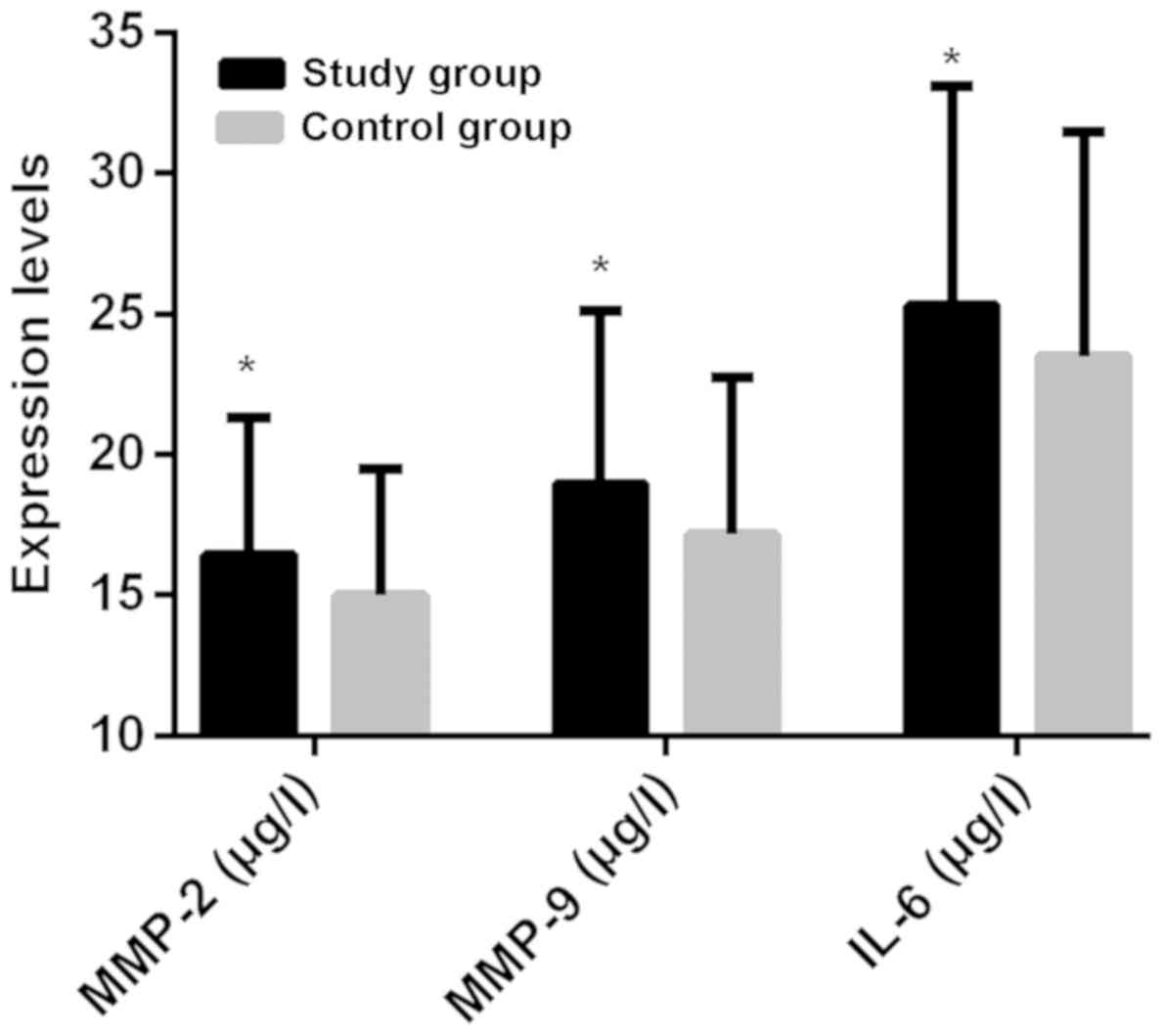
Comparison of the expression levels of
MMP-2, MMP-9 and IL-6 between the study and the control group
The expression levels of MMP-2 in the study and the
control group were 16.45±4.87 and 14.98±4.51 µg/l, respectively.
MMP-2 expression level in the study group was higher than that in
the control group, and the difference was statistically significant
(t=2.960, P=0.003). The expression levels of MMP-9 in the study and
the control group were 18.96±6.16 and 17.15±5.58 µg/l,
respectively. MMP-9 expression level in the study group was higher
than that in the control group, and the difference was
statistically significant (t=2.911, P=0.004). The expression levels
of IL-6 in the study and the control group were 25.31±8.55 and
23.51±7.97 µg/l, respectively. IL-6 expression level in the study
group was higher than that in the control group, and the difference
was statistically significant (t=2.058, P=0.040) (Fig. 1).
Expression levels of MMP-2, MMP-9 and
IL-6 in the patients of the study group with injury degree of
different sclerotins in CT manifestations
i) The levels of MMP-2 in the serum of the patients
whose CT manifestation showed that the ossicular chain was injured
were significantly higher than those in the serum of the patients
whose malleus or incus was injured, and the differences were
statistically significant (t=2.664, P=0.009). MMP-2 levels in the
serum of the patients whose ossicular chain was injured were
significantly higher than those in the serum of the patients with
intact ossicular chain, and the differences were statistically
significant (t=7.140, P<0.001). Also, the levels of MMP-2 in the
serum of the patients whose malleus or incus was injured were
higher than those in the serum of the patients with intact
ossicular chain, and the differences were statistically significant
(t=4.949, P<0.001).
ii) MMP-9 levels in the serum of the patients whose
CT manifestation showed that ossicular chain was injured were
significantly higher than those in the serum of the patients whose
malleus or incus was injured, and the differences were
statistically significant (t=3.252, P=0.002). The levels of MMP-9
in the serum of the patients whose ossicular chain was injured were
significantly higher than those in the serum of the patients with
intact ossicular chain, and the differences were statistically
significant (t=6.352, P<0.001). Also, the levels of MMP-9 in the
serum of the patients whose malleus or incus was injured were
higher than those in the serum of the patients with intact
ossicular chain, and the differences were statistically significant
(t=3.033, P=0.003).
iii) IL-6 levels in the serum of the patients whose
CT manifestation showed that ossicular chain was injured were
significantly higher than those in the serum of the patients, whose
malleus or incus was injured, and the differences were
statistically significant (t=3.431, P=0.001). The levels of IL-6 in
the serum of the patients whose ossicular chain was injured were
significantly higher than those in the serum of the patients with
intact ossicular chain, and the differences were statistically
significant (t=5.839, P<0.001). Moreover, the levels of IL-6 in
the serum of the patients whose malleus or incus was injured were
higher than those in the serum of the patients with intact
ossicular chain, and the differences were statistically significant
(t=2.646, P=0.009) (Table II).
| Table II.Expression levels of MMP-2, MMP-9 and
IL-6 in patients of the study group with injury degree of different
sclerotins in CT manifestations. |
Table II.
Expression levels of MMP-2, MMP-9 and
IL-6 in patients of the study group with injury degree of different
sclerotins in CT manifestations.
| Item | Ossicular chain is
intact (n=58) | Malleus or incus is
injured (n=61) | Ossicular chain is
injured (n=57) | F value | P-value |
|---|
| MMP-2 (µg/l) | 11.64±4.57 | 15.76±4.51a |
18.19±5.25a,b | 27.640 | <0.001 |
| MMP-9 (µg/l) | 16.29±5.41 |
19.35±7.38a |
23.64±6.92a,b | 17.790 | <0.001 |
| IL-6 (mg/l) | 19.57±8.39 |
23.92±9.48a |
30.61±11.65a,b | 18.080 | <0.001 |
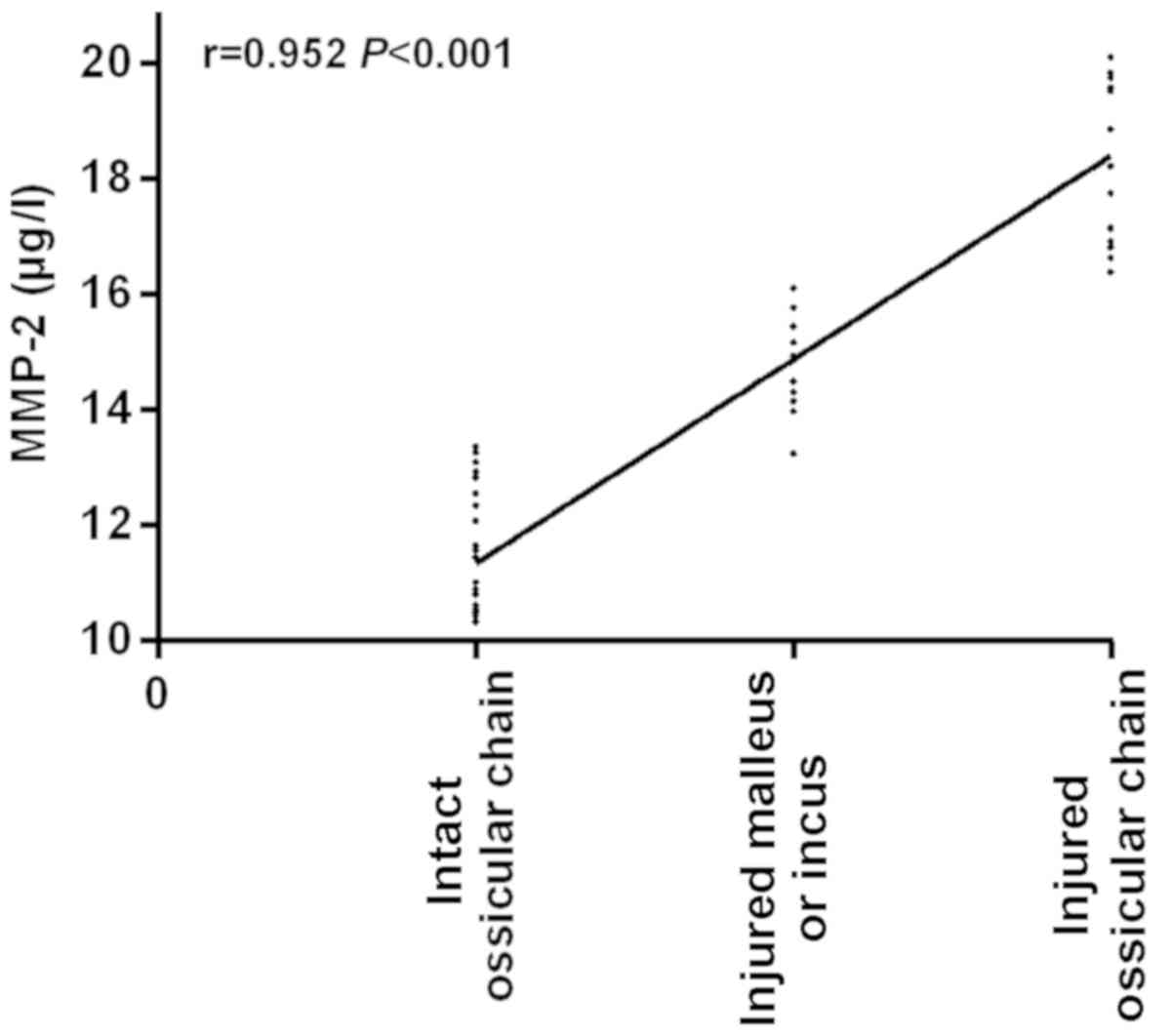
Correlation analysis between the
injury degree of different sclerotins in CT manifestations of the
patients in the study group and MMP-2
Spearman's correlation analysis showed that MMP-2
expression is positively correlated with the injury degree of
different sclerotins in CT manifestations of the patients in the
study group (r=0.952, P<0.001; Fig.
2).
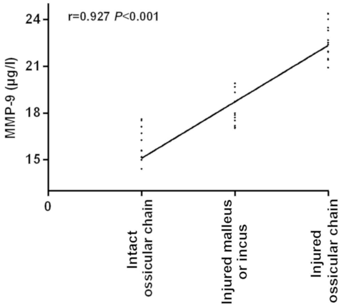
Correlation analysis between the
injury degree of different sclerotins in CT manifestations of the
patients in the study group and MMP-9
The injury degree of different sclerotins in CT
manifestations of the patients in the study group was positively
correlated with the expression levels of MMP-9 (r=0.927,
P<0.001), as shown in Fig. 3.
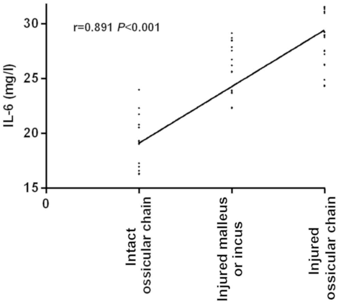
Correlation analysis between the
injury degree of different sclerotins in CT manifestations of the
patients in the study group and IL-6
An obvious positive correlation was observed between
the injury degree of different sclerotins in CT manifestations of
the patients in the study group and IL-6 (r=0.891, P<0.001), as
shown in Fig. 4.
Discussion
Cholesteatomatous otitis media is a type of
cholesteatoma disease, whose morbidity accounts for 0.5–1.8% of the
morbidity of all brain tumors (14).
In most patients, it occurs after adulthood. The peak age of
patients who suffer from this disease ranges from 40 to 45 years
old, and patients aging from 25 to 50 years old correspond to the
72% of patients with this disease (15,16). A
large number of researches have reported that if cholesteatomatous
otitis media is not treated on time, the cholesteatoma will
continue to increase, compress sclerotins and be absorbed, and the
auditory ossicle will be injured. If bacteria enter into cranium,
serious intracranial complications will occur, which may even lead
to the patients death (17–19). There are no obvious specific symptoms
in the early stage of cholesteatomatous otitis media, and its
symptoms are easily confused with the clinical symptoms of patients
with simple otitis media. Also, the primary site of
cholesteatomatous otitis media is often concealed and extensive,
which brings some difficulties to clinical diagnosis (20).
This study analyzed the clinical data of 176
patients with cholesteatomatous otitis media and 181 patients with
simple otitis media. The expression levels of MMP-2, MMP-9 and IL-6
in the patients of the two groups were analyzed and compared, and
their correlation with the injury degree of different sclerotins in
CT manifestations was investigated. Firstly, it was found that the
expression levels of MMP-2 and MMP-9 in the serum of patients with
cholesteatomatous otitis media were higher than those in the
patients with simple otitis media. Also, the levels of MMP-2 and
MMP-9 in the serum of the patients whose ossicular chain was
injured were significantly higher than those in the serum of the
patients whose malleus or incus was injured and significantly
higher than those in the serum of the patients with intact
ossicular chain; and MMP-2 and MMP-9 levels in the serum of the
patients whose malleus or incus was injured were significantly
higher than those in the serum of the patients with intact
ossicular chain. Moreover, the injury degree of different
sclerotins was found to be significantly and positively correlated
with MMP-2 and MMP-9 and the differences were statistically
significant. MMP-2 and MMP-9 are the main proteases involved in the
degradation of extracellular matrix and basement membrane in
vitro and in vivo, they facilitate the release of
angiogenic factors in the body, resulting in the proliferation of
cholesteatoma epithelial cells and the injury of sclerotins
(21,22). As the injury degree of sclerotins
increases, the expression levels of MMP-2 and MMP-9 also increase,
so MMP-2 and MMP-9 are closely related to the occurrence and
development of cholesteatoma (23).
According to the study report of Banerjee et al (24), the expression levels of MMP-2 and
MMP-9 in patients with extensive cholesteatoma are significantly
higher than those in patients with localized cholesteatoma, in
consistency with the above results.
In the present study, the expression levels of IL-6
in the serum of patients with cholesteatomatous otitis media were
found to be higher than those of patients with simple otitis media.
Also, IL-6 levels in the serum of patients whose ossicular chain
was injured were significantly higher than those in the serum of
patients whose malleus or incus was injured and those in the serum
of patients with intact ossicular chain. The levels of IL-6 in the
serum of patients whose malleus or incus was injured were also
significantly higher than those in the serum of patients with
intact ossicular chain. The injury degree of different sclerotins
was significantly and positively correlated with IL-6 and the
differences were statistically significant. As known, tissue is
stimulated after patients get the disease, releasing a large number
of inflammatory factors in the reaction mechanism of the patients'
body. Macrophages, B and T lymphocytes can further induce the
cascade reaction of cytokines and induce the production of other
cytokines and chemokines, as well as stimulate the release of IL-6
and cause a series of inflammatory chain reactions (25,26).
According to the reports of Liu et al (27), IL-6 plays a key role in the
pathogenesis of cholesteatoma, which further supports the results
of our experiment.
In this study, due to the limited medical resources
in Changxing People's Hospital, the number of the selected subjects
was small. A further limitation is the differences in the response
of patients with different ages after anesthesia and surgery. Thus,
longer-term follow-up surveys for the subjects in this study, will
be conducted to confirm the findings of the present study.
In summary, MMP-2, MMP-9 and IL-6 are highly
expressed in the serum of patients with cholesteatomatous otitis
media, and are significantly and positively correlated with the
severity thereof. This is of great significance for the prevention
and treatment of cholesteatomatous otitis media, and may be
beneficial for clinic.
Acknowledgements
Not applicable.
Funding
No funding was received.
Availability of data and materials
The datasets used and/or analyzed during the present
study are available from the corresponding author on reasonable
request.
Authors' contributions
YW and XT performed ELISA. WS and YL recorded and
analyzed the CT examination results. YW wrote the manuscript. All
authors read and approved the final manuscript.
Ethics approval and consent to
participate
The study was approved by the Ethics Committee of
Changxing People's Hospital (Huzhou, China). Patients who
participated in this research had complete clinical data. Signed
written informed consents were obtained from the patients and/or
guardians.
Patient consent for publication
Not applicable.
Competing interests
The authors declare that they have no competing
interests.
References
|
1
|
Abramson M and Huang CC: Localization of
collagenase in human middle ear cholesteatoma. Laryngoscope.
87:771–791. 1977. View Article : Google Scholar : PubMed/NCBI
|
|
2
|
Yung M, Tono T, Olszewska E, Yamamoto Y,
Sudhoff H, Sakagami M, Mulder J, Kojima H, İncesulu A, Trabalzini
F, et al: EAONO/JOS joint consensus statements on the definitions,
classification and staging of middle ear cholesteatoma. J Int Adv
Otol. 13:1–8. 2017. View Article : Google Scholar : PubMed/NCBI
|
|
3
|
Morita Y, Yamamoto Y, Oshima S, Takahashi
K and Takahashi S: Pediatric middle ear cholesteatoma: The
comparative study of congenital cholesteatoma and acquired
cholesteatoma. Eur Arch Otorhinolaryngol. 273:1155–1160. 2016.
View Article : Google Scholar : PubMed/NCBI
|
|
4
|
Xie S, Wang X, Ren J and Liu W: The role
of bone resorption in the etiopathogenesis of acquired middle ear
cholesteatoma. Eur Arch Otorhinolaryngol. 274:2071–2078. 2017.
View Article : Google Scholar : PubMed/NCBI
|
|
5
|
Glikson E, Yousovich R, Mansour J, Wolf M,
Migirov L and Shapira Y: Transcanal endoscopic ear surgery for
middle ear cholesteatoma. Otol Neurotol. 38:e41–e45. 2017.
View Article : Google Scholar : PubMed/NCBI
|
|
6
|
Locketz GD, Li PM, Fischbein NJ,
Holdsworth SJ and Blevins NH: Fusion of computed tomography and
PROPELLER diffusion-weighted magnetic resonance imaging for the
detection and localization of middle ear cholesteatoma. JAMA
Otolaryngol Head Neck Surg. 142:947–953. 2016. View Article : Google Scholar : PubMed/NCBI
|
|
7
|
Lingam RK and Bassett P: A meta-analysis
on the diagnostic performance of non-echoplanar diffusion-weighted
imaging in detecting middle ear cholesteatoma: 10 years on. Otol
Neurotol. 38:521–528. 2017. View Article : Google Scholar : PubMed/NCBI
|
|
8
|
Rogha M, Hashemi SM, Mokhtarinejad F,
Eshaghian A and Dadgostar A: Comparison of preoperative temporal
bone CT with intraoperative findings in patients with
cholesteatoma. Iran J Otorhinolaryngol. 26:7–12. 2014.PubMed/NCBI
|
|
9
|
Robert S, Gicquel T, Victoni T, Valença S,
Barreto E, Bailly-Maître B, Boichot E and Lagente V: Involvement of
matrix metalloproteinases (MMPs) and inflammasome pathway in
molecular mechanisms of fibrosis. Biosci Rep. 36:362016. View Article : Google Scholar
|
|
10
|
Kim M, Park SC, Baek I and Chun J:
Large-scale evaluation of experimentally determined DNA G+C
contents with whole genome sequences of prokaryotes. Syst Appl
Microbiol. 38:79–83. 2015. View Article : Google Scholar : PubMed/NCBI
|
|
11
|
Schmidt-Arras D and Rose-John S: IL-6
pathway in the liver: From physiopathology to therapy. J Hepatol.
64:1403–1415. 2016. View Article : Google Scholar : PubMed/NCBI
|
|
12
|
Rysz J, Banach M, Stolarek RA, Pasnik J,
Cialkowska-Rysz A, Koktysz R, Piechota M and Baj Z: Serum matrix
metalloproteinases MMP-2 and MMP-9 and metalloproteinase tissue
inhibitors TIMP-1 and TIMP-2 in diabetic nephropathy. J Nephrol.
20:444–452. 2007.PubMed/NCBI
|
|
13
|
Trinidade A, Page JC and Dornhoffer JL:
Therapeutic mastoidectomy in the management of noncholesteatomatous
chronic otitis media: Literature review and cost analysis.
Otolaryngol Head Neck Surg. 155:914–922. 2016. View Article : Google Scholar : PubMed/NCBI
|
|
14
|
Tono T, Sakagami M, Kojima H, Yamamoto Y,
Matsuda K, Komori M, Hato N, Morita Y and Hashimoto S: Staging and
classification criteria for middle ear cholesteatoma proposed by
the Japan Otological Society. Auris Nasus Larynx. 44:135–140. 2017.
View Article : Google Scholar : PubMed/NCBI
|
|
15
|
Matsuda K, Tono T, Kojima H, Yamamoto Y,
Sakagami M, Mishiro Y, Hinohira Y and Okuno T: Practicality
analysis of the staging system proposed by the Japan Otological
Society for acquired middle ear cholesteatoma: A multicenter study
of 446 surgical cases in Japan. Auris Nasus Larynx. 45:45–50. 2018.
View Article : Google Scholar : PubMed/NCBI
|
|
16
|
Rutkowska J, Özgirgin N and Olszewska E:
Cholesteatoma definition and classification: A literature review. J
Int Adv Otol. 13:266–271. 2017. View Article : Google Scholar : PubMed/NCBI
|
|
17
|
Suzuki H, Ikezaki S, Imazato K, Koizumi H,
Ohbuchi T, Hohchi N and Hashida K: Partial mastoid obliteration
combined with soft-wall reconstruction for middle ear
cholesteatoma. Ann Otol Rhinol Laryngol. 123:571–575. 2014.
View Article : Google Scholar : PubMed/NCBI
|
|
18
|
Blanco P, González F, Holguín J and Guerra
C: Surgical management of middle ear cholesteatoma and
reconstruction at the same time. Colomb Med (Cali). 45:127–131.
2014.PubMed/NCBI
|
|
19
|
Zhu Z, Hong Y, Wang Y, He G and Ye S: The
significance of keratinocyte in hyperproliferation of middle ear
cholesteatoma. Lin Chung Er Bi Yan Hou Tou Jing Wai Ke Za Zhi.
30:139–143. 2016.(In Chinese). PubMed/NCBI
|
|
20
|
Akkari M, Gabrillargues J, Saroul N,
Pereira B, Russier M, Mom T and Gilain L: Contribution of magnetic
resonance imaging to the diagnosis of middle ear cholesteatoma:
Analysis of a series of 97 cases. Eur Ann Otorhinolaryngol Head
Neck Dis. 131:153–158. 2014. View Article : Google Scholar : PubMed/NCBI
|
|
21
|
Suchozebrska-Jesionek D, Szymański M,
Kurzepa J, Gołabek W and Stryjecka-Zimmer M: Gelatinolytic activity
of matrix metalloproteinases 2 and 9 in middle ear cholesteatoma. J
Otolaryngol Head Neck Surg. 37:628–632. 2008.PubMed/NCBI
|
|
22
|
Schmidt M, Grünsfelder P and Hoppe F:
Up-regulation of matrix metalloprotease-9 in middle ear
cholesteatoma - correlations with growth factor expression in vivo?
Eur Arch Otorhinolaryngol. 258:472–476. 2001. View Article : Google Scholar : PubMed/NCBI
|
|
23
|
Zhu W, Xie Y and Wang P: Expression of
matrix metalloproteinasa-2,9 in cholesteatoma and middle ear
cancer. Zhonghua Er Bi Yan Hou Ke Za Zhi. 36:119–122. 2001.(In
Chinese). PubMed/NCBI
|
|
24
|
Banerjee AR, James R and Narula AA: Matrix
metalloproteinase-2 and matrix metalloproteinase-9 in cholesteatoma
and deep meatal skin. Clin Otolaryngol Allied Sci. 23:345–347.
1998. View Article : Google Scholar : PubMed/NCBI
|
|
25
|
Kuczkowski J, Sakowicz-Burkiewicz M,
Iżycka-Świeszewska E, Mikaszewski B and Pawełczyk T: Expression of
tumor necrosis factor-α, interleukin-1α, interleukin-6 and
interleukin-10 in chronic otitis media with bone osteolysis. ORL J
Otorhinolaryngol Relat Spec. 73:93–99. 2011. View Article : Google Scholar : PubMed/NCBI
|
|
26
|
Kumari N, Dwarakanath BS, Das A and Bhatt
AN: Role of interleukin-6 in cancer progression and therapeutic
resistance. Tumour Biol. 37:11553–11572. 2016. View Article : Google Scholar : PubMed/NCBI
|
|
27
|
Liu W, Xie S, Chen X, Rao X, Ren H, Hu B,
Yin T, Xiang Y and Ren J: Activation of the IL-6/JAK/STAT3
signaling pathway in human middle ear cholesteatoma epithelium. Int
J Clin Exp Pathol. 7:709–715. 2014.PubMed/NCBI
|