Introduction
Avascular necrosis of the femoral head (ANFH) is one
of the most diagnosed osteoarthritic diseases worldwide (1), it is a chronic disease that typically
leads to destruction of the hip joint in patients aged 30–60. It
has been estimated that there are 10,000-20,000 new cases of ANFH
each year in the USA (2–5). ANFH may be induced by a number of risk
factors, including vascular compression, hypertension, thrombosis,
alcoholism, chemotherapy and excessive glucocorticoid treatment;
however, the pathophysiology of ANFH remains unclear (6–8). Without
effective and prompt treatment, progression of the disease may
worsen joint dysfunction and even lead to permanent disability
(6). The majority of patients
exhibit dysfunction of the hip joints within 1–4 years of disease
development (9). Once the articular
surface of the femur collapses, treatment is extremely challenging
and usually ineffective.
Mesenchymal stem cells (MSCs) are adult stem cells
that are capable of self-renewal and divergence into multiple
lineages, including bone, cartilage, adipose tissue, muscle, tendon
and stroma (10). The ability of
MSCs to overcome cellular senescence demonstrates a resolution to
prolong life span and supports the study of MSC biogenesis on ANFH.
MSCs have been isolated from almost every tissue or organ of the
human body, including fat, bone marrow, blood, umbilical cord,
placenta, lung, skin and skeletal muscle (11). MSC based treatments are regarded as
novel methods of ANFH therapy due to the differentiation potential
of MSCs and the ease with which they are prepared (12).
Salvia miltiorrhiza (danshen) is a widely
used Chinese herbal medicine (13).
Danshen may hold promise as a sensitizing agent for chemotherapy
and radiotherapy to enhance the cytotoxic effects of anti-cancer
agents (14). A number of previous
studies have reported that danshen performs a variety of biological
activities, including antithrombous, antiplatelet aggregation,
moluscicide, antioxidant, antiviral and antitumor actions (14–17).
However, the underlying mechanisms behind how the active components
function still require clarification (15). The present study was performed to
investigate the potential therapeutic effect of danshen in
ANFH.
The aim of the present study was to establish a
rabbit model of methylprednisolone (MP)-induced ANFH using the
Shwartzman response and to explore the mechanism of MSCs migration
to the necrotic area of the femoral head. The local Shwartzman
response has traditionally been produced in rabbits using an
intradermal injection of endotoxin followed by an intravenous
injection of an agent which activates neutrophils 24 h later
(16). Initially, pathological
changes in the femoral head region were detected using imaging and
histopathology techniques, including magnetic resonance imaging
(MRI) and hematoxylin and eosin (H&E), Sudan III and
phosphotungstic acid hematoxylin (PATH) staining, then the number
of MSCs in the femoral head necrotic area was detected. The mRNA
expression of monocyte chemoattractant protein-1 (MCP-1) and
stromal cell-derived factor-1 (SDF-1) was measured using reverse
transcription polymerase chain reaction (RT-PCR). The expression of
bone morphogenetic protein-2 (BMP-2) and vascular endothelial
growth factor (VEGF) was also measured to determine the ability of
danshen in combination with MSCs to promote bone re-ossification
and revascularization for the treatment of ANFH. These results were
analyzed and the feasibility of clinical treatment using danshen
combined with MSCs transplantation for ANFH was evaluated.
Materials and methods
Experimental animals
A total of 72 male New Zealand rabbits (age, 24
weeks; weight, 2.5±0.2 kg) were purchased from the Experimental
Animal Center of Zhejiang University of Traditional Chinese
Medicine (Hangzhou, China). The animals were bred and maintained
under a 12 h light/dark cycle with ad libiutm access to food
and water. Room temperature and relative humidity were set at
25±3°C and 60±15%, respectively. All procedures and animal care
were approved by the Institutional Animal Care (Hangzhou, China)
and Use Ethical Review Committee of Wenzhou Medical University
(Wenzhou, China).
Isolation, culture, identification and
labeling of MSCs
MSCs from rabbits were isolated and cultured as
previously described (17). Briefly,
bone marrow was extracted from the rabbit femur and purified by
density gradient centrifugation (1,600 × g; 4°C; 25 min) using
Percoll solution (Sangon Biotech Co., Ltd., Shanghai, China). The
cells were cultured in Dulbecco's Modified Eagle medium (Gibco;
Thermo Fisher Scientific, Inc., Waltham, MA, USA) with 10% fetal
bovine serum (Gibco) and incubated at 37°C in humidified air with
5% CO2 for 24 h. Non-adherent cells were discarded when
the medium was refreshed, which was performed every 3–4 days.
Following culturing for three passages in vitro, the cells
were detached and incubated with the anti-rabbit antibodies, CD34
(1:300; cat. no. bs-2042R) and CD44 (1:300; cat. no. bs-0521R) at
4°C overnight. All antibodies were purchased from BIOSS (Beijing,
China). Cells were then observed under an inverted fluorescence
microscope following immunofluorescence staining at a magnification
of ×200. Following 3–4 passages, the MSCs were harvested and used
for cell implantation. Cultured MSCs were labeled with
5-bromo-2-deoxyuridine (BrdU; 10 mg/l; Sigma-Aldrich; Merck KGaA,
Darmstadt, Germany) overnight at 37°C according to the
manufacturer's protocol prior to transplantation. Following
labeling, the cells were washed five times with PBS to remove all
excess unbound BrdU. The MSCs were subsequently harvested
(~1×10⁷ cells per implantation) and resuspended in IMDM
media for further use.
Establishment of a rabbit model of
ANFH
A rabbit model of ANFH was established according to
the Shwartzman response method with modifications: 20 µg/kg
Escherichia coli endotoxin (LPS) was utilized instead of the
50 µg/kg dose utilized in the literature (18). Healthy New Zealand rabbits were
separated randomly into the normal (n=3) and model groups (n=7; 4
of 7 rabbits succumbed). A total of 20 µg/kg LPS (Sigma-Aldrich)
was injected through an ear vein to induce bone necrosis and this
process was repeated after 24 h. Following the second
administration of LPS, 20 mg/kg MP (Pharmacia and Upjohn; Pfizer
Inc, NY, USA) was injected through the gluteus muscle three times
with an interval of 24 h between each dose.
MRI
All rabbits were examined using a GE Signa EXCITE
1.5T MRI machine (GE Healthcare, Chicago, IL, USA). At 3 weeks an
orthogonal head coil was placed on the rabbits while they were
anesthetized, with its center located on the femoral head. The
layer thickness was 3 mm and the field of view was 240
mm2. The changes in femoral head signal and the
surrounding soft tissue were observed.
Histopathological assay
The rabbits were anesthetized using intravenous
administration with 3% sodium pentobarbital (30 mg/kg) in an ear
vein and the animals were subsequently sacrificed via air embolism.
The left femoral head was divided into two parts along the coronal
plane of the central hole and fixed in 10% formaldehyde solution
(Sangon Biotech Co., Ltd., Shanghai, China) at 4°C for 48 h,
decalcified for 3 months with 0.27 mol/l EDTA-2Na (Sangon Biotech
Co., Ltd.) at 25°C with pH 7.4. H&E and Sudan III (performed on
5 µm and 20 µm sections obtained from the same part of the femoral
head, respectively, at room temperature for 15 min), and PATH
staining (performed on 7 µm sections at room temperature for 24 h;
all Sigma-Aldrich; Merck KGaA) was performed to observe
histopathological changes using a light microscope (Olympus
Corporation, Tokyo, Japan). Following H&E staining, the number
of empty bone cells were counted at a magnification, ×400 to
calculate the empty bone trap rate. Following Sudan III staining,
the fat embolism number (number of blood vessels with fat
embolization in the femoral head cartilage) was counted by eye
using a microscope at magnification, ×200. Following PATH staining,
the thrombosis number was observed and the thrombosis rate was
calculated (thrombosis number/total blood vessels).
RT-PCR
The rabbits in the model group were sacrificed at 1,
2 and 3 weeks following the final injection of MP. RNA was
extracted from the right femoral head using a Qiagen RNeasy Mini
kit, which included the RNA buffer (cat. no. 74104; Qiagen, Inc.,
Valencia, CA, USA) after the bone was milled (SPEX 6870 Freezer
Mill; SPEX SamplePrep, Metuchen, NJ, USA) in liquid nitrogen.
Samples were obtained from the femoral heads of the normal group
and animals were sacrificed at the same time points as the model
group. The sequences of rabbit MCP-1, SDF-1, BMP, VEGF and β-actin
genes were obtained from GenBank (https://www.ncbi.nlm.nih.gov/nuccore). The primer
sequences used are listed in Table
I. The corresponding primers were synthesized by Sangon Biotech
Co., Ltd. (Shanghai, China).
 | Table I.Primer sequences for reverse
transcription-quantitative polymerase chain reaction. |
Table I.
Primer sequences for reverse
transcription-quantitative polymerase chain reaction.
| Gene | Direction | Sequence
(5′-3′) |
|---|
| MCP-1 | F |
CCGCCAGGTGGGCTAATA |
|
| R |
AGCAAGCAGAGCGAGGGT |
| SDF-1 | F |
CCCACCATCTACTCCATCA |
|
| R |
GAAATCGGGAATAGTCAGC |
| VEGF | F |
ATGGCAGAAGAAGGAGACA |
|
| R |
GCCCTGGTGAGGTTTGAT |
| BMP-2 | F |
GGAAGAACTGCCAGAAAC |
|
| R |
GACCTGCTAATCCTCACG |
| β-actin | F |
TCCTGCGTCTGGACCTGG |
|
| R |
GCCCGACTCGTCATACTCC |
Following RNA extraction from the right femoral head
a PrimeScript™ 1st Strand cDNA Synthesis kit (Takara Bio, Inc.,
Otsu, Japan) was used to reverse transcribe the RNA into cDNA
according to the manufacturers' protocol. RT-PCR was performed
according to the following thermocycling conditions: 94°C for 3
min, followed by 40 cycles of 94°C for 30 sec, 52°C for 30 sec and
72°C for 20 sec. The relative quantification of each gene mRNA was
calculated relative to the β-actin mRNA.
Immunohistochemistry assay
The 24 model rabbits were randomly divided into the
following four groups (n=6 in each): Danshen (cat. no.
Z33020177-2009; lot no. 0908213; Chiatai Qingchun Bao
Pharmaceutical Co. Ltd., Hangzhou China) injection group (2 ml per
rabbit), the MSCs group (1×10⁷ cells per rabbit), the
danshen combined with MSCs group (2 ml danshen + 1×10⁷
MSCs per rabbit) and the model group, which were administered
physiological saline (2 ml per rabbit). The BrdU-labeled MSCs were
extracted from rabbits through density gradient centrifugation and
adherence screening at the third generation as described above.
BrdU has been previously confirmed to have no inhibiting effect on
MSC proliferation (19). The rabbits
were administered their respective treatments in two sides of the
femoral artery at 4 weeks following the final injection of MP. All
rabbits were administered penicillin 3 days later to prevent
infection following treatment. At 3 and 6 weeks following
treatment, 3 rabbits from each group were sacrificed.
Immunohistochemistry analysis was used to determine the protein
expression of VEGF and BMP-2 and the number of MSCs labeled with
BrdU to evaluate the ability of MSCs to migrate to the necrotic
area. Femoral head samples were fixed in 10% formaldehyde for 48 h
at room temperature, cut into 3–4 µm sections and embedded in
paraffin (Sangon Biotech Co., Ltd.). For VEGF, BMP-2 and BrdU
immunohistochemical staining, femoral head sections were
deparaffinized in xylene and dehydrated with graded ethanol. After
washing with distilled water, tissue peroxidase was blocked using
3.0% hydrogen peroxide in methanol for 10 min at room temperature.
For antigen retrieval using citric acid buffer, slides were heated
at 120°C for 20 min and then cooled for 15 min at room temperature.
Following washing with PBS solution, slides were incubated with
VEGF (1:100; cat no. SC-7269; Santa Cruz Biotechnology, Inc.,
Dallas, TX, USA), BMP-2 (1:100; cat no. SAB1411278) and BrdU
(1:100; cat. no. B2531; both Sigma-Aldrich; Merck KGaA) antibodies
at 37°C for 1 h. Samples were then washed with PBS in triplicate
and incubated with anti-mouse horseradish peroxidase (HRP)
conjugated immunoglobulin G (IgG; cat. no. 7076) or anti-rabbit
HRP-conjugated IgG antibodies (cat. no. 7074; both CST Biological
Reagents Co., Ltd., Shanghai, China) diluted (1:2,000) by Antibody
Diluent Reagent Solution (cat. no. 1956331A; Thermo Fisher
Scientific, Inc.) for 30 min at 37°C. Any immune reaction was
detected using 3,3′-diaminobenzidine. Sections were then
counterstained at room temperature for 10 min using Meyer's
hematoxylin, dehydrated and mounted. The known positive samples
were then utilized as positive controls. Following
immunohistochemical staining using the Histostain-Plus kit (Mai Bio
Co, Ltd., China), the cell number was counted under a light
microscope at magnification, ×400.
Statistical analysis
All statistical analyses were performed using SPSS
software, version 13 (SPSS, Inc., Chicago, IL, USA). Data are
presented as the mean ± standard deviation and all tests were
performed in triplicate. Statistical comparisons between two groups
were made using Student's t-test. One-way analysis of variance
followed by a post hoc Tukey's test was used to analyze differences
among multiple groups. P<0.05 was considered to indicate a
statistically significant difference.
Results
MRI
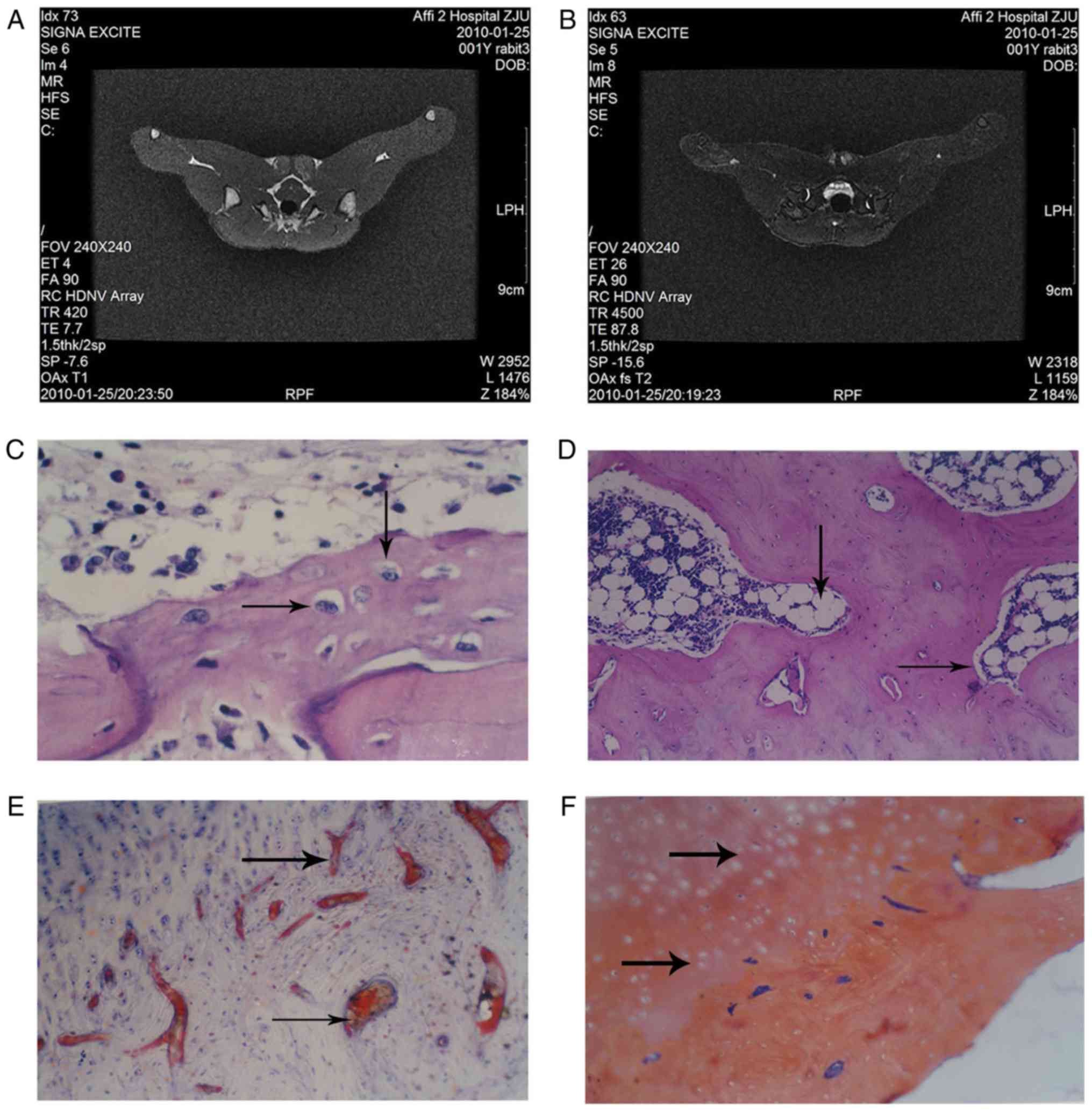
There was no clear abnormality observed in the
normal group femoral heads (Fig.
1A). In T2 weighted imaging, MRI revealed a normal joint space
of the femoral head in the rabbit model and an infusion in the
joint cavity. The right femoral head exhibited scattered spots in
the medium-high signal region and inhomogeneous low signal mixing.
MRI images of the model group revealed that the femoral head was
not smooth (Fig. 1B).
Histopathology results
H&E staining revealed that in the normal group,
the trabecular bone was complete and arranged, the bone marrow was
rich in hematopoietic cells and osteoblasts were observed on the
surface of the trabecular bone (Fig.
1C). Cancellous bone near the femoral neck had undergone bone
necrosis at week 4 in the model group; this was accompanied by
neutrophil aggregation (Fig.
1D).
Sudan III staining demonstrated that there was no
notable steatosis or lipid embolism in the liver and kidneys in the
model group (Fig. 1E). Lipid
droplets filled with cellular fluid were observed and the nucleus
appeared misshapen under pressure. In the normal group, the lipid
droplets in the bone cells were not clear, the fat cells in the
bone marrow were smaller compared with the model group and the
hematopoietic cells were observed in the voids of the bone marrow
(data not shown).
The model group exhibited a large number of fibrin
embolus in the femoral head cartilage, which was observed by PATH
staining (Fig. 1F). The presence of
fibrin embolus was less common in the control group (data not
shown).
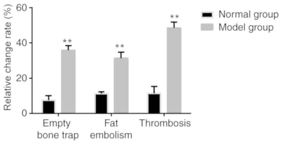
The empty bone rate, fat embolism rate and
thromboembolism rate were calculated according to the
aforementioned results and are presented in Fig. 2. The empty bone, fat embolism and
thromboembolism rates were all significantly increased in the model
group compared with the normal group (P<0.05).
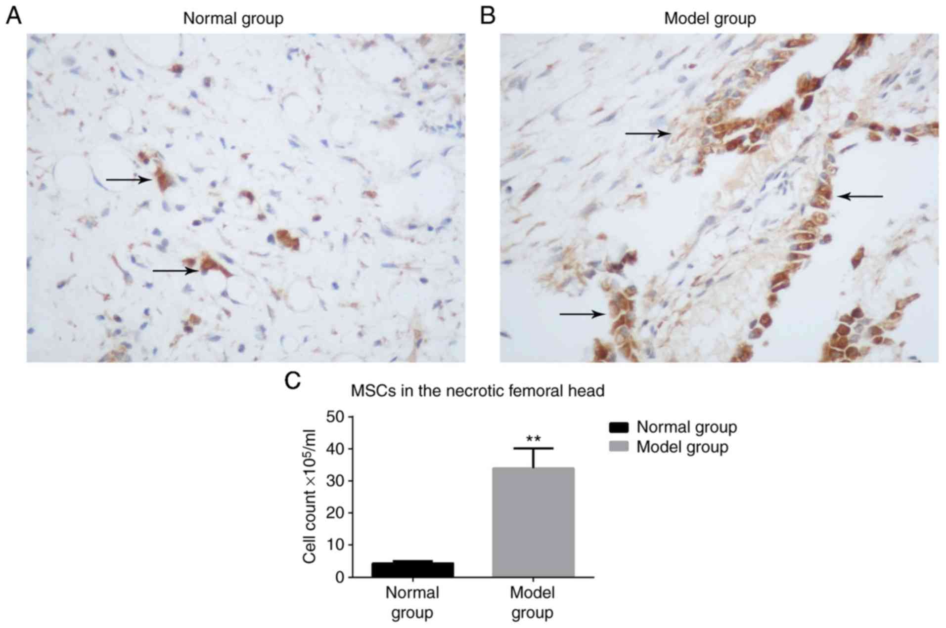
BrdU-labeled MSCs are increased in the
model group compared with the normal group
At 3 weeks, few BrdU-labeled MSCs were detected in
the normal group (Fig. 3A), whereas
a greater number of BrdU-labeled MSCs were observed in the model
group (Fig. 3B). The nuclei were
stained brown. There was a significantly higher number of MSCs
observed in the model group compared with the normal group
(P<0.05; Fig. 3C).
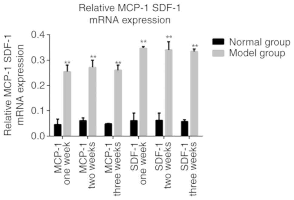
Expression of MCP-1 and SDF-1 are
significantly increased in the model group
RT-PCR revealed that the chemokine expression of
MCP-1 and SDF-1 was significantly increased in the model group
compared with the normal group at all time points measured
(P<0.05; Fig. 4). These results
suggest that in the necrotic femoral head MCP-1 and SDF-1 mRNA
expression was increased and continued for an extended period of
time.
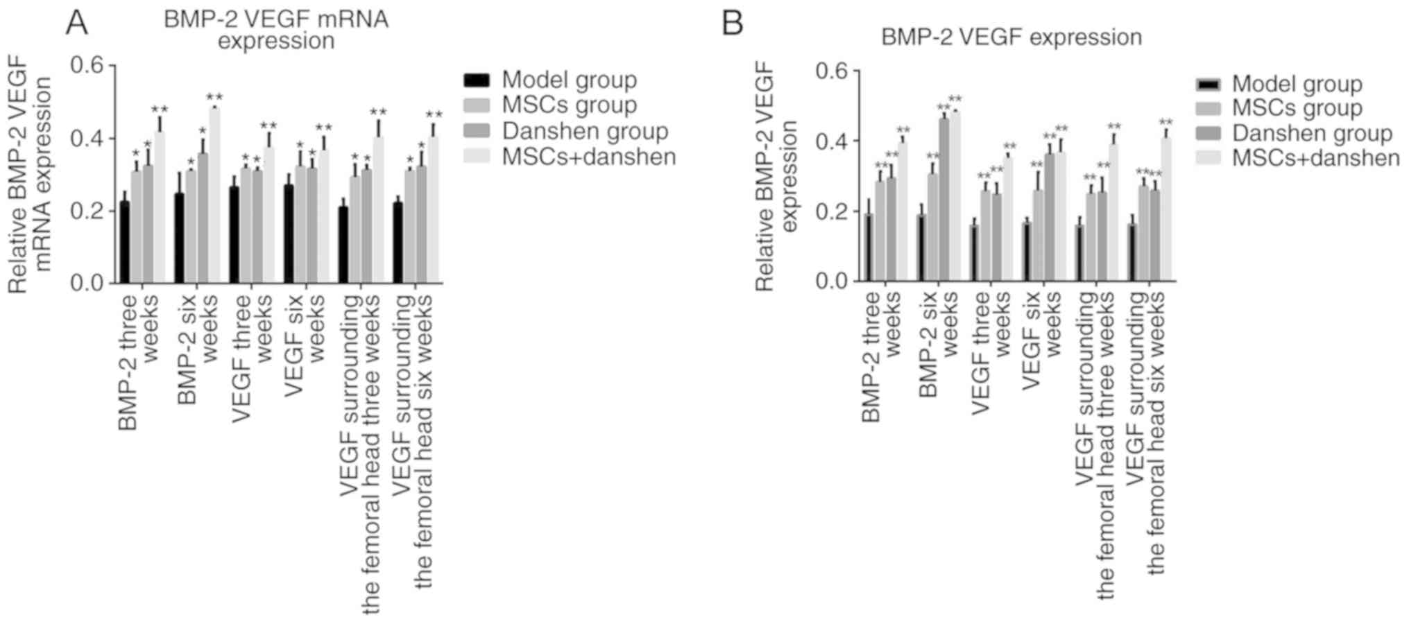
Expression of BMP-2 and VEGF
At 3 weeks following the final administration of
MSCs and danshen, the mRNA expression of BMP-2 and VEGF in the
femoral head and surrounding area was significantly increased in
the groups treated with MSCs, danshen, or a combination of the two
compared with the model group (P<0.05; Fig. 5), Similar results were observed at 6
weeks. There was no significant difference observed between the
results at 3 weeks and the results at 6 weeks. These results
suggest that the expression of BMP-2 and VEGF mRNA is increased by
treatment with danshen or MSCs, but this difference is not
time-dependent. The group that received MSC in combination with
danshen demonstrated a significant increase in the level of BMP-2
compared with the single treatment groups (P<0.05). The mRNA
expression results were consistent with the immunohistochemistry
results.
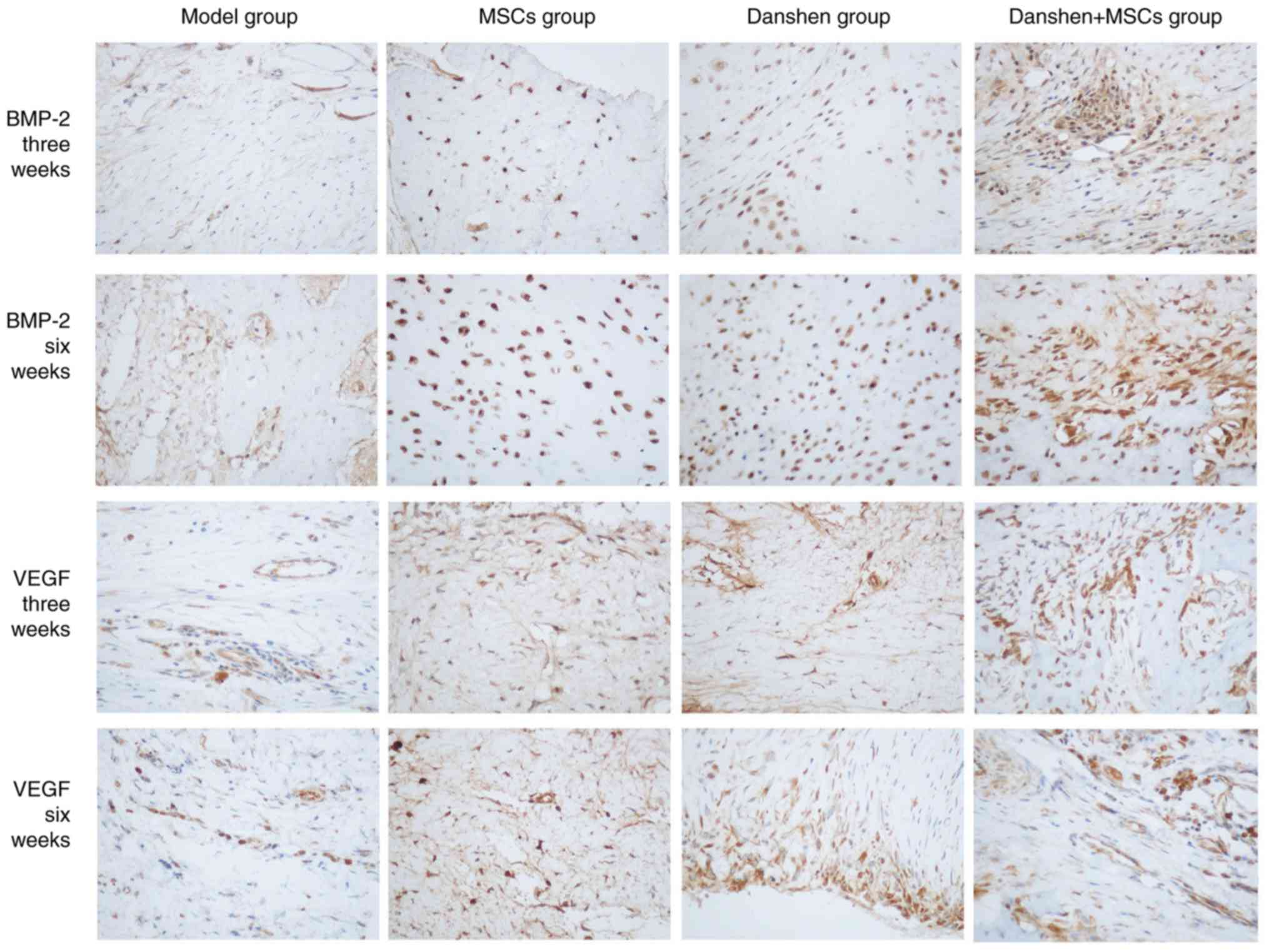
Protein expression of MCP-2 and VEGF
in the femoral head
Immunohistochemistry staining was performed on
tissue samples from each treatment group at 3 and 6 weeks following
treatment (Fig. 6) The
immunohistochemistry results revealed that the protein expression
rates of BMP-2 and VEGF in the femoral head at 3 and 6 weeks
following treatment were significantly higher in all groups treated
with MSCs and/or danshen compared with the model group (P<0.05;
Fig. 5B). However there was no
significant difference between the BMP-2 and VEGF expression in all
of the groups at week 3 compared with week 6. This suggests that
the expression of BMP-2 and VEGF may be increased by the injection
of danshen and MSCs, either individually or in combination, but
this is not time-dependent. The positive expression rate of BMP-2
and VEGF in the MSCs and danshen group was significantly increased
compared with that of the MSCs or danshen only groups (P<0.05).
At week 6 the MSCs with danshen group had a significantly increased
BMP-2 expression compared with week 3 (P<0.05), however this was
not observed for VEGF expression. This suggests that the effect of
danshen combined with MSCs was higher compared with a single
administration of either danshen or MSCs and that BMP-2 expression
increased over an extended period of time.
Discussion
The rabbit model of hormone-induced ANFH has been
used to study ANFH for a number of years. At present, there are
many different types of model available to study ANFH including the
serological necrosis model and Shwartzman response model (18–21). As
ANFH is not reversible it is important to develop novel therapeutic
agents for its early diagnosis and treatment. Previously, hormones
have been used to induce ANFH with limited success (22). In the present study, the improved
Shwartzman response model (18) was
used to develop ANFH via administration of MP following LPS, which
induced experimental femoral head necrosis with a low mortality
rate. These aforementioned improved models are of great value in
the future study of the ANFH. In clinical practice, MRI is the most
accurate imaging method available and is particularly useful to
diagnose ANFH at its early stage when changes are only visible in
the bone marrow (23). In the
present study MRI scans revealed an edema and a spot-like high
level signal in the right femoral head. These results are
consistent with the image characteristics of ANFH (24). The histopathological methodologies
used in the present study, including H&E, Sudan Ш and PATH
staining revealed that the rabbit model of ANFH was successfully
established.
Chemokines are small molecular weight proteins (8–13
kDa) and are categorized into four different families (CC, CXC,
CX3C and C) based on the presence of NH2 terminal
cysteine motifs (24). Chemokine
receptors are typical G protein-coupled transmembrane proteins
(24). When the associated chemokine
ligand binds with the chemokine receptor, Gα1 and Gβ-γ subunits
dissociate and activate phosphatidylinositol 3-kinase and small Rho
guanosine triphosphatase, which leads to cellular calcium influx
(25). Chemokine receptors are
typically expressed by leukocyte subsets and immune cells generally
carry different sets of chemokine receptors (26). The roles of chemokines relevant to
orthopedic implant debris includes pro-inflammatory cytokine
production, pyroptosis, apoptosis, angiogenesis and collagen
production, which act together to product aseptic bone reabsorption
around implants (27). In the
present study RT-PCR revealed that the expression of MCP-1 and
SDF-1 mRNA was significantly increased in the model group compared
with the normal group, which may be associated with the repair of
the femoral head as a previous study revealed that MCP-1 mRNA and
SDF-1 may attract MSCs to the necrotic area by chemotaxis and then
use the MSCs to repair injury (25).
However, the specific repair mechanisms require further study to be
fully understood.
Osteoblasts, osteoclasts and mesenchymal stem cells
secrete BMP-2 and VEGF for bone growth and repair. Previous studies
have demonstrated that the combined use of BMP-2 and VEGF is
superior to their use alone, as they have a synergistic effect in
promoting bone regeneration and vascularization (28–30). In
the present study, immunohistochemical detection and RT-PCR
revealed that BMP-2 and VEGF had a significantly increased
expression in groups administered the combined treatment of danshen
and MSCs compared with the single treatment groups. The combined
use of danshen and MSCs may have synergistic effects in promoting
the attachment, proliferation, osteogenic and angiogenic activities
of MSCs. It was observed that treatment with MSCs or danshen
separately induced a significant increase in BMP-2 and VEGF
expression compared with the model group and the danshen and MSCs
group had significantly increased expression of BMP-2 and VEGF
compared with the single treatment groups. This indicates that the
effect of danshen combined with MSCs on femoral artery intervention
was better than danshen or MSCs treatment alone. The role of BMP-2
and VEGF in promoting bone and vessel formation in vivo may
be due to its induction of the proliferation and osteogenic and
angiogenic differentiation in bone MSCs. High expression of BMP-2
and VEGF may accelerate re-vascularization and re-ossification in
patients with ANFH.
Clinicians have suggested that an ideal treatment
should consider pain relief, preservation or restoration of the
integrity of the femoral head and prevention of deterioration of
the hip (31). Several treatment
methods and procedures have been performed to try and cure ANFH,
including core decompression (32–34),
electrical stimulation (35,36), transtrochanteric rotational osteotomy
(37), non-vascularized structural
grafting (38,39) and a vascularized bone graft (40,41).
However, none of these offer a complete cure (42,43) and
to the best of our knowledge there is no treatment currently
available that effectively reduces painful necrotic bone without
damaging native hip geometry. Therefore, the development of a
therapeutic treatment that is low risk, minimally invasive, low
cost, easily applied and effective is currently of major focus in
the orthopedic field. Due to developments in tissue engineering,
stem cell transplantation has become a good alternative for tissue
repair and is an applicable methodology for ANFH due to the ability
of MSCs to differentiate into osteoblasts, chondrocytes, muscle and
fat cells (44). To improve the
therapeutic effect and reduce trauma, the present study treated
ANFH with the co-transplantation of MSCs and danshen injection. The
aim of gene therapy for osteonecrosis disease is bone and vessel
regeneration and previous studies have confirmed that bone and
vessel regeneration is mediated by BMP-2 and VEGF, which are
critical for bone repair (28,45).
Previous studies have reported that BMP-2 and VEGF may promote
self-repair of necrotic femoral heads (46,47), in
the present study, it was observed that MSCs combined with Danshen
promoted BMP-2 and VEGF expression, which is regarded as a critical
factor to promote revascularization and re-ossification.
In the present study it was confirmed that LPS and
MP induce ANFH in a rabbit model. Additionally, following the
administration of MSCs and danshen into the femoral artery, a
significantly increased number of BrdU-labeled MSCs were observed
and there was a significant increase in the expression of MCP-1 and
SDF-1, BMP-2 and VEGF compared with the groups without treatment.
When transferred to a clinical setting, these results suggest that
MSCs combined with danshen may be used as a novel approach to treat
ANFH, as they increase the expression of BMP-2 and VEGF in the
necrotic bone area and therefore accelerate the process of
revascularization and re-ossification.
Acknowledgements
Not applicable.
Funding
The present study was supported by Wenzhou Medical
University (grant no. 2008YB011).
Availability of data and materials
The datasets used and/or analyzed during the current
study are available from the corresponding author on reasonable
request.
Authors' contributions
YW and CW designed the present study, and CZ, JW and
YH were responsible for data access and analysis. All authors
collaborated to interpret results and develop the manuscript.
Ethics approval and consent to
participate
All procedures and animal care were approved by the
Institutional Animal Care and Use Ethical Review Committee of
Wenzhou Medical University.
Patient consent for publication
Not applicable.
Competing interests
The authors declare that they have no competing
interests.
References
|
1
|
Zhao D, Cui D, Wang B, Tian F, Guo L, Yang
L, Liu B and Yu X: Treatment of early stage osteonecrosis of the
femoral head with autologous implantation of bone marrow-derived
and cultured mesenchymal stem cells. Bone. 50:325–330. 2012.
View Article : Google Scholar : PubMed/NCBI
|
|
2
|
Ohzono K, Saito M, Takaoka K, Ono K, Saito
S, Nishina T and Kadowaki T: Natural history of nontraumatic
avascular necrosis of the femoral head. J Bone Joint Surg Br.
73:68–72. 1991. View Article : Google Scholar : PubMed/NCBI
|
|
3
|
Arlet J: Nontraumatic avascular necrosis
of the femoral head. Past, present, and future. Clin Orthop Relat
Res. 277:12–21. 1992.
|
|
4
|
Mankin HJ: Nontraumatic necrosis of bone
(osteonecrosis). N Engl J Med. 326:1473–1479. 1992. View Article : Google Scholar : PubMed/NCBI
|
|
5
|
Roshan A and Ram S: The neglected femoral
neck fracture in young adults: Review of a challenging problem.
Clin Med Res. 6:33–39. 2008. View Article : Google Scholar : PubMed/NCBI
|
|
6
|
Ciapetti G, Granchi D, Fotia C, Savarino
L, Dallari D, Del Piccolo N, Donati DM and Baldini N: Effects of
hypoxia on osteogenic differentiation of mesenchymal stromal cells
used as a cell therapy for avascular necrosis of the femoral head.
Cytotherapy. 18:1087–1099. 2016. View Article : Google Scholar : PubMed/NCBI
|
|
7
|
Tabatabaee RM, Saberi S, Parvizi J,
Mortazavi SM and Farzan M: Combining Concentrated autologous bone
marrow stem cells injection with core decompression improves
outcome for patients with early-stage osteonecrosis of the femoral
head: A comparative study. J Arthroplasty. 30 (Suppl 9):S11–S15.
2015. View Article : Google Scholar
|
|
8
|
Song HM, Wei YC, Li N, Wu B, Xie N, Zhang
KM, Wang SZ and Wang HM: Effects of Wenyangbushen formula on the
expression of VEGF, OPG, RANK and RANKL in rabbits with
steroid-induced femoral head avascular necrosis. Mol Med Rep.
12:8155–8161. 2015. View Article : Google Scholar : PubMed/NCBI
|
|
9
|
Rajpura A, Wright AC and Board TN: Medical
management of osteonecrosis of the hip: A review. Hip Int.
21:385–392. 2011. View Article : Google Scholar : PubMed/NCBI
|
|
10
|
Zhai L, Sun N, Zhang B, Liu ST, Zhao Z,
Jin HC, Ma XL and Xing GY: Effects of focused extracorporeal shock
waves on bone marrow mesenchymal stem cells in patients with
avascular necrosis of the femoral head. Ultrasound Med Biol.
42:753–762. 2016. View Article : Google Scholar : PubMed/NCBI
|
|
11
|
Kim N and Cho SG: Clinical applications of
mesenchymal stem cells. Korean J intern Med. 28:387–402. 2013.
View Article : Google Scholar : PubMed/NCBI
|
|
12
|
Wen Q, Zhou C, Luo W, Zhou M and Ma L:
Pro-osteogenic effects of fibrin glue in treatment of avascular
necrosis of the femoral head in vivo by hepatocyte growth
factor-transgenic mesenchymal stem cells. J Transl Med. 12:1142014.
View Article : Google Scholar : PubMed/NCBI
|
|
13
|
Ye YT, Zhong W, Sun P, Wang D, Wang C, Hu
LM and Qian JQ: Apoptosis induced by the methanol extract of
salvia miltiorrhiza bunge in non-small cell lung cancer
through PTEN-mediated inhibition of PI3K/Akt pathway. J
Ethnopharmacol. 200:107–116. 2017. View Article : Google Scholar : PubMed/NCBI
|
|
14
|
Park CH, Shin SH, Lee EK, Kim DH, Kim MJ,
Roh SS, Yokozawa T and Chung HY: Magnesium lithospermate B from
salvia miltiorrhiza bunge ameliorates aging-induced renal
inflammation and senescence via NADPH oxidase-mediated reactive
oxygen generation. Phytother Res. 31:721–728. 2017. View Article : Google Scholar : PubMed/NCBI
|
|
15
|
Zhou L, Zuo Z and Chow MS: Danshen: An
overview of its chemistry, pharmacology, pharmacokinetics, and
clinical use. J Clin Pharmacol. 45:1345–1359. 2005. View Article : Google Scholar : PubMed/NCBI
|
|
16
|
Argenbright LW and Barton RW: The
Shwartzman response: A model of ICAM-1 dependent vasculitis. Agents
Actions. 34:208–210. 1991. View Article : Google Scholar : PubMed/NCBI
|
|
17
|
Xia CS, Zuo AJ, Wang CY and Wang YZ:
Isolation of rabbit bone marrow mesenchymal stem cells using
density gradient centrifugation and adherence screening methods.
Minerva Medica. 104:519–525. 2013.PubMed/NCBI
|
|
18
|
Yamamoto T, Hirano K, Tsutsui H, Sugioka Y
and Sueishi K: Corticosteroid enhances the experimental induction
of osteonecrosis on rabbits with Shwartzman reaction. Clin Orthop
Relat Res. 316:235–243. 1995. View Article : Google Scholar
|
|
19
|
Saeed H, Abdallah BM, Ditzel N,
Catala-Lehnen P, Qiu W, Amling M and Kassem M:
Telomerase-deficiency-related bone loss is caused by intrinsic
impairment of mesenchymal stem cell (MSC) functions and increased
osteoclastogenesis due to pro-inflammatory micro-environment. Bone.
47:S392010. View Article : Google Scholar
|
|
20
|
Wang D, Wang G, Liu M, Sun L, Zong W,
Jiang H, Zhang H, Li H, Gong J and Sun S: A novel animal model of
osteonecrosis of the femoral head induced using a magnetic
resonance imaging-guided argon-helium cryotherapy system. Exp Ther
Med. 7:1525–1528. 2014. View Article : Google Scholar : PubMed/NCBI
|
|
21
|
Wen Q, Ma L, Chen YP, Yang L, Luo W and
Wang XN: A rabbit model of hormone-induced early avascular necrosis
of the femoral head. Biomed Environ Sci. 21:398–403. 2008.
View Article : Google Scholar : PubMed/NCBI
|
|
22
|
Nowak DA and Yeung J: Steroid-induced
osteonecrosis in dermatology: A review. J Cutan Med Surg.
19:358–360. 2015. View Article : Google Scholar : PubMed/NCBI
|
|
23
|
Li Z, Liao W, Zhao Q, Liu M, Xia W, Yang Y
and Shao N: Angiogenesis and bone regeneration by allogeneic
mesenchymal stem cell intravenous transplantation in rabbit model
of avascular necrotic femoral head. J Surg Res. 183:193–203. 2013.
View Article : Google Scholar : PubMed/NCBI
|
|
24
|
Bartonicek J, Vavra J and Bartoska R:
Operative treatment of avascular necrosis of the femoral head after
slipped capital femoral epiphysis. Arch Orthop Trauma Surg.
131:497–502. 2011. View Article : Google Scholar : PubMed/NCBI
|
|
25
|
Zlotnik A and Yoshie O: Chemokines: A new
classification system and their role in immunity. Immunity.
12:121–127. 2000. View Article : Google Scholar : PubMed/NCBI
|
|
26
|
Fritz EA, Glant TT, Vermes C, Jacobs JJ
and Roebuck KA: Chemokine gene activation in human bone
marrow-derived osteoblasts following exposure to particulate wear
debris. J Biomed Mater Res A. 77:192–201. 2006. View Article : Google Scholar : PubMed/NCBI
|
|
27
|
Marra F and Tacke F: Roles for chemokines
in liver disease. Gastroenterology. 147:577–594, e571. 2014.
View Article : Google Scholar : PubMed/NCBI
|
|
28
|
Wasmuth HE, Tacke F and Trautwein C:
Chemokines in liver inflammation and fibrosis. Semin Liver Dis.
30:215–225. 2010. View Article : Google Scholar : PubMed/NCBI
|
|
29
|
Zhang C, Wang KZ, Qiang H, Tang YL, Li Q,
Li M and Dang XQ: Angiopoiesis and bone regeneration via
co-expression of the hVEGF and hBMP genes from an adeno-associated
viral vector in vitro and in vivo. Acta Pharmacol
Sin. 31:821–830. 2010. View Article : Google Scholar : PubMed/NCBI
|
|
30
|
Young S, Patel ZS, Kretlow JD, Murphy MB,
Mountziaris PM, Baggett LS, Ueda H, Tabata Y, Jansen JA, Wong M and
Mikos AG: Dose effect of dual delivery of vascular endothelial
growth factor and bone morphogenetic protein-2 on bone regeneration
in a rat critical-size defect model. Tissue Eng Part A.
15:2347–2362. 2009. View Article : Google Scholar : PubMed/NCBI
|
|
31
|
Zhang C, Ma J, Li M, Li XH, Dang XQ and
Wang KZ: Repair effect of coexpression of the hVEGF and hBMP genes
via an adeno-associated virus vector in a rabbit model of early
steroid-induced avascular necrosis of the femoral head. Transl Res.
166:269–280. 2015. View Article : Google Scholar : PubMed/NCBI
|
|
32
|
Aldridge JM III and Urbaniak JR: Avascular
necrosis of the femoral head: Role of vascularized bone grafts.
Orthop Clin North Am. 3813–22. (v)2007. View Article : Google Scholar : PubMed/NCBI
|
|
33
|
Fairbank AC, Bhatia D, Jinnah RH and
Hungerford DS: Long-term results of core decompression for
ischaemic necrosis of the femoral head. J Bone Joint Surg Br.
77:42–49. 1995. View Article : Google Scholar : PubMed/NCBI
|
|
34
|
Koo KH, Kim R, Ko GH, Song HR, Jeong ST
and Cho SH: Preventing collapse in early osteonecrosis of the
femoral head. A randomised clinical trial of core decompression. J
Bone Joint Surg Br. 77:870–874. 1995. View Article : Google Scholar : PubMed/NCBI
|
|
35
|
Mont MA, Carbone JJ and Fairbank AC: Core
decompression versus nonoperative management for osteonecrosis of
the hip. Clin Orthop Relat Res. 324:169–178. 1996. View Article : Google Scholar
|
|
36
|
Aaron RK, Lennox D, Bunce GE and Ebert T:
The conservative treatment of osteonecrosis of the femoral head. A
comparison of core decompression and pulsing electromagnetic
fields. Clin Orthop Relat Res. 249:209–218. 1989.
|
|
37
|
Bassett CA, Schink-Ascani M and Lewis SM:
Effects of pulsed electromagnetic fields on Steinberg ratings of
femoral head osteonecrosis. Clin Orthop Relat Res. 246:172–185.
1989.
|
|
38
|
Sugioka Y, Hotokebuchi T and Tsutsui H:
Transtrochanteric anterior rotational osteotomy for idiopathic and
steroid-induced necrosis of the femoral head. Indications and
long-term results. Clin Orthop Relat Res. 277:111–120. 1992.
|
|
39
|
Boettcher WG, Bonfiglio M and Smith K:
Non-traumatic necrosis of the femoral head. II. Experiences in
treatment. J Bone Joint Surg Am. 52:322–329. 1970. View Article : Google Scholar : PubMed/NCBI
|
|
40
|
Buckley PD, Gearen PF and Petty RW:
Structural bone-grafting for early atraumatic avascular necrosis of
the femoral head. J Bone Joint Surg Am. 73:1357–1364. 1991.
View Article : Google Scholar : PubMed/NCBI
|
|
41
|
Kirschenbaum IH, Vernace JV, Booth RE Jr,
Balderston RA and Rothman RH: Total hip arthroplasty for
osteonecrosis. Semin Arthroplasty. 2:234–240. 1991.PubMed/NCBI
|
|
42
|
Katz RL, Bourne RB, Rorabeck CH and McGee
H: Total hip arthroplasty in patients with avascular necrosis of
the hip. Follow-up observations on cementless and cemented
operations. Clin Orthop Relat Res. 281:145–151. 1992.
|
|
43
|
Plakseychuk AY, Kim SY, Park BC,
Varitimidis SE, Rubash HE and Sotereanos DG: Vascularized compared
with nonvascularized fibular grafting for the treatment of
osteonecrosis of the femoral head. J Bone Joint Surg Am 85-A.
589–596. 2003. View Article : Google Scholar
|
|
44
|
González Della Valle A, Bates J, Di Carlo
E and Salvati EA: Failure of free vascularized fibular graft for
osteonecrosis of the femoral head: A histopathologic study of 6
cases. J Arthroplasty. 20:331–336. 2005. View Article : Google Scholar : PubMed/NCBI
|
|
45
|
Duran JM, Makarewich CA, Sharp TE,
Starosta T, Zhu F, Hoffman NE, Chiba Y, Madesh M, Berretta RM, Kubo
H and Houser SR: Bone-derived stem cells repair the heart after
myocardial infarction through transdifferentiation and paracrine
signaling mechanisms. Circ Res. 113:539–552. 2013. View Article : Google Scholar : PubMed/NCBI
|
|
46
|
Zhang HX, Zhang XP, Xiao GY, Hou Y, Cheng
L, Si M, Wang SS, Li YH and Nie L: In vitro and in vivo evaluation
of calcium phosphate composite scaffolds containing BMP-VEGF loaded
PLGA microspheres for the treatment of avascular necrosis of the
femoral head. Mater Sci Eng C Mater Biol Appl. 60:298–307. 2016.
View Article : Google Scholar : PubMed/NCBI
|
|
47
|
Street J, Bao M, deGuzman L, Bunting S,
Peale FV Jr, Ferrara N, Steinmetz H, Hoeffel J, Cleland JL,
Daugherty A, et al: Vascular endothelial growth factor stimulates
bone repair by promoting angiogene-sis and bone turnover. Proc Natl
Acad Sci USA. 99:9656–9661. 2002. View Article : Google Scholar : PubMed/NCBI
|