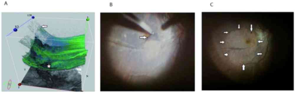
|
1
|
Phillips CI: Retinal detachment at the
posterior pole. Br J Ophthalmol. 42:749–753. 1958. View Article : Google Scholar : PubMed/NCBI
|
|
2
|
Lim SJ, Kwon YH, Kim SH, You YS and Kwon
OW: Vitrectomy and internal limiting membrane peeling without gas
tamponade for myopic foveoschisis. Graefes Arch Clin Exp
Ophthalmol. 250:1573–1577. 2012. View Article : Google Scholar : PubMed/NCBI
|
|
3
|
Ellabban AA, Tsujikawa A, Matsumoto A,
Yamashiro K, Oishi A, Ooto S, Nakata I, Akagi-Kurashige Y, Miyake
M, Elnahas HS, et al: Three-dimensional tomographic features of
dome-shaped macula by swept-source optical coherence tomography. Am
J Ophthalmol. 155:320–328.e2. 2013. View Article : Google Scholar : PubMed/NCBI
|
|
4
|
Zheng B, Chen Y, Chen Y, Zhao Z, Zhang Z,
Zheng J, You Y, Wang Q and Shen L: Vitrectomy and internal limiting
membrane peeling with perfluoropropane tamponade or balanced saline
solution for myopic foveoschisis. Retina. 31:692–701. 2011.
View Article : Google Scholar : PubMed/NCBI
|
|
5
|
Wong TY, Ohno-Matsui K, Leveziel N, Holz
FG, Lai TY, Yu HG, Lanzetta P, Chen Y and Tufail A: Myopic
choroidal neovascularisation: Current concepts and update on
clinical management. Br J Ophthalmol. 99:289–296. 2015. View Article : Google Scholar : PubMed/NCBI
|
|
6
|
Todorich B, Scott IU, Flynn HW Jr and
Chang S: Macular retinoschisis associated with pathologic myopia.
Retina. 33:678–683. 2013. View Article : Google Scholar : PubMed/NCBI
|
|
7
|
Meng B, Zhao L, Yin Y, Li H, Wang X, Yang
X, You R, Wang J, Zhang Y, Wang H, et al: Internal limiting
membrane peeling and gas tamponade for myopic foveoschisis: A
systematic review and meta-analysis. BMC Ophthalmol. 17:166–176.
2017. View Article : Google Scholar : PubMed/NCBI
|
|
8
|
Faghihi H, Hajizadeh F and Riazi-Esfahani
M: Optical coherence tomographic findings in highly myopic eyes. J
Ophthalmic Vis Res. 5:110–121. 2010.PubMed/NCBI
|
|
9
|
García-Ben A, Blanco MJ, Piñeiro A, Mera
P, Rodriguez-Alvarez MX and Capeans C: Relationship between macular
bending and foveoschisis in myopic patients. Optom Vis Sci.
91:497–506. 2014. View Article : Google Scholar : PubMed/NCBI
|
|
10
|
Mateo C, Gómez-Resa MV, Burés-Jelstrup A
and Alkabes M: Surgical outcomes of macular buckling techniques for
macular retinoschisis in highly myopic eyes. Saudi J Ophthalmol.
27:235–239. 2013. View Article : Google Scholar : PubMed/NCBI
|
|
11
|
García-Layana A, García-Arumí J,
Ruiz-Moreno JM, Arias-Barquet L, Cabrera-López F and Figueroa MS: A
review of current management of vitreomacular traction and macular
hole. J Ophthalmol. 2015:8096402015. View Article : Google Scholar : PubMed/NCBI
|
|
12
|
Gómez-Resa M, Burés-Jelstrup A and Mateo
C: Myopic traction maculopathy. Dev Ophthalmol. 54:204–212. 2014.
View Article : Google Scholar : PubMed/NCBI
|
|
13
|
Zhang T, Zhu Y, Jiang CH and Xu GZ:
Long-term follow-up of vitrectomy in patients with pathologic
myopic foveoschisis. Int J Ophthalmol. 10:277–284. 2017.PubMed/NCBI
|
|
14
|
Kumagai K, Furukawa M, Ogino N and Larson
E: Factors correlated with postoperative visual acuity after
vitrectomy and internal limiting membrane peeling for myopic
foveoschisis. Retina. 30:874–880. 2010. View Article : Google Scholar : PubMed/NCBI
|
|
15
|
Figueroa MS, Ruiz-Moreno JM, Gonzalez del
Valle F, Govetto A, de la Vega C, Plascencia RN, Contreras I and
Medina JL: Long-term outcomes of 23-gauge pars plana vitrectomy
with internal limiting membrane peeling and gas tamponade for
myopic traction maculopathy: A prospective study. Retina.
35:1836–1843. 2015. View Article : Google Scholar : PubMed/NCBI
|
|
16
|
Arevalo JF, Berrocal MH, Arias JD and
Banaee T: Minimally invasive vitreoretinal surgery: Is sutureless
vitrectomy the future of vitreoretinal surgery? J Ophthalmic Vis
Res. 6:136–144. 2011.PubMed/NCBI
|
|
17
|
Fujii GY, De Juan E Jr, Humayun MS, Chang
TS, Pieramici DJ, Barnes A and Kent D: Initial experience using the
transconjunctival sutureless vitrectomy system for vitreoretinal
surgery. Ophthalmology. 109:1814–1820. 2002. View Article : Google Scholar : PubMed/NCBI
|
|
18
|
Ohta K, Sato A, Senda N and Fukui E:
Comparisons of foveal thickness and slope after macular hole
surgery with and without internal limiting membrane peeling. Clin
Ophthalmol. 12:503–510. 2018. View Article : Google Scholar : PubMed/NCBI
|
|
19
|
VanderBeek BL and Johnson MW: The
diversity of traction mechanisms in myopic traction maculopathy. Am
J Ophthalmol. 153:93–102. 2012. View Article : Google Scholar : PubMed/NCBI
|
|
20
|
Hwang JU, Joe SG, Lee JY, Kim JG and Yoon
YH: Microincision vitrectomy surgery for myopic foveoschisis. Br J
Ophthalmol. 97:879–884. 2013. View Article : Google Scholar : PubMed/NCBI
|
|
21
|
Ho TC, Yang CM, Huang JS, Yang CH, Yeh PT,
Chen TC, Ho A and Chen MS: Long-term outcome of foveolar internal
limiting membrane nonpeeling for myopic traction maculopathy.
Retina. 34:1833–1840. 2014. View Article : Google Scholar : PubMed/NCBI
|
|
22
|
Shimada N, Sugamoto Y, Ogawa M, Takase H
and Ohno-Matsui K: Fovea-sparing internal limiting membrane peeling
for myopic traction maculopathy. Am J Ophthalmol. 154:693–701.
2012. View Article : Google Scholar : PubMed/NCBI
|
|
23
|
Qi Y, Duan AL, Meng X and Wang N:
Vitrectomy without inner limiting membrane peeling for macular
retinoschisis in highly myopic eyes. Retina. 36:953–956. 2016.
View Article : Google Scholar : PubMed/NCBI
|
|
24
|
Taniuchi S, Hirakata A, Itoh Y, Hirota K
and Inoue M: Vitrectomy with or without internal limiting membrane
peeling for each stage of myopic traction maculopathy. Retina.
33:2018–2025. 2013. View Article : Google Scholar : PubMed/NCBI
|
|
25
|
Faes L, Bodmer NS, Bachmann LM, Thiel MA
and Schmid MK: Diagnostic accuracy of the Amsler grid and the
preferential hyperacuity perimetry in the screening of patients
with age-related macular degeneration: Systematic review and
meta-analysis. Eye (Lond). 28:788–796. 2014. View Article : Google Scholar : PubMed/NCBI
|
|
26
|
Tranos P, Koukoula S, Charteris DG,
Perganda G, Vakalis A, Asteriadis S, Georgalas I and Petrou P: The
role of internal limiting membrane peeling in epiretinal membrane
surgery: A randomised controlled trial. Br J Ophthalmol.
101:719–724. 2017. View Article : Google Scholar : PubMed/NCBI
|
|
27
|
Alkabes M and Mateo C: Macular buckle
technique in myopic traction maculopathy: A 16-year review of the
literature and a comparison with vitreous surgery. Graefes Arch
Clin Exp Ophthalmol. May. 256:863–877. 2018.
|
|
28
|
Ikuno Y, Sayanagi K, Soga K, Oshima Y,
Ohji M and Tano Y: Foveal anatomical status and surgical results in
vitrectomy for myopic foveoschisis. Jpn J Ophthalmol. 52:269–276.
2008. View Article : Google Scholar : PubMed/NCBI
|
|
29
|
Elwan MM, Abd Elghafar AE, Hagras SM, Abou
Samra WA and Saleh SM: Long-term outcome of internal limiting
membrane peeling with and without foveal sparing in myopic
foveoschisis. Eur J Ophthalmol. 29:69–74. 2019. View Article : Google Scholar : PubMed/NCBI
|
|
30
|
Sayanagi K, Ikuno Y and Tano Y:
Reoperation for persistent myopic foveoschisis after primary
vitrectomy. Am J Ophthalmol. 141:414–417. 2006. View Article : Google Scholar : PubMed/NCBI
|
|
31
|
Seppey C and Wolfensberger TJ: Vitrectomy
with fovea-sparing internal limiting membrane peeling for myopic
foveoschisis. Klin Monbl Augenheilkd. 234:497–500. 2017. View Article : Google Scholar : PubMed/NCBI
|
|
32
|
Mii M, Matsuoka M, Matsuyama K, Otsu Y and
Nishimura T: Favorable anatomic and visual outcomes with 25-gauge
vitrectomy for myopic foveoschisis. Clin Ophthalmol. 8:1837–1844.
2014. View Article : Google Scholar : PubMed/NCBI
|
|
33
|
Spaide RF and Fisher Y: Removal of
adherent cortical vitreous plaques without removing the internal
limiting membrane in the repair of macular detachments in highly
myopic eyes. Retina. 25:290–295. 2005. View Article : Google Scholar : PubMed/NCBI
|
|
34
|
Ikuno Y, Sayanagi K, Ohji M, Kamei M, Gomi
F, Harino S, Fujikado T and Tano Y: Vitrectomy and internal
limiting membrane peeling for myopic foveoschisis. Am J Ophthalmol.
137:719–724. 2004. View Article : Google Scholar : PubMed/NCBI
|
|
35
|
Liu HJ and Bi XJ: Vitrectomy combined with
internal limiting membrane peeling for treating foveoschisis in
high myopia. Int Eye Sci. 10:1871–1872. 2014.
|
|
36
|
Kwok AK, Lai TY and Yip WW: Vitrectomy and
gas tamponade without internal limiting membrane peeling for myopic
foveoschisis. Br J Ophthalmol. 89:1180–1183. 2005. View Article : Google Scholar : PubMed/NCBI
|
|
37
|
Sanisoglu H, Sevim MS, Aktas B, Sevim S
and Nohutcu A: Outcomes of 23-gauge pars plana vitrectomy and
internal limiting membrane peeling with brilliant blue in macular
hole. Clin Ophthalmol. 5:1177–1183. 2011.PubMed/NCBI
|
|
38
|
Shukla D, Kalliath J, Neelakantan N,
Naresh KB and Ramasamy K: A comparison of brilliant blue G, trypan
blue, and indocyanine green dyes to assist internal limiting
membrane peeling during macular hole surgery. Retina. 31:2021–2025.
2011. View Article : Google Scholar : PubMed/NCBI
|
|
39
|
Farah ME, Maia M, Penha FM and Rodrigues
EB: The use of vital dyes during vitreoretinal
surgery-chromovitrectomy. Dev Ophthalmol. 55:365–375. 2016.
View Article : Google Scholar : PubMed/NCBI
|
|
40
|
Yonekawa Y, Abbey AM, Shah AR, Thomas BJ
and Capone A Jr: Endoilluminator phototoxic maculopathy associated
with combined ICG-assisted epiretinal membrane and internal
limiting membrane peeling. Clin Ophthalmol. 8:2501–2506. 2014.
View Article : Google Scholar : PubMed/NCBI
|
|
41
|
Badaró E, Moraes-Filho M, Maia M, Penha
FM, Novais EA, Souza-Lima RA, Hirai F, Meyer CH, Farah ME and
Rodrigues EB: Retinal biocompatibility of brilliant blue g with
deuterated water for chromovitrectomy. J Ophthalmic Vis Res.
9:204–209. 2014.PubMed/NCBI
|
|
42
|
Januschowski K, Mueller S, Spitzer MS,
Schramm C, Doycheva D, Bartz-Schmidt KU and Szurman P: Evaluating
retinal toxicity of a new heavy intraocular dye, using a model of
perfused and isolated retinal cultures of bovine and human origin.
Graefes Arch Clin Exp Ophthalmol. 250:1013–1022. 2012. View Article : Google Scholar : PubMed/NCBI
|
|
43
|
Enaida H, Hisatomi T, Hata Y, Ueno A, Goto
Y, Yamada T, Kubota T and Ishibashi T: Brilliant blue G selectively
stains the internal limiting membrane/brilliant blue G-assisted
membrane peeling. Retina. 26:631–636. 2006. View Article : Google Scholar : PubMed/NCBI
|
|
44
|
Zhu SQ, Zheng LY, Pan AP, Yu AY, WangQ M
and Xue AQ: The efficacy and safety of posterior scleral
reinforcement using genipin cross-linked sclera for macular
detachment and retinoschisis in highly myopic eyes. Br J
Ophthalmol. 5:210–216. 2016.
|
|
45
|
Zhu Z, Ji X, Zhang J and Ke G: Posterior
scleral reinforcement in the treatment of macular retinoschisis in
highly myopic patients. Clin Exp Ophthalmol. 37:660–663. 2009.
View Article : Google Scholar : PubMed/NCBI
|