Introduction
Viral myocarditis (VMC), as one of the most common
cardiac infectious diseases, is mainly caused by myocardial cells
infected with the virus, causing localized or diffuse inflammation
of the myocardium. The virus causes direct damage to the myocardial
cells and stimulates the immune response, thus causing sustained
damage to the myocardial tissues (1,2).
Coxsackie B3 virus (CVB3) is the most important virus that causes
myocarditis. CVB3 can induce oxidative stress response and
apoptosis in a few weeks in the pathogenesis of VMC, resulting in
arrhythmia, myocardial failure and may eventually lead to sudden
death, but special treatment of VMC has not been found as yet
(3,4).
Ubiquitin-proteasome system (UPS), as an important
ATP-dependent protein control system in eukaryotic cells, not only
participates in physiological processes such as apoptosis,
inflammatory response and intracellular signaling, but also plays
an important role in maintaining cell homeostasis (5–7). In
recent years, studies have found that UPS not only plays an
important role in the inflammatory response of various diseases,
but also plays a key role in the occurrence and development of
various viral infectious diseases (8,9). As an
aldehyde peptide proteasome inhibitor that can inhibit the activity
of chyme protein, MG-132 also has a protective effect on the
occurrence of inflammatory reactions in many diseases (10). As a cytokine with a variety of
biological effects produced by macrophages, tumor necrosis factor-α
(TNF-α) has been shown to play an important role in the
inflammatory response of VMC (11).
However, transforming growth factor-β1 (TGF-β1) is an initiating
factor synthesized from the extracellular matrix of collagen
fibers, and it is also one of the many factors leading to the
occurrence of viral myocarditis (11).
At present, few studies have reported the role of
UPS in the inflammatory reaction of VMC, so we explored the effect
of ubiquitin-proteasome inhibitor MG-23 on the expression levels of
serum TNF-α and TGF-β1 in CVB mice, in order to understand the
inflammatory mechanism of VMC.
Materials and methods
Experimental animals and
materials
A total of 80 healthy male SPF grade mice aged 6
weeks and weighing 25.1±5.53 g were selected. All the mice were
purchased from the animal experiment center of Zhejiang province,
with the production license of SCXK (Zhejiang) 2011-0166. The mice
were fed in a plastic box with bedding material on a solid bottom
with the temperature of 22°C and the relative humidity between 50
and 65%, 12 h day-night rhythm was normal, and they were free to
eat and drink.
The study was approved by the Ethics Committee of
Central Hospital of Zibo (Zibo, China). The CVB3 virus was
purchased from Cell Signaling Inc. at a titer of 100 TCID50 (50%
tissue culture infective dose) /0.1 ml. Mg-132 was purchased from
Calbiochem Inc. at a concentration of 0.75 mg/kg.
Grouping and modeling
Twenty mice were randomly selected as the blank
group, and they were kept in routine feeding without any
intervention. The remaining 60 mice were then randomly divided into
the VMC, MG-132 and control group, each containing 20 mice. Mice in
the control group were intraperitoneally injected with 0.1 ml PBS
(phosphate buffersaline) at 0.1 mmol/l, mice in the VMC and MG-132
group were intraperitoneally injected with 0.1 ml diluent of CVB3
at 100 TCID50/0.1 ml, and mice in the MG-132 group were
intraperitoneally injected with 0.75 mg/kg MG-132 the day after the
injection of the CVB3 virus. They were continuously injected for 7
days. Mortality rates of each group were recorded and compared on
day 8 of modeling, and then peripheral blood and heart samples of
the remaining mice were taken for subsequent detection.
Pathological examination
Hearts of mice were fixed with 10% formaldehyde and
dehydrated routinely, then embedded with paraffin and sectioned.
After sectioning, the pathological changes of mouse myocardial
tissues were observed under light microscope (Olympus Corp.) and
the pathological score of the myocardial tissues was evaluated. The
judgement scores were as follows: 1 point, when the proportion of
inflammatory cell infiltration and myocardial necrosis was <25%,
2 points, when the proportion of inflammatory cell infiltration and
myocardial necrosis was between 25 and 50%, 3 points, when the
proportion of inflammatory cell infiltration and myocardial
necrosis was between 51 and 75%, 4 points, when the proportion of
inflammatory cell infiltration and myocardial necrosis was
>75%.
Expression levels of mRNA in TNF-α and
TGF-β1 detected by RT-qPCR
First, the TRIzol reagent (purchased from Applide
Invitrogen, Inc.) was added into the myocardial tissue of mice to
extract the total RNA in the blood, and the concentration and
quality of total RNA were detected by ultraviolet spectrophotometer
(purchased from Shanghai Yuanxi Instrument Co., Ltd.). Total RNA (2
µl) was used to compound cDNA in strict accordance with the
instructions of minScript reverse transcription kit (Takara Bio,
Inc.). Synthetic cDNA (2 µl) was used for qPCR (RT-qPCR kit was
purchased from Takara). Total RNA (2 μl) was added to a
microcentrifuge tube, then 11 μl of DEPC H2O was
added. A total of 12–18 μl of 10 μM Oligo (dT) was
added, mixed and heated at 70°C for 10 min. After ice bath for 1
min, 2 μl 10X PCR buffer, 2 μl 25 mM
MgCl2, 1 μl 10 mM dNTP mix and 2 μl 0.1 M
DTT were added, mixed and incubated for 3 min at 42°C. A total of 1
μl of Superscript II was added, incubated at 42°C for 30
min, and heated at 70°C for 10 min. Ice bath was followed for 5
min. The qPCR reaction system was: 2 µl reverse transcription
product, 0.5 µl upstream primer, 0.5 µl downstream primer, 2X SYBR
Green PCR Master Mix 10 µl, and sterilized and deionized water
complemented to 20 µl. Reaction conditions were: 95°C for 2 min,
95°C for 50 sec, 60°C for 45 sec, extension at 72°C for 30 sec, and
for a total of 40 cycles. The expression levels of TNF-α mRNA and
TGF-β1 mRNA were detected with glyceraldehyde-3-phosphate
dehydrogenase (GAPDH) as the internal reference. The primer
sequences are shown in Table I (the
primers were synthesized and designed by Shanghai Gemma Company),
and the experiment was repeated 3 times. The result was analyzed
with 2ΔΔ−Cq method (12).
 | Table I.Sequences of the primers. |
Table I.
Sequences of the primers.
| Factor | Upstream primer | Downstream
primer |
|---|
| TNF-α |
5′-CCACGCTCTTCTGTCTACTGA-3′ |
5′-AAGGTACAACCCATCGGCTG-3′ |
| TGF-β1 |
5′-CCAACTATTGCTTCAGCTCCA-3′ |
5′-GTGTCCAGGCTCCAAATGT-3′ |
| GAPDH |
5′-GGTTGTCTCCTGCGACTTCA-3′ |
5′-TGGTCCAGGGTTTCTTACTCC-3′ |
Expression levels of TNF-α protein and
TGF-β1 protein in myocardial tissues measured by western blot
analysis
Cardiac muscle tissues (100 g) of mice were taken
and RIPA was added lysate to extract the total protein. Then the
extracted protein was boiled at 100°C for 5 min to denaturate, then
separated with 10% SDS-PAGE after being cooled, transferred to the
PVDF membrane, and sealed with 5% of skim milk at room temperature
for 1 h. Then the primary rabbit anti-mice polyclonal antibodies of
TNF-α (1:500), TGF-β1 (1:500) and β-actin (1:1,000) (cat. nos.
17590-1-AP, 21898-1-AP, 14395-1-AP; ProteinTech Group, Inc.) were
added at 4°C for incubation overnight, then the secondary goat
anti-rabbit polyclonal antibody (dil, 1:500; cat. no. SA00001-2;
ProteinTech Group, Inc.) at room temperature for 1 h incubation,
finally ECL developer was used to develop color.
Observation indicators
i) Eight-day survival rates of mice in each group
were recorded and compared. ii) The pathological score in
myocardial tissues of mice in each group was evaluated and
compared. iii) The expression levels of TNF-α mRNA and TGF-β1 mRNA
in myocardial tissues of mice in each group were compared. iv)
TNF-α protein and TGF-β1 protein in myocardial tissues of mice in
each group were compared. v) Correlation analysis was performed
between the pathological score and the protein expression levels of
TNF-α and TGF-β1 in myocardial tissues of mice, and the protein
expression levels of TNF-α and TGF-β1 in myocardial tissues.
Statistical analysis
SPSS 20.0 (IBM Corp., Armonk, NY, USA) was used for
statistical analysis of the experimental data. The Chi-square test
was used to compare the enumeration data. Kaplan-Meier curve was
used for survival analysis. Measurement data were expressed as mean
± standard deviation. t-test was used for comparison between the
two groups and univariate analysis of variance was used for
multigroup comparison. LSD/t-test was used for postoperative
comparison. Correlation was analyzed by Pearson's correlation
analysis. GraphPad Prism 6 software (Hangzhou Aimeilv Biotechnology
Co., Ltd.) was used to draw the experimental images. P<0.05 was
considered to indicate a statistically significant difference.
Results
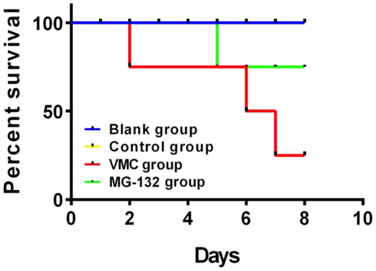
Comparison of survival rates of mice
in each group
The survival rate of the blank group and the control
group at day 8 was 100%. A total of 11 mice in the VMC group died
at day 8, with a survival rate of 45%. A total of 5 mice in the
MG-132 group died at day 8, with a survival rate of 75%. The 8-day
survival rates of the blank group and control group were
significantly higher than those of the VMC group and MG-132 group,
but the 8-day survival rate of mice in the MG-132 group was
significantly higher than that of the VMC group (P<0.05)
(Fig. 1).
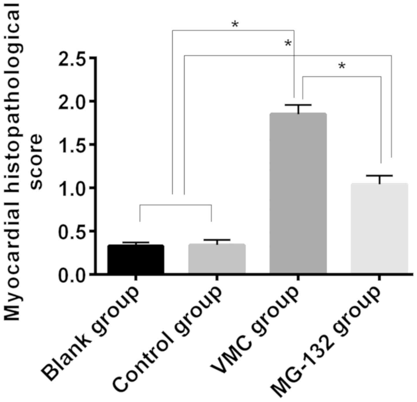
Histopathological scores of myocardial
tissues of mice in each group
There was no inflammatory cell infiltration in
myocardial tissues of mice in blank or control group. There was a
large amount of inflammatory cell infiltration in myocardial
tissues of mice in VMC group. The inflammatory cells in the
myocardial tissues of mice in MG-132 group were significantly
reduced compared with those in VMC group. The myocardial
histopathological scores of mice in blank and the control group
were respectively 0.33±0.04 and 0.34±0.06, the myocardial
histopathological score of mice in the VMC group was 1.85±0.11, and
was 1.04±0.10 in the MG-132 group. The myocardial histopathological
scores of mice in the blank and the control group were
significantly lower than those in VMC and MG-132 group, and
myocardial histopathological score of mice in MG-132 was
significantly lower than that in VMC group (P<0.05) (Fig. 2).
Expression levels of mRNA of TNF-α and
TGF-β1 in myocardial tissues of mice in each group
There was no significant difference in expression
levels of TNF-α mRNA and TGF-β1 mRNA between the blank group and
the control group (P>0.05), but were significantly lower than
those in the VMC group and the MG-132 group. However, expression
levels of TNF-α mRNA and TGF-β1 mRNA in myocardial tissues of
MG-132 group were significantly lower than those of the VMC group
(P<0.05) (Table II).
| Table II.Expression levels of TNF-α mRNA and
TGF-β1 mRNA in myocardial tissues of mice in each group. |
Table II.
Expression levels of TNF-α mRNA and
TGF-β1 mRNA in myocardial tissues of mice in each group.
| Factor | Blank group
(n=20) | Control group
(n=20) | VMC group (n=9) | MG-132 group
(n=15) | F value | P-value |
|---|
| TNF-α |
0.41±0.05a,b |
0.41±0.05a,b | 1.83±0.13 |
1.09±0.12a | 779.6 | <0.001 |
| TGF-β1 |
0.51±0.06a,b |
0.52±0.07a,b | 1. 94±0.31 |
1.27±0.24a | 204.0 | <0.001 |
Expression levels of TNF-α protein and
TGF-β1 protein in myocardial tissues of mice in each group
There was no significant difference in the
expression levels of TNF-α protein and TGF-β1 protein between blank
and control group (P>0.05), but both were significantly lower
than those of VMC group and the MG-132 group. However, expression
levels of TNF-α and TGF-β1 protein in myocardial tissues of mice in
the MG-132 group were significantly lower than those in the VMC
group (P<0.05) (Table III).
| Table III.Expression levels of TNF-α protein and
TGF-β1 protein in myocardial tissues of mice in each group. |
Table III.
Expression levels of TNF-α protein and
TGF-β1 protein in myocardial tissues of mice in each group.
| Factor | Blank group
(n=20) | Control group
(n=20) | VMC group (n=9) | MG-132 group
(n=15) | F value | P-value |
|---|
| TNF-α (ng/l) |
1.15±0.59a,b |
1.14±0.62a,b | 2.87±0.65 |
1.84±0.58a | 21.36 | <0.001 |
| TGF-β1(ng/ml) |
1.05±0.41a,b | 1.
03±0.42a,b | 2.14±0.61 |
1.57±0.59a | 14.07 | <0.001 |
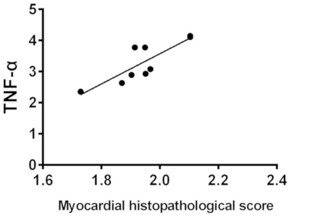
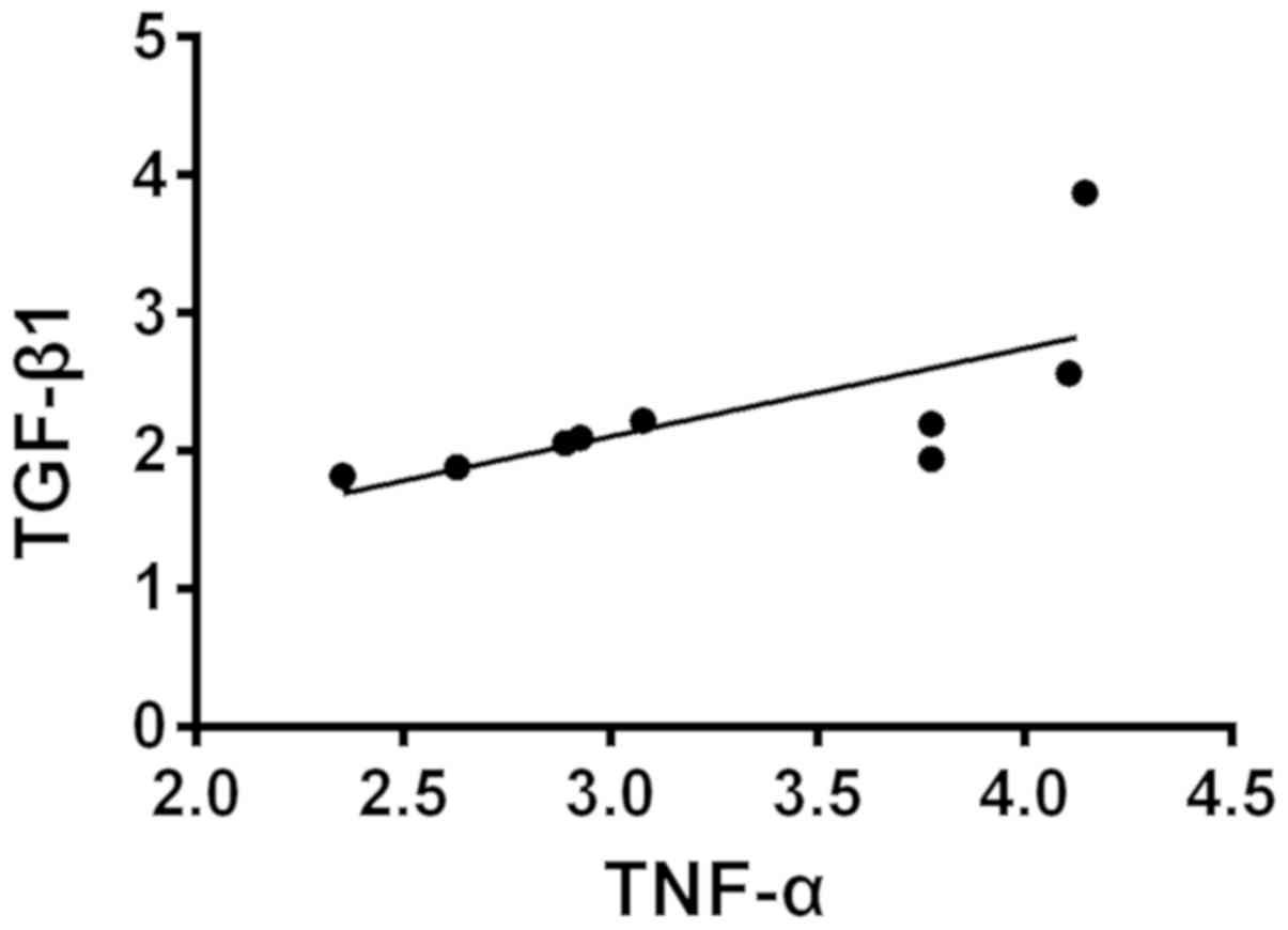
Correlation analysis of the
pathological score in myocardial tissues and expression levels of
TNF-α protein and TGF-β1 protein in myocardial tissues of mice and
the protein expression levels between TNF-α and TGF-β1 in
myocardial tissues of VMC mice
The expression levels of TNF-α protein and TGF-β1
protein in myocardial tissues were positively correlated with the
pathological score of myocardial tissues (r=0.843, P<0.05;
r=0.763, P<0.05), and there was a positive correlation between
expression levels of TNF-α protein and TGF-β1 protein of VMC mice
in myocardial tissues (r=0.672, P<0.05) (Figs. 3–5).
Discussion
As a viral infectious disease, VMC currently has no
specific effective treatment in clinical practice, thus, VMC is one
of the most challenging diseases in the diagnosis and treatment of
cardiovascular field at present (13). Besides, there are also great
controversies regarding the pathogenesis of VMC, most of which
support cytokine theory and immune theory (14). As an important protein quality
control system in eukaryotic cells, UPS has been found to play a
very important role in various viral infectious diseases in recent
years. Specifically, through UPS, viruses can replicate and lead to
oxidative stress in host cells and eventually cause cell damage
(15–17). For example, studies (18) have found that the normal UPS pathway
is involved in the replication of vaccinia virus. However, there
are few studies on UPS in VMC, and no detailed description of its
mechanism has been made.
In our study, the effects of UPS inhibitor MG-132
intervention on myocardial cells, TNF-α and TGF-β1 in VMC mice were
investigated. The results showed that the mortality of the VMC
group was significantly higher than that of the blank group,
control group and MG-132 group (P<0.05), suggesting VMC mice had
higher mortality rates, but the mortality of VMC mice can be
significantly reduced after the intervention of UPS inhibitor
MG-132. In addition, the expression levels of myocardial
histopathological scores, mRNA and protein of TNF-α and TGF-β1 in
the blank group and the control group were significantly lower than
those in the VMC group and the MG-132 group, and the myocardial
histopathological scores, mRNA and protein of TNF-α and TGF-β1 in
the MG-132 group were significantly lower than those in the VMC
group (P<0.05). This suggests that the intervention of UPS
inhibitor MG-132 can effectively improve the inflammatory
infiltration in myocardial tissues of VMC mice and reduce the
expression levels of inflammatory factors in myocardial tissues.
UPS plays an important role in inflammatory response. For example,
a previous study (19) found that
UPS was involved in the response injury of hepatitis B coronavirus
to a certain extent and could effectively alleviate the occurrence
of inflammatory response.
Other studies (20)
have shown that UPS inhibitor MG-132 can inhibit the AKT and ERK
pathways, thereby alleviating the inflammatory response and
inhibiting the progression of heart failure. Although these studies
did not directly confirm our conclusions, they can also show that
the intervention of UPS inhibitor MG-132 does have a certain
alleviating effect on the inflammatory response. Therefore,
previous studies (21) suggested
that inhibition of the process of heart failure by UPS inhibitors
may be achieved by reducing oxidative stress. However, some studies
(22) have obtained different
results when different UPS inhibitors were applied in the
intervention of ischemic cardiomyopathy, and the reason is not
clear at present. We speculate that it may be because UPS
inhibitors are involved in different molecular reactions, so they
have different effects on inflammatory reactions or oxidative
stress reactions. Finally, we analyzed the correlation between the
pathological score in myocardial tissues of mice and the protein
expression levels of TNF-α and TGF-β1 in myocardial tissues, as
well as the protein expression levels between TNF-α and TGF-β1 in
myocardial tissues of VMC mice. The results showed that the
expression levels of TNF-α and TGF-β1 proteins in myocardial
tissues were positively correlated with the pathological score in
myocardial tissues of mice, and the expression levels of TNF-α
protein and TGF-β1 protein in myocardial tissues of VMC mice were
also positively correlated. Previous studies (23) have shown that the regulation of TGF-β
can effectively stimulate the release of TNF-α in human monocytes.
Although it is not a study of the myocardial tissues, it also
confirms our conclusion.
In recent years, the role of UPS in cardiovascular
diseases has been gradually recognized and discovered (24). After the establishment of VMC mouse
model, we also found that the UPS inhibitor MG-132 can
significantly alleviate the myocardial injury of VMC mice, reduce
the expression levels of inflammatory factors in myocardial
tissues, and improve the survival rate of mice. UPS inhibitor
MG-132 may be a new treatment scheme for VMC. However, our study
also has some shortcomings. We did not further explore the
mechanism of action between TNF-α and TGF-β1, or describe in detail
how MG-132 reduced the inflammatory response in VMC mice. This will
be further explored in our subsequent experiments.
Acknowledgements
Not applicable.
Funding
No funding was received.
Availability of data and materials
The datasets used and/or analyzed during the present
study are available from the corresponding author on reasonable
request.
Authors' contributions
HZ, JY and HS performed RT-qPCR. YZ and JW were
responsible for western blot analysis. JZ and BM contributed to
analysis of the observation indexes. HZ wrote the manuscript. All
authors read and approved the final manuscript.
Ethics approval and consent to
participate
The study was approved by the Ethics Committee of
Central Hospital of Zibo (Zibo, China).
Patient consent for publication
Not applicable.
Competing interests
The authors declare that they have no competing
interests.
References
|
1
|
Pal VK, Bandyopadhyay P and Singh A:
Hydrogen sulfide in physiology and pathogenesis of bacteria and
viruses. IUBMB Life. 70:393–410. 2018. View
Article : Google Scholar : PubMed/NCBI
|
|
2
|
Guo YB, Zhou LW, Chen FL and Zou N:
Effects and related mechanism of overexpression of human
thioredoxin on the inflammatory response in mice with viral
myocarditis. Zhonghua Xin Xue Guan Bing Za Zhi. 46:444–449.
2018.(In Chinese). PubMed/NCBI
|
|
3
|
Cao SS, Luo KL and Shi L: Endoplasmic
reticulum stress interacts with inflammation in human diseases. J
Cell Physiol. 231:288–294. 2016. View Article : Google Scholar : PubMed/NCBI
|
|
4
|
Huang Z, Rose AH and Hoffmann PR: The role
of selenium in inflammation and immunity: From molecular mechanisms
to therapeutic opportunities. Antioxid Redox Signal. 16:705–743.
2012. View Article : Google Scholar : PubMed/NCBI
|
|
5
|
Crawford LJ and Irvine AE: Targeting the
ubiquitin proteasome system in haematological malignancies. Blood
Rev. 27:297–304. 2013. View Article : Google Scholar : PubMed/NCBI
|
|
6
|
Ao N, Chen Q and Liu G: The small
molecules targeting ubiquitin-proteasome system for cancer therapy.
Comb Chem High Throughput Screen. 20:403–413. 2017. View Article : Google Scholar : PubMed/NCBI
|
|
7
|
Schlossarek S, Frey N and Carrier L:
Ubiquitin-proteasome system and hereditary cardiomyopathies. J Mol
Cell Cardiol. 71:25–31. 2014. View Article : Google Scholar : PubMed/NCBI
|
|
8
|
Treuer AV and Gonzalez DR: Nitric oxide
synthases, S-nitrosylation and cardiovascular health: From
molecular mechanisms to therapeutic opportunities (Review). Mol Med
Rep. 11:1555–1565. 2015. View Article : Google Scholar : PubMed/NCBI
|
|
9
|
Kasaikina MV, Hatfield DL and Gladyshev
VN: Understanding selenoprotein function and regulation through the
use of rodent models. Biochim Biophys Acta. 1823:1633–1642. 2012.
View Article : Google Scholar : PubMed/NCBI
|
|
10
|
Radwan M, Wilkinson DJ, Hui W, Destrument
AP, Charlton SH, Barter MJ, Gibson B, Coulombe J, Gray DA, Rowan
AD, et al: Protection against murine osteoarthritis by inhibition
of the 26S proteasome and lysine-48 linked ubiquitination. Ann
Rheum Dis. 74:1580–1587. 2015. View Article : Google Scholar : PubMed/NCBI
|
|
11
|
Sun XH, Fu J and Sun DQ: Halofuginone
alleviates acute viral myocarditis in suckling BALB/c mice by
inhibiting TGF-β1. Biochem Biophys Res Commun. 473:558–564. 2016.
View Article : Google Scholar : PubMed/NCBI
|
|
12
|
Livak KJ and Schmittgen TD: Analysis of
relative gene expression data using real-time quantitative PCR and
the 2 (-Delta Delta C(T)) method. Methods. 25:402–408. 2001.
View Article : Google Scholar : PubMed/NCBI
|
|
13
|
Reisz JA, Barrett AS, Nemkov T, Hansen KC
and D'Alessandro A: When nature's robots go rogue: Exploring
protein homeostasis dysfunction and the implications for
understanding human aging disease pathologies. Expert Rev
Proteomics. 15:293–309. 2018. View Article : Google Scholar : PubMed/NCBI
|
|
14
|
Stein EA, Pinkert S, Becher PM, Geisler A,
Zeichhardt H, Klopfleisch R, Poller W, Tschöpe C, Lassner D,
Fechner H, et al: Combination of RNA interference and virus
receptor trap exerts additive antiviral activity in coxsackievirus
B3-induced myocarditis in mice. J Infect Dis. 211:613–622. 2015.
View Article : Google Scholar : PubMed/NCBI
|
|
15
|
Maiese K, Hou J, Chong ZZ and Shang YC: A
fork in the path: Developing therapeutic inroads with FoxO
proteins. Oxid Med Cell Longev. 2:119–129. 2009. View Article : Google Scholar : PubMed/NCBI
|
|
16
|
Maiese K, Chong ZZ, Hou J and Shang YC:
The ‘O’ class: Crafting clinical care with FoxO transcription
factors. Adv Exp Med Biol. 665:242–260. 2009. View Article : Google Scholar : PubMed/NCBI
|
|
17
|
Maiese K, Chong ZZ, Shang YC and Hou J:
Clever cancer strategies with FoxO transcription factors. Cell
Cycle. 7:3829–3839. 2008. View Article : Google Scholar : PubMed/NCBI
|
|
18
|
Teale A, Campbell S, Van Buuren N, Magee
WC, Watmough K, Couturier B, Shipclark R and Barry M:
Orthopoxviruses require a functional ubiquitin-proteasome system
for productive replication. J Virol. 83:2099–2108. 2009. View Article : Google Scholar : PubMed/NCBI
|
|
19
|
Zhang H, Diab A, Fan H, Mani SK, Hullinger
R, Merle P and Andrisani O: PLK1 and HOTAIR accelerate proteasomal
degradation of SUZ12 and ZNF198 during hepatitis B virus-induced
liver carcinogenesis. Cancer Res. 75:2363–2374. 2015. View Article : Google Scholar : PubMed/NCBI
|
|
20
|
Lee H, Park J, Kim EE, Yoo YS and Song EJ:
Proteasome inhibitors attenuated cholesterol-induced cardiac
hypertrophy in H9c2 cells. BMB Rep. 49:270–275. 2016. View Article : Google Scholar : PubMed/NCBI
|
|
21
|
Maiese K, Chong ZZ, Shang YC and Hou J:
FoxO proteins: Cunning concepts and considerations for the
cardiovascular system. Clin Sci (Lond). 116:191–203. 2009.
View Article : Google Scholar : PubMed/NCBI
|
|
22
|
Magesh S, Chen Y and Hu L: Small molecule
modulators of Keap1-Nrf2-ARE pathway as potential preventive and
therapeutic agents. Med Res Rev. 32:687–726. 2012. View Article : Google Scholar : PubMed/NCBI
|
|
23
|
Sionov RV, Vlahopoulos SA and Granot Z:
Regulation of Bim in health and disease. Oncotarget. 6:23058–23134.
2015. View Article : Google Scholar : PubMed/NCBI
|
|
24
|
Pagan J, Seto T, Pagano M and Cittadini A:
Role of the ubiquitin proteasome system in the heart. Circ Res.
112:1046–1058. 2013. View Article : Google Scholar : PubMed/NCBI
|