Background
PSS is a chronic inflammatory, multi-systemic
autoimmune disease (1) that may
involve various organs, but commonly results in renal damage
(40–50% of cases) (2). Although the
condition may be life-threatening in severe cases, the early
clinical symptoms of renal lesions in such cases are frequently
atypical, and may hence be overlooked by clinicians. The present
study reports on the clinical and pathological features, as well as
the laboratory and immunofluorescence data of a patient who
initially presented with chronic renal insufficiency, but was
finally diagnosed with PSS.
Case presentation
A 32-year-old female presented at our hospital The
Second Affiliated Hospital of Guangzhou University of Chinese
Medicine (Guangzhou, China) in June 2017 with a 1-year history of
elevated serum creatinine levels and a 6-month history of mild pain
in the waist and leg. For >6 months prior to admission, the
patient did not experience any symptoms/complaints including fever,
dizziness, headache, cough, expectoration, nausea, vomiting,
abdominal pain, diarrhea, hematuria, frothy urine, eyelid, facial
or lower extremity edema. The patient had undergone a routine
medical examination in June 2018 and ignored the result of blood
creatinine levels of 229.0 µmol/l (normal range, 53–98 µmol/l).
Thereafter, the patient was admitted to the First Affiliated
Hospital of Guangzhou University of Traditional Chinese Medicine
(Guangzhou, China) in August 2017 for low back pain, and the
laboratory test results indicated a creatinine level of 242.0
µmol/l, urea levels of 11.2 mmol/l (2.86–7.14 mmol/l), parathyroid
hormone levels of 73.1 pmol/l (1–10.5 pmol/l), and total
cholesterol level of 6.3 mmol/l (3–5.2 mmol/l); the other
symptoms/complaints were similar to those observed previously.
Furthermore, the presence of kidney stones was noted in a physical
examination 1 year previously. The patient had no history of
tuberculosis, type 2 diabetes, steroid use, traditional homeopathic
remedies or herbal medications.
On admission to our hospital, the patient weighed
47.0 kg, had a body height of 160.0 cm and a body mass index of
18.3 kg/m2. The patient's blood pressure and pulse were
recorded as 123/77 mmHg and 86 beats/min. The patient was mildly
anemic, but did not have any goiter or clinically palpable lymph
nodes. Furthermore, no gynecomastia, striae or evidence of pruritus
was noted. The patient's visual field was normal and no papilledema
was detected. The other physical findings were unremarkable.
The laboratory test results on admission and during
the follow-up period are presented in Table I. Twenty-four-hour urine output
monitoring indicated stable fluctuations from 2,200 to 3,100 ml,
and a urine osmolarity of 169.0 mOsm/kg H2O (normal
range, 280.0–310.0 mOsm/kg H2O) was noted. Based on
these results, tests for antinuclear antibodies (ANA; 1:100
granular pattern), along with other tumor, rheumatic disease and
immune system markers, were performed, but the results were
negative. Thus, the patient was initially diagnosed with severe
chronic interstitial nephritis and chronic kidney disease stage IV.
As the patient had a long history of xerostomia and dry eye
syndrome, the presence of Sjogren's syndrome was suspected. To
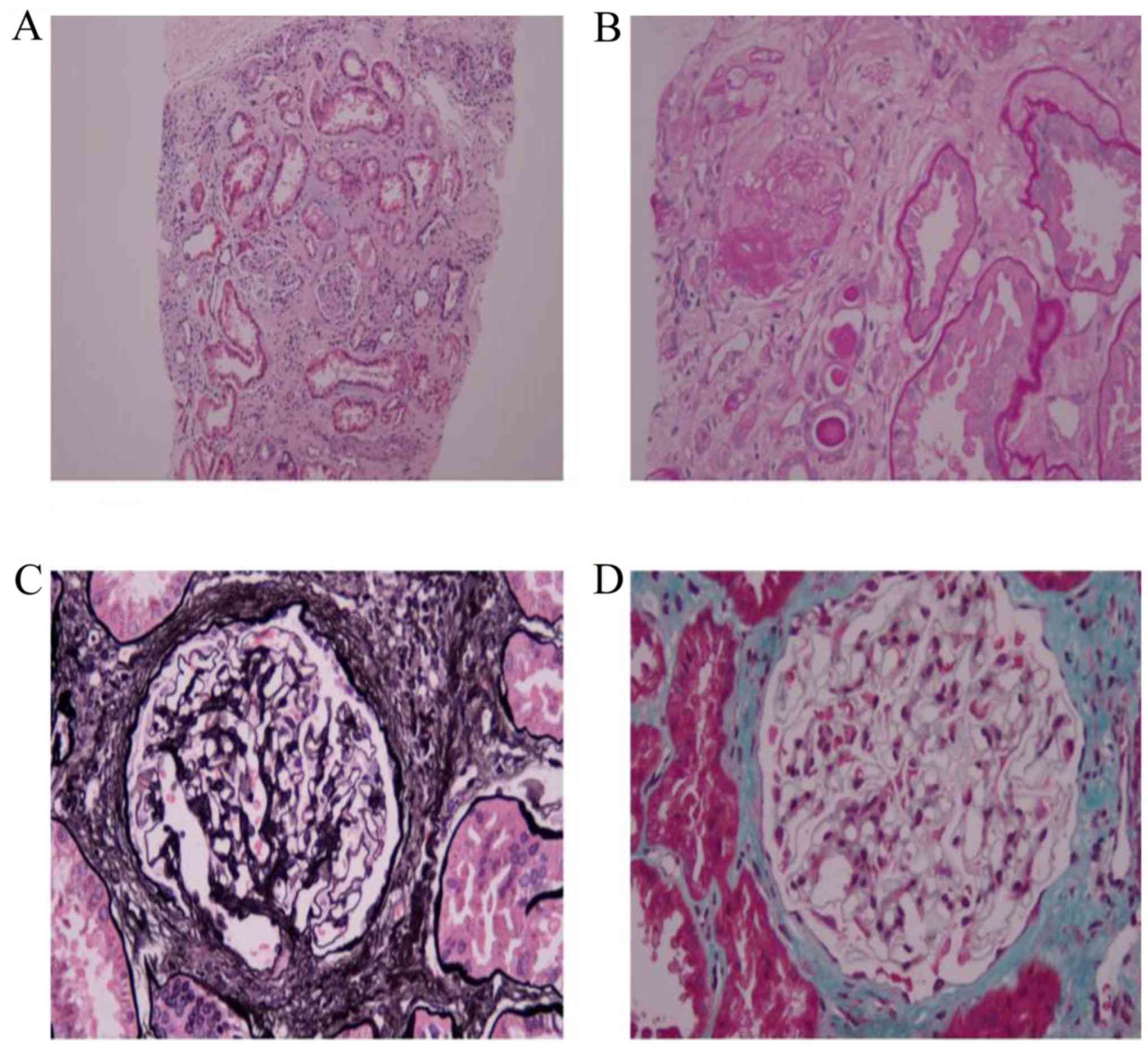
confirm this hypothesis, a kidney biopsy was performed. Routine
clinical examination by specialist including comprehensive light
microscopy, immunofluorescence and electron microscopy examination
indicated the presence of severe chronic interstitial nephritis
(Figs. 1–3), consistent with the biochemical
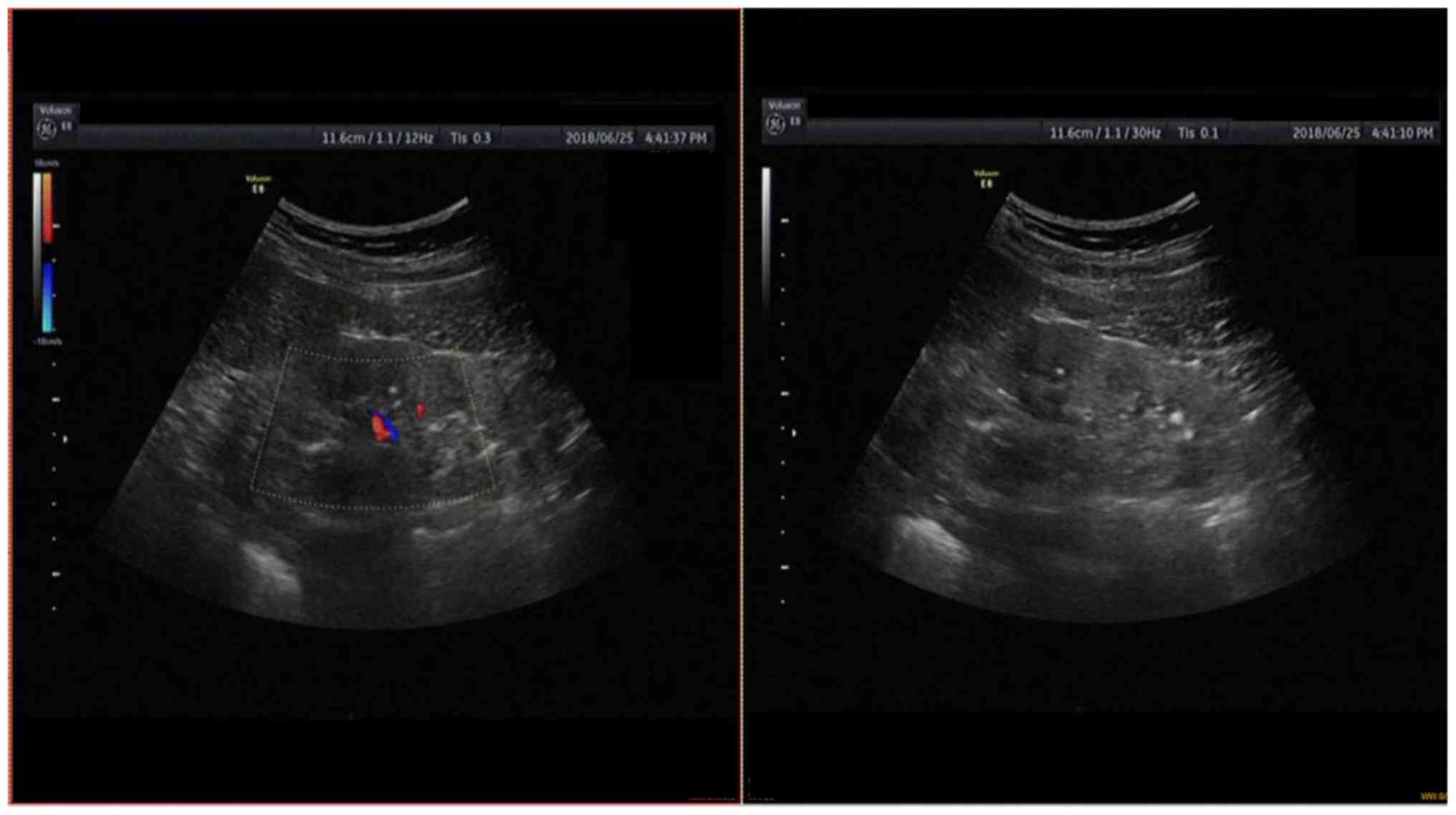
indicators. Furthermore, kidney ultrasonography was performed
(Fig. 4), which indicated an
abnormal echo of the bilateral kidneys (consistent with sonographic
changes in chronic kidney disease), as well as multiple calculi or
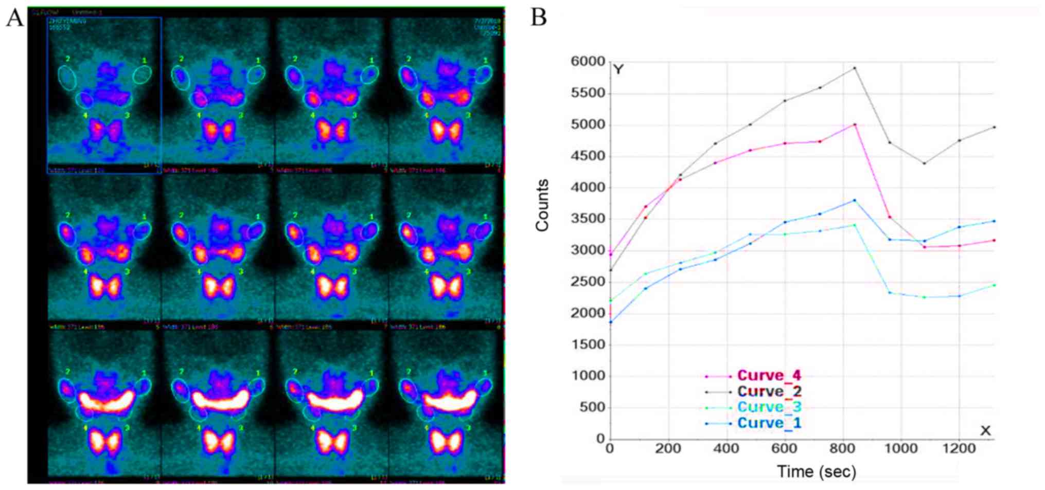
calcifications in the bilateral kidneys. In addition, salivary
gland scintigraphy indicated a decrease in left parotid uptake
tracer function, as well as impairment of secretion and excretion.
However, no abnormal uptake, secretion or excretion were observed
in the right parotid gland and bilateral submandibular glands
(Fig. 5).
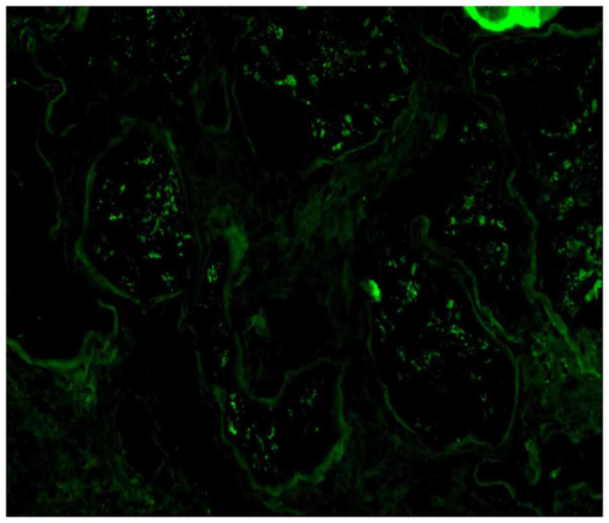 | Figure 1.Immunofluorescence to clarify
diagnosis. magnification, ×400. κ, negative; λ, negative; amyloid
A, negative; fibrin, negative; albumin, reabsorbed droplets visible
in the renal tubules; IgG1, negative; IgG2, negative; IgG3,
negative; IgG4, negative; phospholipase A2 receptor, negative;
hepatitis B surface antigen, negative; hepatitis B e antigen,
negative; hepatitis B core antigen, negative; C4d, glomeruli in the
kidney (−); thrombospondin type 1 domain containing 7A,
negative. |
 | Table I.Laboratory results at presentation and
during the follow-up period. |
Table I.
Laboratory results at presentation and
during the follow-up period.
| Clinical
parametera | First admission | 1 Month | 2 Months | 3 Months | 6 Months | 9 Months | 12 Months |
|---|
| Blood |
|
|
|
|
|
|
|
|
Hemoglobin, g/l (115–150) | 105 | 93 | 110 | 107 | 106 | 107 | 131 |
| White
blood cells, ×109/l (4–10) | 10.2 | 6.0 | 13.8 | 11.4 | 14.4 | 15.4 | 11.0 |
|
Platelets, ×109/l
(125–350) | 320 | 204 | 320 | 246 | 283 | 288 | 245 |
|
Creatinine, µmol/l | 221.3 | 172.4 | 231.8 | 180.7 | 160.0 | 171.4 | 178.7 |
| eGFR,
ml/min/1.73 m2 (CKD-EPI) | 24.6 | 33.2 | 23.2 | 31.4 | 36.4 | 33.4 | 31.8 |
| K, mmol/l
(3.50–5.30) | 3.77 | 3.2 | 3.3 | 4.3 | 3.0 | 4.0 | 3.8 |
| Na,
mmol/l (137.0–147.0) | 135.5 | 139.9 | 143.3 | 141.6 | 142.0 | 143.0 | 142 |
| Cl,
mmol/l (96.0–108.0) | 106.9 | 110.9 | 105.9 | 102.4 | 103.4 | 104.4 | 103.8 |
|
PO4, mmol/l
(0.85–1.51) | 1.4 | – | – | – | 1.2 | 1.4 | 1.1 |
| Ca,
mmol/l (2.11–2.52) | 2.3 | 2.1 | 2.1 | 2.3 | 2.1 | 2.4 | 2.2 |
| MG,
mmol/l (0.75–1.02) | 0.9 | 0.8 | – | – | 0.6 | 0.8 | 0.7 |
| PTH,
pg/ml (15–65) | 475.1 | – | – | – | 371.5 | – | 267.4 |
| ANA
(Speckled) | +(1:100) |
|
|
| +(1:100) | – | +(1:100) |
| dsDNA
(0.0–20.0) | 13.5 |
|
|
| 21.8 | – | 14.9 |
| Urine |
|
|
|
|
|
|
|
| Ca,
mmol/24 h (0.00–6.20) | 1.04 |
|
|
|
|
|
|
| Protein,
mg/24 h (<150) | 453.6 |
|
|
| 383.4 |
| 118.4 |
| ALB,
mg/24 h (<30) | 160.2 |
|
|
| 119.4 |
| 90.7 |
| UPro:UCr
mg/g (0.00–100.00) | 2,990.3 | 412.1 |
|
| 385.2 | 581.0 | 121.7 |
| ALBU:CrU
mg/gCr (0.00–30.00) | 1,797.3 | 109.1 |
|
| 122.9 | 203.7 | 82.3 |
| Beta-2
microglobulin, mg/l (<0.4) | 4.0 | – | – | – | – | – | 3.7 |
| PH
(4.5–8.0) | 6.0 |
| 6.0 | 7.0 | 6.0 | 5.5 | 7.0 |
| SG
(1.003–1.030) | 1.01 | 1.00 | 1.01 | 1.01 | 1.00 | 1.00 | 1.01 |
Based on these results, the patient was diagnosed
with severe chronic interstitial nephritis, stage IV chronic kidney
disease, PSS and anemia due to chronic kidney disease. To address
the PSS-associated chronic interstitial nephritis, the patient was
subjected to continuous treatment with half-dose glucocorticoid.
Given the presence of PSS, initial treatment involved prednisone
(0.5 mg/kg/day), with subsequent slow tapering to 0.5–0.25
mg/kg/day, followed by addition of cyclophosphamide at doses of 800
mg/month over the next 6 months. A 12-month follow-up examination
indicated significant improvement (Table
I).
Discussion
PSS is a common condition involving various organs.
The detection of autoimmune diseases with the exocrine gland as the
target organ has improved with the advancements in detection
methods and a better understanding of the disease (3). Accordingly, the rate of early detection
and diagnosis in cases with PSS has also significantly increased.
In particular, kidney damage as a result of PSS has been receiving
an increasing amount of attention (4). PSS-associated renal damage may be
asymptomatic or may present only as an electrolyte disorder.
Furthermore, as the clinical manifestations of the disease in these
patients are varied (including proteinuria, simple hematuria,
combined hematuria and proteinuria, or nephrotic syndrome and renal
insufficiency), and the prognosis is good, only a small number of
clinical studies have focused on this condition thus far (5). Although the incidence of renal damage
in PSS was previously thought to be low, recent studies indicated a
rate of as high as 33.5% (2,6,7). Most
cases exhibit renal tubular dysfunction, particularly involving the
distal renal tubule. Prominent manifestations include distal renal
tubular acidosis, renal diabetes insipidus and urinary
concentration dysfunction, followed by glomerulonephritis and
partial renal insufficiency (8). As
there are no uniform diagnostic criteria for assessing renal damage
in PSS patients with mild or no symptoms, the condition is
overlooked in most of such cases. Only a small number of such
patients exhibit renal failure at the time of visit, and hence,
early diagnosis is particularly important.
It is recommended that patients with PSS are
screened at least twice a year, including urine protein, urine pH,
urine osmotic pressure, serum creatinine, glomerular filtration
rate and electrolyte levels (9). The
European League Against Rheumatism Sjogren's syndrome disease
activity index may be used to assess renal disease activity during
follow-up (10). Renal biopsy should
be promptly performed, if required, in such cases, and the cause
and extent of renal lesions may be determined via renal pathology
examinations (11).
With regard to treatment, individualized therapy may
be suitable, based on the patient's clinical manifestation and the
type of renal damage. At present, treatment may be divided into
local replacement therapy and systemic treatment (4,12). The
present case was managed by low-dose hormone therapy and regular
follow-up. The renal function was stable and did not deteriorate
during follow-up, which indicated that the treatment was effective.
However, it may be necessary to perform further detailed studies in
order to better understand the disease mechanism, and large-sample
multicenter randomized controlled trials should be performed to
assess the efficacy and safety of drugs including hormones and
immunosuppressants in these patients with different types of renal
damage.
In the clinical setting, patients with kidney stones
and renal insufficiency are frequently encountered, and it is
likely that kidney dysfunction has led to stone obstruction in
these cases. However, it is recommended that clinicians should also
consider systemic diseases, including tumors, rheumatism and immune
diseases, in such patients. Accordingly, careful assessment of the
relevant medical history may help improve the diagnostic accuracy
and avoid kidney failure.
Acknowledgements
Not applicable.
Funding
No funding was received.
Availability of data and materials
The data used for the preparation of the manuscript,
including all relevant raw data, are freely available to any
scientist wishing to use them for non-commercial purposes, without
breaching participant confidentiality. Available from the
corresponding author upon reasonable request.
Authors' contributions
YY, BZ and JH prepared the manuscript, made
substantial contributions to the design of the work and to revise
it critically for important intellectual content. XJ and DD
critically reviewed the manuscript for important intellectual
content and approved the final manuscript. All authors read and
approved the final manuscript.
Ethics approval and consent to
participate
The patient consented to participate and provided
written informed consent.
Patient consent for publication
The patient agreed to the publication of their data
and images.
Competing interests
The authors declare that they have no competing
interests.
References
|
1
|
Nilsson AM, Tufvesson E, Hesselstrand R,
Olsson P, Wollmer P and Mandl T: Increased B-cell activating
factor, interleukin-6, and interleukin-8 in induced sputum from
primary Sjögren's syndrome patients. Scand J Rheumatol. 48:149–156.
2019. View Article : Google Scholar : PubMed/NCBI
|
|
2
|
Jain A, Srinivas BH, Emmanuel D, Jain VK,
Parameshwaran S and Negi VS: Renal involvement in primary Sjögren's
syndrome: A prospective cohort study. Rheumatol Int. 38:2251–2262.
2018. View Article : Google Scholar : PubMed/NCBI
|
|
3
|
Holdgate N and St Clair EW: Recent
advances in primary Sjögren's syndrome. F1000Res. 5(pii): F1000
Faculty Rev. 14122016. View Article : Google Scholar
|
|
4
|
Brito-Zerón P and Ramos-Casals M; EULAR-SS
task force group, : Advances in the understanding and treatment of
systemic complications in Sjögren's syndrome. Curr Opin Rheumatol.
26:520–527. 2014. View Article : Google Scholar : PubMed/NCBI
|
|
5
|
Flores-Chavez A, Kostov B, Solans R,
Fraile G, Maure B, Feijoo-Massó C, Rascón FJ, Pérez-Alvarez R,
Zamora-Pasadas M, García-Pérez A, et al: Severe, life-threatening
phenotype of primary Sjögren's syndrome: Clinical characterisation
and outcomes in 1580 patients (GEAS-SS Registry). Clin Exp
Rheumatol. 36 (Suppl 112):S121–S129. 2018.
|
|
6
|
Zou Y, Ling G, Tian J, Chen J and Ge Y:
Research progress in renal injury relevant to primary Sjögren's
syndrome. Zhong Nan Da Xue Xue Bao Yi Xue Ban. 43:320–326. 2018.(In
Chinese). PubMed/NCBI
|
|
7
|
Weiner SM: Renal involvement in connective
tissue diseases. Dtsch Med Wochenschr. 143:89–100. 2018.(In
German). PubMed/NCBI
|
|
8
|
Narayan R, Abdulla MC, Alungal J and
Krishnadas NC: Distal renal tubular acidosis in Sjögren's syndrome.
Saudi J Kidney Dis Transpl. 29:470–473. 2018. View Article : Google Scholar : PubMed/NCBI
|
|
9
|
Liu X, Li X, Li X, Li Z, Zhao D, Liu S,
Zhang M, Zhang F, Zhu P, Chen J, et al: The efficacy and safety of
total glucosides of peony in the treatment of primary Sjögren's
syndrome: A multi-center, randomized, double-blinded,
placebo-controlled clinical trial. Clin Rheumatol. 38:657–664.
2019. View Article : Google Scholar : PubMed/NCBI
|
|
10
|
Kobayashi I, Okura Y, Ueki M, Tozawa Y,
Takezaki S, Yamada M and Ariga T: Evaluation of systemic activity
of pediatric primary Sjögren's syndrome by EULAR Sjögren's syndrome
disease activity index (ESSDAI). Mod Rheumatol. 29:130–133. 2019.
View Article : Google Scholar : PubMed/NCBI
|
|
11
|
Jasiek M, Karras A, Le Guern V, Krastinova
E, Mesbah R, Faguer S, Jourde-Chiche N, Fauchais AL, Chiche L,
Dernis E, et al: A multicentre study of 95 biopsy-proven cases of
renal disease in primary Sjögren's syndrome. Rheumatology (Oxford).
56:362–370. 2017.PubMed/NCBI
|
|
12
|
Meiners PM, Vissink A, Kroese FG,
Spijkervet FK, Smitt-Kamminga NS, Abdulahad WH, Bulthuis-Kuiper J,
Brouwer E, Arends S and Bootsma H: Abatacept treatment reduces
disease activity in early primary Sjögren's syndrome (open-label
proof of concept ASAP study). Ann Rheum Dis. 73:1393–1396. 2014.
View Article : Google Scholar : PubMed/NCBI
|