Introduction
Biomedical imaging can be classified into magnetic
resonance, near-infrared (NIR) and ionizing radiation (x-rays and
γ-rays) based on electromagnetic spectrum used (1,2). Due to
the spectral characteristics, NIR fluorescent imaging is of
interest for two main reasons: i) The auto-fluorescent background
of biological cells can be significantly reduced under NIR
excitation (3–5); and ii) greater tissue penetration can
be realized owning to lower levels of NIR light scattering compared
with visible light (6,7). Therefore, NIR fluorescent imaging
offers a unique approach for the visualization of physiological
activities in living biological systems, and has become a powerful
tool for diagnostic and therapeutic applications in biology and
medicine (8–11). A number of highly fluorescent probes
based on the NIR region (600–900 nm) have been developed to date
(12–15); however, only a few were approved for
determining hepatic function, cardiac output and liver blood flow,
as well as in ophthalmic angiography by the US Food and Drug
Administration (16–18). As one of the NIR fluorescent probes
authorized by the FDA, indocyanine green (ICG) shows no apparent
cytotoxicity and few side effects, and can be widely applied to
test hepatic output, cardiac function and to perform ophthalmologic
angiography. ICG was also used as a potential photosensitizer
(19–22). Nevertheless, its limitations such as
low quantum yield, optical instability in vivo and
unrestrained leakage in blood vessels have limited its applications
(23). Numerous research groups are
still attempting to design and synthesize improved ICG derivatives
for in vivo imaging (24,25).
The aim of the present study was to introduce a
unique NIR fluorescent probe with high water solubility and good
optical stability. The IR787 probe synthetized in the present study
contained a sulfonic acid group, which was previously shown to
improve water solubility when used in a biological (26), and tricarbocyanine dye, which was
previously used as an NIR fluorescent dye due to its high
extinction coefficient (27). The
results of the current study suggest that the IR787 probe could
provide good optical stability when it was applied to biological
systems.
Materials and methods
Materials and instruments
Proton nuclear magnetic resonance (1H
NMR) was conducted using the Bruker Avance ACF-300/500 MHz
instrument (Bruker Corporation) in DMSO-d6 or
CDCl3 (Tedia Company, Inc.). Tetramethylsilane (TMS,
Sigma-Aldrich; Merck KGaA) was used as an internal standard. The
1H NMR data were calculated based on TMS results and
coupling constants (J values) were expressed in Hz. Molecular
weight was obtained using a 6500 Q-TOF mass analyzer (Agilent
Technologies, Inc.). Potassium
2,3,3-trimethyl-3H-indole-5-sulfonate (compound 1), 3-fluorobenzyl
bromide (compound 2),
2-chloro-1--formyl-3-hydroxymethylenecyclohexene (compound 4) and
ICG were purchased from Sigma Aldrich; Merck KGaA. The human lung
adenocarcinoma epithelial cell line A549 was obtained from the
American Type Culture Collection. Unless otherwise stated, all
reagents were purchased from commercial suppliers and were used
without further purification. Solvents were purified and dried by
standard methods and distilled prior to use when necessary. All
synthesis was performed under an inert atmosphere at 25°C,
magnetically stirred, and monitored by thin-layer chromatography
(TLC) on silica gel GF-254 glass plates.
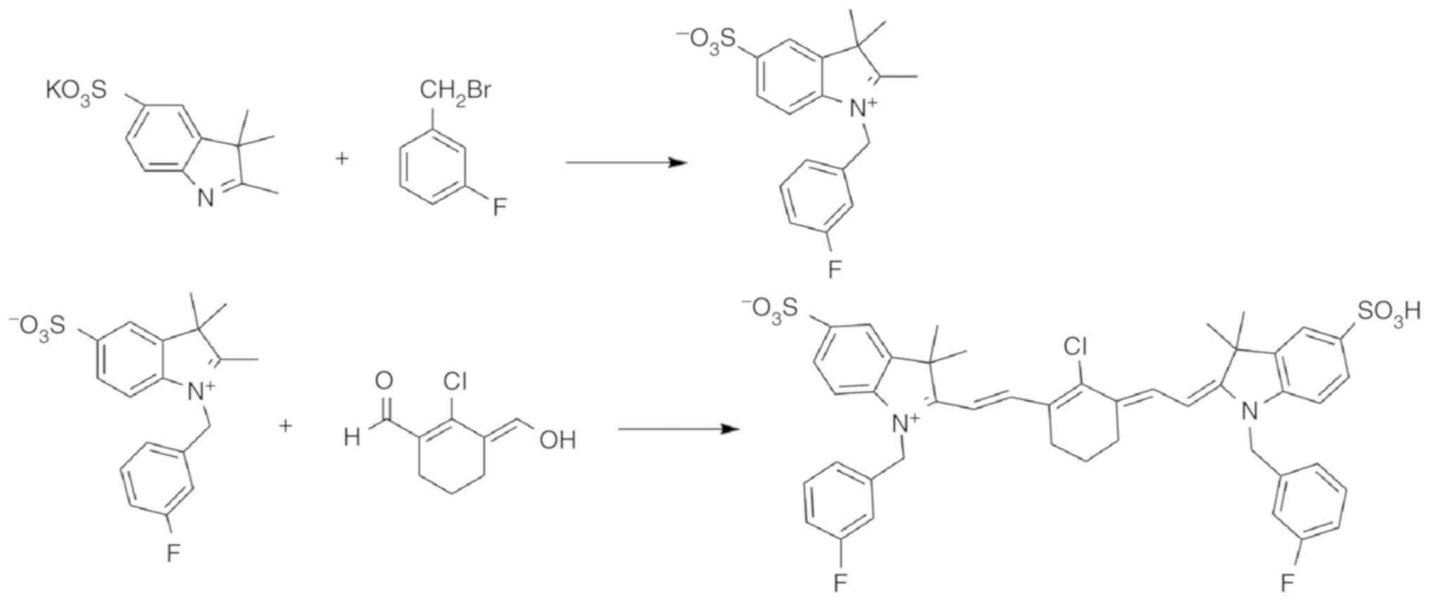
Synthesis of IR787. Synthesis of
1-(3-fluorobenzyl)-2,3,3-trimethyl-3H-indol-1-ium-5-sulfonate
The synthetic route of IR787 was shown in Fig. 1. A mixture of compound 1 (1.24 g;
4.47 mmol), and compound 2 (0.10 g; 5.81 mmol) and toluene
(Shanghai Chemical Reagents Institute Co., Ltd.; 12 ml) was
refluxed with stirring under argon at 90°C for 14 h. The reaction
progress was monitored using TLC [EA/MeOH, 2:1 v/v; Rf value
(retention factor)=0.75]. The resulting mixture was cooled down to
25°C and the resulting precipitate was isolated by vacuum
filtration and then vacuum-concentrated. The crude residue obtained
was purified by column chromatography on silica gel using ethyl
acetate/methanol (Shanghai Chemical Reagents Institute Co., Ltd.),
which yielded a pink solid, 1-(3-fluorobenzyl)-2,3,3-trimethyl
−3H-indol-1-ium-5-sulfonate (compound 3; 1.31 g; yield, 84.4%).
Compound 3 (0.96 g; 2.76 mmol), compound 4 (0.24 g;
1.38 mmol) and anhydrous sodium acetate (Shanghai Chemical Reagents
Institute Co. Ltd.; 0.23 g; 2.76 mmol) were dissolved in 20 ml
acetic anhydride (Shanghai Chemical Reagents Institute Co., Ltd.).
The solution was refluxed with stirring at 75°C for 4 h. After the
reaction, a green solid was produced and purified by column
chromatography on silica gel using DCM/MeOH (10:1 v/v;
Rf=0.30) as an eluent. The resulting green solid was
IR787, and its recovery rate was 33.1% (0.38 g).
Absorption spectra
The absorption spectra of IR787 (1 ml; 0.01 mM) and
ICG (1 ml; 0.01 mM) were measured with the UV-3600 UV visible
spectrophotometer (Shimadzu Corporation) within the wavelength
range of 600–900 nm at 25°C with methanol as the solvent.
Fluorescence spectra
IR787 and ICG were dissolved in methanol as stock
solutions for fluorescence spectral analysis. The stock solutions
were diluted to 0.01 mM in methanol solution. The mixture was
incubated for 3 min at 25°C before measurement and detected using
fluorescence spectroscopy. Fluorescence intensity was measured at
an excitation wavelength of 826 nm.
Cell culture
Human lung adenocarcinoma epithelial cell line A549
was incubated routinely in RPMI-1640 medium supplemented with 10%
(v/v) fetal calf serum, 1% penicillin and 1% streptomycin in an
atmosphere of 5% CO2 and 95% air at 37°C in T25 cell
culture flasks. Cells were split in 1:3 ratios at 80%
confluence.
Cytotoxicity assay
The cytotoxicity of probe IR787 toward A549 cells
was determined in 96-well microplates with ICG as control (28,29).
A549 cells were cultured as described above. Exponentially growing
A549 cells were harvested and seeded in 96-well plates. After a 24
h incubation, cells were treated with probe concentrations from
10–120 µM for 24 h. The plates were incubated for 4 h after the
addition of 10 µl MTT solution (5 mg/ml). The supernatant was
subsequently removed and 100 µl DMSO was added to dissolve the
formazan crystals. The absorbance was measured with a plate reader
(Victor3™; PerkinElmer, Inc.) at a wavelength of 490 nm.
All the experiments were performed in quintuplicate, and data are
presented as the mean ± SD.
Confocal imaging
The A549 cells were transferred to glass-bottom
culture dishes and incubated with 10 µM IR787 or 10 µM ICG in
FBS-free RPMI-1640 medium (Nanjing KeyGen Biotech Co., Ltd.) at
37°C for 30 min. The medium was then removed, and A549 cells were
washed thrice with PBS buffer (pH 7.4). Florescent images were
captured using a confocal microscope with an objective lens
(magnification, ×200). The excitation wavelengths were 787 and 805
nm for IR787 and ICG, respectively.
Statistical analysis
All experiments had at least three replicates for
every tested group. Statistical analysis was performed using SPSS
software (version 20.0; IBM Corp.). Cytotoxicity data and probe
selectivity data were expressed as the mean ± SD. Differences were
tested for statistical significance using a t-test. P<0.05 was
considered to indicate a statistically significant difference.
Results
Characterization of IR787
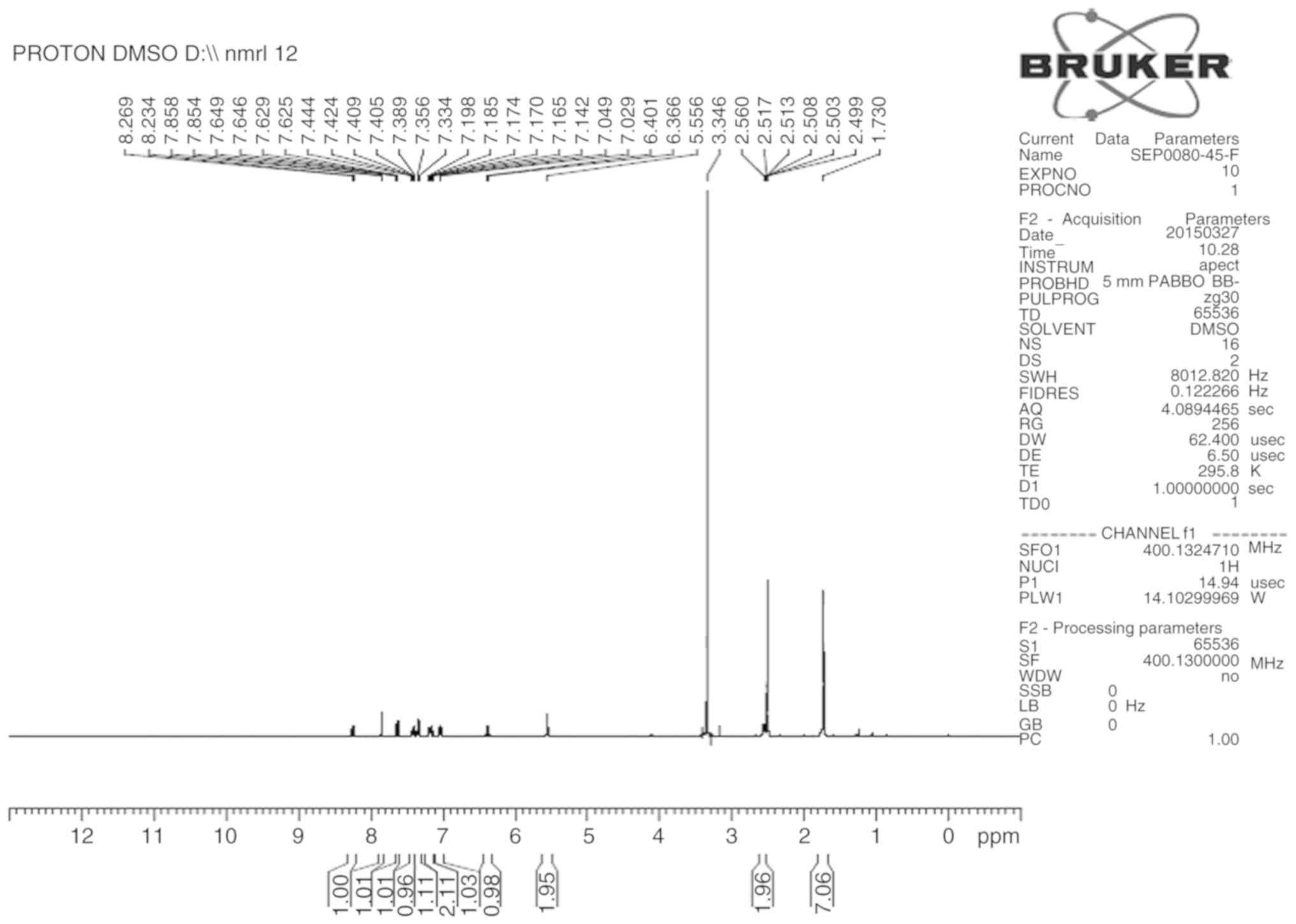
The structure of IR787 was confirmed via
1H-NMR and MS. 1H-NMR (400 MHz,
DMSO-d6): δ (ppm)=1.73 (s, 14H), 2.54–2.57 (t, J=4Hz,
4H), 5.50 (s, 4H), 6.36–6.40 (d, J=14Hz, 2H), 7.02–7.04 (d, J=8Hz,
2H), 7.14–7.19 (m, 4H), 7.33–7.35 (d, J=8.4Hz, 2H), 7.38–7.44 (m,
4H), 7.625–7.629 (d, J=1.6Hz, 1H), 7.646–7.650 (d, J=1.6Hz, 1H),
7.85 (s, 2H), 8.23–8.26 (d, J=14Hz, 2H). TOF-MS m/z: 829.3
[M-H+]−. 1H NMR and MS spectra of
IR787 were shown in Figs. 2 and
3.
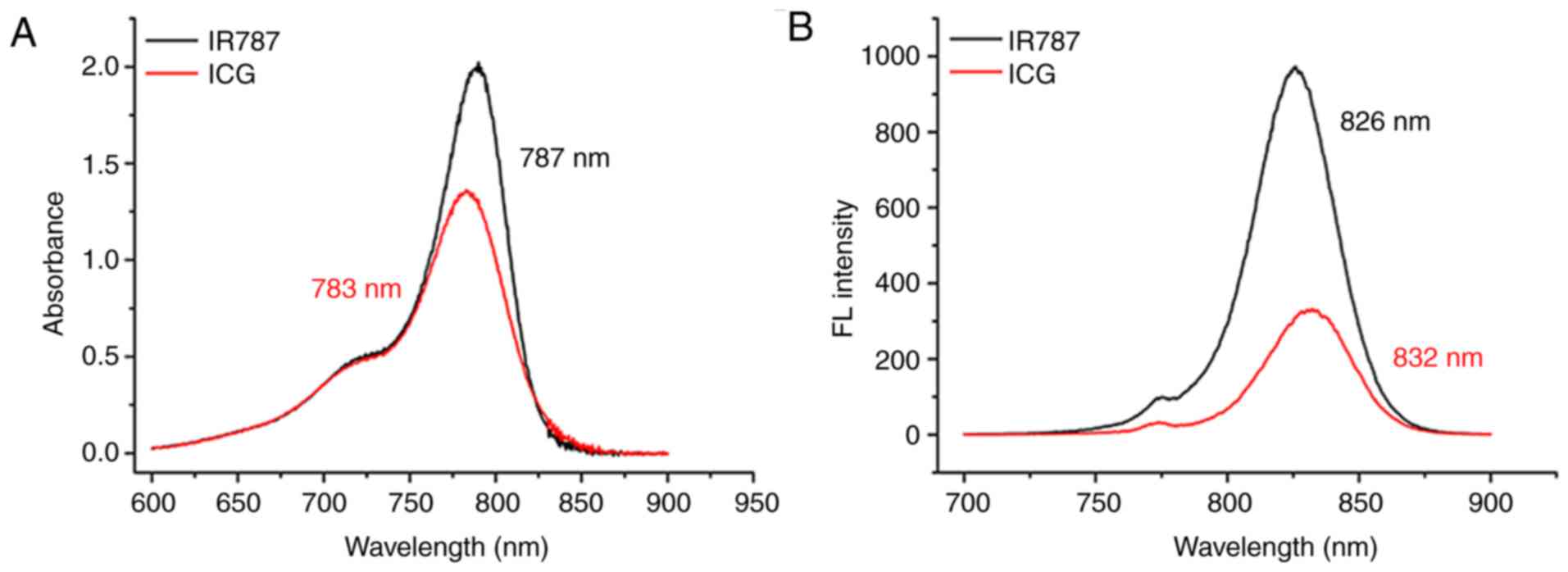
Absorption and fluorescence spectra of
IR787
The absorption and emission spectra of IR787 and ICG
were examined in methanol solution. The maximal absorption
wavelengths of IR787 and ICG were 787 and 783 nm, respectively
(Fig. 4). The results showed that
IR787 exhibited a stronger blue excitation peak than ICG. The
maximal emission wavelengths of IR787 and ICG were 826 and 832 nm,
respectively, and both exhibited a similar emission peak in
methanol solution (Fig. 4). These
results suggested that IR787 may be a promising potential NIR probe
for biological systems.
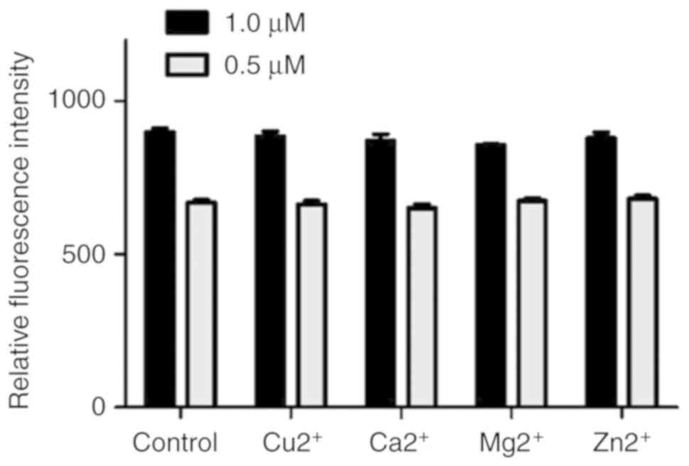
Test for probe selectivity
Considering the complexity of the intracellular
environment, an additional test was performed to determine whether
Cu2+, Ca2+, Mg2+ and
Zn2+ ions were potential interferents to the
photostability of IR787. IR787 was used to determine whether the
ions were potential interferents. The relative fluorescence
intensity of IR787 (1 and 0.5 µM) at 787 nm in the absence or
presence of 200 µM Cu2+, Ca2+,
Mg2+ and Zn2+ ions in 40 mM HEPES buffer
solution at pH 6.50 (excitation wavelength=789 nm) indicated that
the effect of these ions on the measurement was negligible, and the
probe had good photostability (Fig.
5).
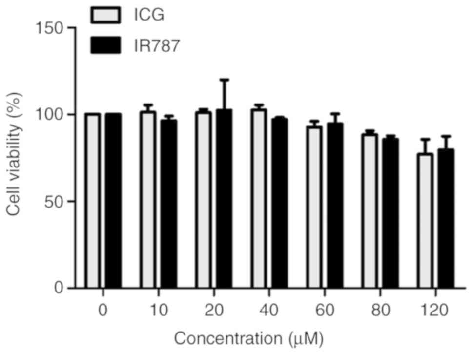
Cytotoxicity assay
The biocompatibility of probes in living cells is an
important consideration (30,31). In
the current study, the ICG and IR787 probes were evaluated in
vitro to determine any cytotoxicity in A549 cells using the MTT
method. The MTT assay was performed with probe concentrations of
10–120 µM. The results indicated that compared with ICG treatment,
the viability of A549 cells treated with IR787 was not
significantly different at any treatment concentration.
Furthermore, A549 cells retained >75% of the control group
viability even when treated with 120 µM ICG or IR787 for 24 h
(Fig. 6). Cytotoxicity assay results
revealed that the viability of A549 cells was not significantly
affected by the probes used in the current study, indicating that
the IR787 probe was safe for in vitro cell imaging.
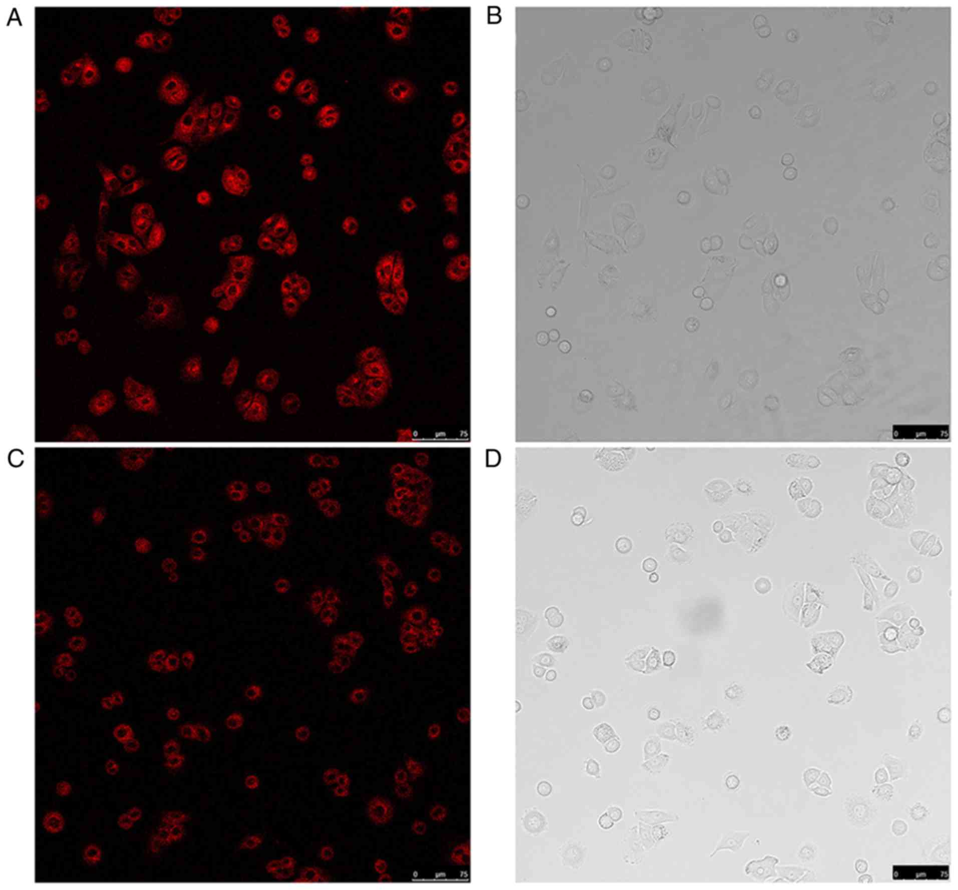
In vitro cell imaging
Due to the low cytotoxicity of IR787, confocal
fluorescence imaging was performed to study its applicability for
living cells. A549 cells were inoculated in 10 µM IR787 or ICG for
30 min. The total time of fluorescence stability (Fig. 7) was 40 min. The distributions of
IR787 and ICG in A549 cells were observed under a fluorescence
microscope. As shown in Fig. 7A and
C, IR787 and ICG could permeate into the membrane with varying
fluorescence intensities. The present results suggested that the
IR-789 had excellent membrane permeability and good
photostability.
Discussion
The application of NIR fluorescent probes to imaging
techniques allows the real-time in vivo cell imaging. In the
present study, a novel NIR fluorescent probe IR787 was designed,
synthesized and its cytotoxicity, optical features and
applicability in living A547 cell imaging were characterized. The
two-step synthetic process of IR787 makes its large-scale
production possible. The stronger excitation and emission
characteristics showed that IR787 had improved optical properties
compared with ICG. The IR787 probe, a derivate of heptamethine
indocyanines, may exhibit a good cytoplasmic localization due to
intracellular accumulation and membrane permeability (32). MTT assay results suggested that IR787
had low cytotoxic effects and the viability of A549 cells was
>75% even when incubated with 120 µM IR787. Further experiments
demonstrated that IR787 could effectively test in living cells.
In conclusion, the current study successfully
developed a novel and simple NIR fluorescent probe for living cell
imaging. Compared with the previously reported NIR fluorescent
probe ICG, the IR787 probe had improved prospects for intracellular
imaging. IR787 is hoped to play an important role in understanding
cell biology, pharmacology, and disease diagnosis. In future work,
this probe will be used as a fluorescent probe with specific
chemically-conjugated ligands to improve its selectivity and
broaden the application potential.
Acknowledgements
Not applicable.
Funding
Not applicable.
Availability of data and materials
All data generated or analyzed during this study are
included in this published article.
Authors' contributions
MW contributed to the conception, design, writing
and revision of the manuscript. YZ and JZ contributed to the
acquisition of data and the analysis of data.
Ethics approval and consent to
participate
Not applicable.
Patient consent for publication
Not applicable.
Competing interests
The authors declare that they have no competing
interests.
References
|
1
|
Pansare VJ, Hejazi S, Faenza WJ and
Prud'Homme RK: Review of long-wavelength optical and NIR imaging
materials: Contrast agents, fluorophores and multifunctional nano
carriers. Chem Mater. 24:812–827. 2012. View Article : Google Scholar : PubMed/NCBI
|
|
2
|
Chan M and Almutairi A: Nanogels as
imaging agents for modalities spanning the electromagnetic
spectrum. Mater Horiz. 3:21–40. 2015. View Article : Google Scholar : PubMed/NCBI
|
|
3
|
Galanzha EI, Shashkov EV, Tuchin VV and
Zharov VP: In vivo multispectral, multiparameter, photoacoustic
lymph flow cytometry with natural cell focusing, label-free
detection and multicolor nanoparticle probes. Cytometry A.
73A:884–894. 2008. View Article : Google Scholar
|
|
4
|
Dip F, Roy M, Lo Menzo E, Simpfendorfer C,
Szomstein S and Rosenthal RJ: Routine use of fluorescent
incisionless cholangiography as a new imaging modality during
laparoscopic cholecystectomy. Surg Endosc. 29:1621–1626. 2015.
View Article : Google Scholar : PubMed/NCBI
|
|
5
|
Kiviharju K, Salonen K, Moilanen U,
Meskanen E, Leisola M and Eerikäinen T: On-line biomass
measurements in bioreactor cultivations: Comparison study of two
on-line probes. J Ind Microbiol Biotechnol. 34:561–566. 2007.
View Article : Google Scholar : PubMed/NCBI
|
|
6
|
Zhang X, Gu YQ and Chen HY: Synthesis of
biocompatible near infrared fluorescence Ag2S quantum dot and
application in bioimaging. J Innov Optic Health Sci. 7:13500592014.
View Article : Google Scholar
|
|
7
|
Sampaio PN, Sales KC, Rosa FO, Lopes MB
and Calado CR: High-throughput FTIR-based bioprocess analysis of
recombinant cyprosin production. J Ind Microbiol Biotechnol.
44:49–61. 2017. View Article : Google Scholar : PubMed/NCBI
|
|
8
|
Yuan L, Lin W, Zhao S, Gao W, Chen B, He L
and Zhu S: A unique approach to development of near-infrared
fluorescent sensors for in vivo imaging. J Am Chem Soc.
134:13510–13523. 2012. View Article : Google Scholar : PubMed/NCBI
|
|
9
|
Jumarie C, Séïde M, Marcocci L,
Pietrangeli P and Mateescu MA: Diamine oxidase from white pea
(Lathyrus sativus) combined with catalase protects the human
intestinal caco-2 cell line from histamine damage. Appl Biochem
Biotechnol. 182:1171–1181. 2017. View Article : Google Scholar : PubMed/NCBI
|
|
10
|
Wang R and Yu C, Yu F, Chen L and Yu C:
Molecular fluorescent probes for monitoring pH changes in living
cells. TrAC Trends Anal Chem. 29:1004–1013. 2010. View Article : Google Scholar
|
|
11
|
Luan LQ, Fang WJ, Liu W, Tian MG, Ni YX,
Chen X, Yu XQ, He J, Yang Y and Li XG: 4-tert-butylphenoxy
substituted phthalocyanine with RGD motif as highly selective
one-photon and two-photon imaging probe for mitochondria and cancer
cell. J Porphyrins Phthalocyanines. 20:1–10. 2016. View Article : Google Scholar
|
|
12
|
Xing J, Zhou G, Sun C, Zhang H, Chen B,
Zong X, Cai J and Ji M: Synthesis and characterization of a novel
near-infrared fluorescent probe for applications in imaging a549
cells. Biotechnol Lett. 38:1–6. 2016. View Article : Google Scholar : PubMed/NCBI
|
|
13
|
Kiyose K, Kojima H, Urano Y and Nagano T:
Development of a ratiometric fluorescent zinc ion probe in
near-infrared region, based on tricarbocyanine chromophore. J Am
Chem Soc. 128:6548–6549. 2006. View Article : Google Scholar : PubMed/NCBI
|
|
14
|
Sun C, Cai J, Chen J, Wu Y, Wang P, Zhou
G, Zong X, Chen B, Lv Y and Ji M: The synthesis of a novel
near-infrared fluorescent probe and its application in imaging of
living cells. Appl Biochem Biotechnol. 175:1644–1650. 2015.
View Article : Google Scholar : PubMed/NCBI
|
|
15
|
Sun C, Wu Y, Cai J, Wang P, Zong X, Zhou
G, Li L and Ji M: Synthesis of a near-infrared fluorescent probe
and its application in imaging of MCF-7 cells. Biotechnol Lett.
36:1203–1207. 2014. View Article : Google Scholar : PubMed/NCBI
|
|
16
|
Liu K, Shang H, Meng F, Liu Y and Lin W: A
novel near-infrared fluorescent platform with good photostability
and the application for a reaction-based Cu (2+) probe in living
cells. Talanta. 147:193–198. 2016. View Article : Google Scholar : PubMed/NCBI
|
|
17
|
Li C, Greenwood TR, Glunde K and Bhujwalla
ZM: Synthesis and characterization of glucosamine-bound
near-infrared probes for optical imaging. Org Lett. 8:3623–3626.
2006. View Article : Google Scholar : PubMed/NCBI
|
|
18
|
Luo S, Zhang E, Su Y, Cheng T and Shi C: A
review of NIR dyes in cancer targeting and imaging. Biomaterials.
32:7127–7138. 2011. View Article : Google Scholar : PubMed/NCBI
|
|
19
|
Lim SY, Hong KH, Kim DI, Kwon H and Kim
HJ: Tunable heptamethine-azo dye conjugate as an NIR fluorescent
probe for the selective detection of mitochondrial glutathione over
cysteine and homocysteine. J Am Chem Soc. 136:7018–7025. 2014.
View Article : Google Scholar : PubMed/NCBI
|
|
20
|
Ziemiński K and Kowalskawentel M: Effect
of different sugar beet pulp pretreatments on biogas production
efficiency. Appl Biochem Biotechno. 181:1211–1227. 2017. View Article : Google Scholar
|
|
21
|
Walther CG, Whitfield R and James DC:
Importance of interaction between integrin and actin cytoskeleton
in suspension adaptation of CHO cells. Appl Biochem Biotechnol.
178:1286–1302. 2016. View Article : Google Scholar : PubMed/NCBI
|
|
22
|
El-Daly SM, Gamal-Eldeen AM, Abo-Zeid MA,
Borai IH, Wafay HA and Abdel-Ghaffar AR: Photodynamic therapeutic
activity of indocyanine green entrapped in polymeric nanoparticles.
Photodiagnosis Photodyn Ther. 10:173–185. 2013. View Article : Google Scholar : PubMed/NCBI
|
|
23
|
Funayama T, Sakane M, Abe T, Hara I, Ozeki
E and Ochiai N: Intraoperative near-infrared fluorescence imaging
with novel indocyanine green-loaded nanocarrier for spinal
metastasis: A preliminary animal study. Open Biomed Eng J. 6:80–84.
2012. View Article : Google Scholar : PubMed/NCBI
|
|
24
|
Shafirstein G, Bäumler W, Hennings LJ,
Siegel ER, Ran F, Moreno MA, Webber J, Jackson C and Griffin RJ:
Indocyanine green enhanced n ear infrared laser treatment of murine
mammary carcinoma. Int J Cancer. 130:1208–1215. 2012. View Article : Google Scholar : PubMed/NCBI
|
|
25
|
Schubert GA, Barth M and Thomé C: The use
of indocyanine green videography for intraoperative localization of
intradural spinal tumors. Spine. 35:E212–E217. 2010. View Article : Google Scholar : PubMed/NCBI
|
|
26
|
Zhou LC, Zhao GJ, Liu JF, Han KL, Wu YK,
Peng XJ and Sun MT: The charge transfer mechanism and spectral
properties of a near-infrared heptamethine cyanine dye in alcoholic
and aprotic solvents. J Photochem Photobiol Chem. 187:305–310.
2007. View Article : Google Scholar
|
|
27
|
Tang B, Liu X, Xu K, Huang H, Yang G and
An L: A dual near-infrared pH fluorescent probe and its application
in imaging of HepG2 cells. Chem Commun (Camb). 41:3726–3728. 2007.
View Article : Google Scholar
|
|
28
|
Seidl K and Zinkernagel AS: The MTT assay
is a rapid and reliable quantitative method to assess
staphylococcus aureus induced endothelial cell damage. J Microbiol
Methods. 92:307–309. 2013. View Article : Google Scholar : PubMed/NCBI
|
|
29
|
Skehan P, Storeng R, Scudiero D, Monks A,
McMahon J, Vistica D, Warren JT, Bokesch H, Kenney S and Boyd MR:
New colorimetric cytotoxicity assay for anticancer-drug screening.
J Natl Cancer Inst. 82:1107–1112. 1990. View Article : Google Scholar : PubMed/NCBI
|
|
30
|
Funayama T, Sakane M, Abe T and Ochiai N:
Photodynamic therapy with indocyanine green injection and
near-infrared light irradiation has phototoxic effects and delays
paralysis in spinal metastasis. Photomed Laser Surg. 30:47–53.
2012. View Article : Google Scholar : PubMed/NCBI
|
|
31
|
Liang Y, Gao W, Peng X, Deng X, Sun C and
He B: Near infrared light responsive hybrid nanoparticles for
synergistic therapy. Biomaterials. 100:76–90. 2016. View Article : Google Scholar : PubMed/NCBI
|
|
32
|
Nakayama A, Bianco AC, Zhang CY, Lowell BB
and Frangioni JV: Quantitation of brown adipose tissue perfusion in
transgenic mice using near-infrared fluorescence imaging. Mol
Imaging. 2:37–49. 2003. View Article : Google Scholar : PubMed/NCBI
|