Introduction
The incidence of type 2 diabetes (T2DM) continues to
increase yearly (1,2). Patients with T2DM are 2-4 times higher
than non-diabetics due to coronary heart disease (CHD) (3). Type 2 diabetes mellitus and coronary
heart disease are common diseases. A growing number of patients
with diabetes mellitus and heart disease require surgery. Due to
recent advances in medical technology, the number of patients with
T2DM that is complicated with CHD, who also undergo surgery, has
increased (4). The selection and
application of perioperative anesthetic drugs in patients with T2DM
is important.
A previous study in rats demonstrated that autophagy
is closely associated with T2DM (5,6) and
myocardial ischemia reperfusion injury (MIRI) (7). Mammalian target of rapamycin (mTOR) is
a key regulator of cell growth, autophagy, translation and survival
(8). The phosphorylated
(p)-mTOR/mTOR signaling pathway serves a role in a variety of cell
autophagy processes (9). LC3II is an
autophagy-related factor, is the most widely studied family
protein, is associated with autophagosome development and
maturation and is used to monitor autophagic activity (10). The selection and application of
perioperative anesthetic drugs in patients with T2DM that is
complicated with coronary heart disease, is becoming increasingly
important. Propofol, a widely used intravenous anesthetic, is
reported to exhibit myocardial protective effects (11). There have been a number of reports
regarding the effect of propofol on myocardial ischemia-reperfusion
injury (12-14).
However, the effect of propofol on diabetes mellitus with
myocardial ischemia-reperfusion injury has not yet been reported.
The underlying mechanism of this interaction is yet to be
determined. The effects of propofol on LC3II, mTOR and p-mTOR
expression in diabetic rats with MIRI has, to the best of our
knowledge, not yet been investigated. In the current study, the
effects of propofol on myocardial ischemia reperfusion in rats with
T2DM is investigated. The present study also investigated whether
LC3II, mTOR and p-mTOR serve roles in T2DM rats with MIRI, and
investigated the role that autophagy serves in this process.
Materials and methods
Preparation of experimental rats
The current study has been examined by the Animal
Welfare and Ethical Committee of Hebei University. A total of 50
healthy Sprague-Dawley male rats (age, 6-8 weeks; weight, 200-220
g) were provided by Hebei Experimental Animal Center [certificate
no. SSCXK (Ji) 2013-1-1003]. Rats were randomly divided into five
groups, with 10 rats in each group. The groups were as follows:
Sham-operation (group CC), ischemia-reperfusion (group CI),
ischemia-reperfusion plus low-dose propofol (group LP),
ischemia-reperfusion plus moderate-dose propofol (group MP) and
ischemia-reperfusion plus high-dose propofol (group HP).
Preparation of type 2 diabetic
rats
T2DM were induced by the following protocol. All
groups were fed a high-sugar, high-fat diet with free access to
drinking water, at 22˚C and 50% humidity, and maintained in a 12/12
h light and dark cycle. Rat feed was provided by Hebei Experimental
Animals Center (Hebei, China). After 8 weeks, streptozotocin (30
mg/kg body weight; Beijing Solarbio Science & Technology Co.,
Ltd.) was injected into the abdomen to establish the T2DM model.
Blood was obtained from the tail vein of rats and fasting glucose
was detected. The model was considered to be successful if fasting
glucose was >14 mol/l (15-18).
Establishment of myocardial
ischemia-reperfusion injury model in rats
Subsequently, rats were anesthetized
intraperitoneally with 1% pentobarbital (40 mg/kg). After
anesthesia, tracheal intubation and mechanical ventilation were
performed to connect the small animal ventilator (Hebei Medical
University Instrument Factory). The femoral vein puncture catheter
was connected using a micro infusion pump for the infusion of
physiological saline or propofol. The right common carotid artery
was connected to a pressure transducer to monitor HR, LVSP and ±
dp/dtmax. Following the stabilization of blood pressure
and HR, rats were placed in the right lateral decubitus position.
The left thoracotomy, scissors and thread were placed below the
left anterior descending coronary artery and polyethylene tubes
were inserted between the myocardium and the ligature.
Rats in the CC group received an intravenous
infusion of physiological (0.9%) saline (3 ml/kg-1 •
h-1) for 10 min, with no ligation. Rats in the CI group
received a 10 min intravenous infusion of normal saline (3
ml/kg-1 • h-1) followed by occlusion of the
left anterior descending coronary artery for 30 min and 2 h of
reperfusion. The LP, MP and HP groups rats received a 10 min
intravenous infusion of propofol (6 mg/kg-1 •
h-1), (12 mg/kg-1 • h-1) and (24
mg/kg-1 • h-1) (equal volume of drugs with
different concentrations), respectively. Each administration was
followed by the occlusion of the left anterior descending coronary
artery for 30 min and 2 h of reperfusion. Euthanasia was performed
2 h after reperfusion to obtain myocardial tissue.
Powerlab/8s (ADInstruments) was used to record heart
rate (HR), left ventricular systolic pressure (LVSP) and the rate
(dP/dtmax) of left ventricular pressure rise in early
systole (± DP/dtmax).
ELISA method
Using an ELISA, cardiac troponin T (cTnT) levels,
superoxide dismutase (SOD) and malondialdehyde (MDA) were measured.
(cTnT ELISA kit; cat. no. E02T0017; SOD ELISA kit; cat. no. a001-1;
MDA ELISA kit; cat. no. E02M0023; all from Shanghai Lanji
Biotechnology Co., Ltd.).
At the time of reperfusion for 2 h, 2 ml blood was
collected from the abdominal aorta of rats in each group and
centrifuged at 1,000 x g/min for 15 min at 4˚C. The supernatant was
taken and stored in refrigerator at -80˚C for testing. All
specimens and kits were rewarmed after the collection of each
group. In the blank microholes of the enzyme standard plate, 100 µl
standard substances were added, 100 µl samples were added and 100
µl PBS (pH 7.0-7.2) was added to the blank control group. A total
of 50 µl enzyme labeling solution was added to each hole (except
the blank control hole). The enzyme standard plate was sealed and
incubated at 37˚C for 1 h at a constant temperature. The seal film
was removed, the liquid discarded and the plate was thoroughly and
dry rinsed. A total of 50 µl chromogenic agent A and 50 µl
chromogenic agent B was added to each well, agitated and mixed in
the dark at 37˚C for 10-15 min. A total of 50 µl termination
solution was added to each well to terminate the reaction (blue
color immediately turns to yellow). After 10 min, the blank well
was adjusted to zero and the absorbance [optical density (OD)
value] of each well was measured in sequence with the wavelength of
450 nm. The linear regression equation of the standard curve was
calculated according to the standard concentration and the
corresponding OD value, and then the corresponding sample
concentration was calculated by the regression equation according
to the OD value of the sample.
Western blotting assay
After anesthesia, the rats' necks were dislocated
and euthanized. The left anterior descending branch was collected
to obtain samples of myocardial tissue and the ultrastructure of
myocardial cells was observed using electron microscopy. Western
blot analysis was used to detect LC3II, mTOR and p-mTOR expression
in myocardial tissue.
Myocardial tissue from the ischemic zone were
solubilized in RIPA lysing buffer [50 mM Tris (pH 7.4), 150 mM
NaCl, 1% NP-40, 0.5% sodium deoxycholate, 1 mM PMSF]. Protein
quantification was carried out by the modified Lowery method.
Protein samples (60 µg) were separated by 10% SDS-PAGE and
transferred to PVDF membranes (Abcam). The membranes were blocked
in 5% skim milk for 2 h at room temperature and incubated with the
primary antibodies (1:1,000) LC3Ⅱ (Abcam; cat. no. ab48394), mTOR
(Abcam; cat. no. ab2732) or p-mTOR (Abcam; cat. no. ab109268)
overnight at 4˚C. Next, the membranes were incubated with secondary
antibodies (horseradish peroxidase-labeled goat anti-mouse IgG;
Abcam; cat. no. ab205719; 1:1,000) at room temperature for 2 h and
were washed three times (5 min/time) with TBST buffer
(0.05%Tween-20 in TBS). GAPDH (ProteinTech Group, Inc.; cat. no.
60004-1-Ig, 1:2,000) was used as an internal reference. Proteins
were visualized using Immobilon ECL Ultra Western HRP Substrate
(Millipore, cat. no. WBULS0500) and the blots were quantified using
BandScan 5.0 software (Glyko, Inc.).
Electron microscopy
After reperfusion for 2 h, the rats were euthanized
and the myocardial ischemic area was taken and cut into 1 mm
pieces. A total of 4% glutaraldehyde solution was used for fixation
at 4˚C for 24 h and washed with 0.1 mol/l PBS 3 times. 1% osmium
acid -1.5% potassium ferricyanide at 4˚C was used to fix the tissue
again for 1.5 h, 0.1 mol/l PBS rinse 3 times. Ethanol-pyruvate was
dehydrated step by step. Anhydrous acetone-epoxy resin 618
embedding agent (1:1) was fixed at 35˚C for 1.5 h. The pure 618
embedding agent was fixed at 35˚C for 12 h, 45˚C for 12 h and 60˚C
for 3 days. Ultrathin sections of 70-80 nm were prepared. Uranium
acetate and lead citrate were dyed at 22˚C for 5 min, then washed
with distilled water. The ultrastructure of myocardium was observed
under transmission electron microscope (Hitachi, Ltd.; model:
h-7500) (magnification, x15,000).
Statistical analyses
SPSS v 16.0 (SPSS, Inc.) was used for all
statistical analyses. Data are presented as the mean ± SD.
Hemodynamics were compared and analyzed within groups and between
groups using multivariate analysis of variance (ANOVA). The level
of cTnT, SOD, MDA, LC3II, mTOR and p-mTOR were compared using a
one-way ANOVA, followed by a post hoc Tukey's test. P<0.05 was
considered to indicate a statistically significant difference. All
experiments were performed in triplicates.
Results
Hemodynamic parameters
No significant differences were exhibited in HR,
LVSP, +dp/dt and -dp/dt, prior to ischemia, in all groups. When
compared with measurements taken prior to ischemia, HR, LVSP,
+dp/dt and -dp/dt significantly decreased following ligation for 30
min, except for the CC group (P<0.05;
Figs. 1-4). Compared with the measurement taken at 30 min after
ligation, HR, LVSP, +dp/dt and -dp/dt significantly decreased at 2
h after reperfusion compared with the before group, except for the
CC group (P<0.05).
HR, LVSP, +dp/dt and -dp/dt were significantly
increased in the LP, MP and HP groups compared with the results at
30 min following ligation and 2 h following reperfusion
(P<0.05). The MP group HR, LVSP, +dp/dt and -dp/dt levels
increased significantly, followed by the LP and HP group
(P<0.05;
Figs. 1-4).
Electron microscopy
At 2 h following reperfusion, ultrastructural
changes of myocardial tissue were observed in all groups using
electron microscopy. The arrangement of myocardial cells in the CC
group was more orderly and no fusion was observed between
mitochondrial cristae and membranes. In the CI group, myocardial
cells were disordered, highly edematous, mitochondrial cristae and
membranes were dissolved and changes in the vacuole were observed.
Compared with the CI group, the myocardial cells in the MP group
were slightly disordered, exhibited mild edema and exhibited partly
dissolved mitochondrial cristae and membrane rupture. The degree of
injury in myocardial cells and mitochondria in the LP and HP group
was between the CI and MP group (Figs.
5-9).
Serum concentrations of cTnT, SOD and
MDA
Following 2 h of reperfusion, compared with the CC
group, SOD significantly decreased in all other groups (P<0.05),
and the cTnT and MDA levels significantly increased (P<0.05;
Table I). Compared with the CI
group, the concentration of SOD in the MP group decreased
significantly (P<0.05) and the concentrations of cTnT and MDA
were decreased (P<0.05). The LP and HP group also exhibited
significant changes in SOD, cTnT and MDA levels (P<0.05;
Table I).
 | Table IcTnT, SOD and MDA concentration of
blood serum of myocardial ischemia reperfusion injury in rats with
diabetes. |
Table I
cTnT, SOD and MDA concentration of
blood serum of myocardial ischemia reperfusion injury in rats with
diabetes.
| Group | cTnT (ng/ml) | SOD (U/ml) | MDA (nmol/ml) |
|---|
| CC | 4.34±1.34 | 160.27±5.15 | 2.75±0.55 |
| CI |
15.26±0.74a |
112.49±7.95a |
5.21±0.24a |
| LP |
12.62±1.00a,b |
122.94±5.38a,b |
4.51±0.20a,b |
| MP |
9.50±1.90a-c |
147.05±7.83a-c |
3.70±0.16a-c |
| HP |
12.40±1.66a,b |
130.01±6.16a,b |
4.40±0.27a,b |
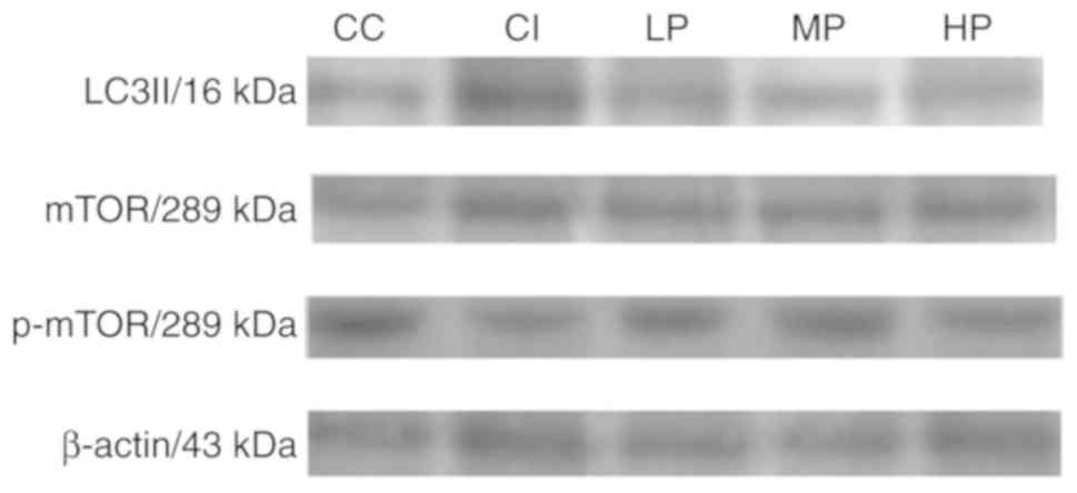
Protein expression of LC3Ⅱ, mTOR and
p-mTOR
Following 2 h of reperfusion, compared with the CC
group, LC3II and mTOR expression was significantly increased in all
other groups (P<0.05), and p-mTOR expression was significantly
decreased (P<0.05). Compared with the CI group, MP group mTOR
expression decreased significantly (P<0.05) and p-mTOR
expression increased significantly (P<0.05), The LP and HP group
also exhibited significant changes in mTOR and p-MTOR expression
(P<0.05; Table II; Fig. 10).
| Table IIThe expression levels of LC3 II and
mTOR/p-mTOR of ischemia-reperfusion myocardium in jury in diabetes
2 rats (n=7, x±s). |
Table II
The expression levels of LC3 II and
mTOR/p-mTOR of ischemia-reperfusion myocardium in jury in diabetes
2 rats (n=7, x±s).
| Group | LC3Ⅱ | mTOR | p-mTOR |
|---|
| CC | 0.30±0.04 | 1.06±0.04 | 0.88±0.02 |
| CI |
1.03±0.12a |
1.41±0.03a |
0.67±0.04a |
| LP |
0.76±0.09a,b |
1.25±0.04a,b |
0.78±0.02a,b |
| MP |
0.51±0.08a-c |
1.18±0.06a-c |
0.84±0.01a-c |
| HP |
0.77±0.13a,b |
1.28±0.02a,b |
0.78±0.05a,b |
Discussion
The polypeptide subunit of troponin complex, cTnT,
is a specific antigen of cardiac myocytes. Elevated serum cTnT can
reflect myocardial structural protein damage. A parallel
relationship exists between cTnT level and the degree of myocardial
cell injury (19). Therefore, cTnT
is used as a specific marker of myocardial injury and due to it
being elevated in patients with coronary artery disease or heart
failure, has also been investigated as a prognostic marker
(20). Among the number of clinical
and biochemical indices that are used for the diagnosis of
myocardial injury, cTnT is considered to be the ‘gold
standard’.
The current study demonstrated that cTnT serum
levels in the LP, MP and HP groups were decreased, and HR and LVSP
levels in the propofol groups were increased, when compared with
the CI group. These results revealed that propofol exhibits
myocardial protective effects on rats with T2DM and the medium dose
was indicated to be most effective.
SOD is a natural oxygen free radical scavenger that
is located in the myocardium and is also an important antioxidant
enzyme. SOD activity can reflect the scavenging ability of the
organism to produce oxygen free radicals. In the process of
myocardial ischemia reperfusion, a large amount of oxygen free
radicals is produced, which leads to a decrease in SOD
concentration and activity (21). A
previous study has demonstrated that SOD content can reflect the
severity of myocardial injury (22).
However, previous reports investigating the beneficial effects of
other plant extracts on I/R induced injury revealed that improved
conditions were due to the antioxidant properties of their
constituents (23-25).
MDA is the most important metabolite of lipid
peroxidation in cardiac myocytes and has been indicated to
cross-link proteins and nucleic acids in cardiomyocytes. During
cardiomyocyte mutation, senescence, denaturation and death, MDA
content has been revealed to increase during myocardial cell
injury. Therefore, the detection of MDA is often used to assess the
extent of myocardial oxidative damage during myocardial
ischemia-reperfusion injury (7).
The results of the current study demonstrated that
SOD concentrations in the LP, MP and HP groups were increased, and
MDA levels in the propofol groups were decreased. These results
suggested that propofol exhibits a myocardial protective effect on
rats with T2DM.
Under normal conditions, 60% of the energy required
for cardiomyocytes to function is gained from fatty acid oxidation.
In ischemia and hypoxia conditions, the energy needed for the
operation of myocardial cells is mainly obtained via the anaerobic
glycolysis of glucose (26). In
patients with diabetes, glucose and lipid metabolism are
disordered, and the use of energy gained from anaerobic glycolysis
is impaired. In patients with diabetes, when myocardial cells are
ischemic, they may suffer from energy supply disorder and their
self-protection ability decreases (27). These results suggest that diabetes
aggravates myocardial injury.
Anesthesia with intravenous propofol is often used
to inhibit lipid peroxidation, improve mitochondrial function
(26), protect the myocardium and
reduce MIRI in rats (27). Propofol
also improves the function of vascular endothelial cells and
promotes anti-apoptotic protein expression, and can therefore
reduce MIRI in T2DM. The influence of propofol on hemodynamics is
associated with the dose quantity and speed of injection. The most
commonly used intravenous maintenance dose is 4-12
mg/kg-1·h-1. A previous study demonstrated
that mean arterial pressure and cardiac output are not
significantly altered by an intravenous infusion of propofol with
8.4-18 mg/kg-1·h-1 (28). Propofol can markedly attenuate
autophagic processes via decreased expression of autophagy-related
proteins in vitro and in vivo. This inhibition can
improve cell survival, which provides a novel explanation for the
pleiotropic effects of propofol that benefit the nervous system
(29).
The current study adopts the traditional preparation
methods of a T2DM model (17,30-32).
A fasting glucose level of 14 mol/l or greater in rats is
considered to indicate a model of T2DM. The results revealed that
HR, LVSP and ± DP/dtmax were reduced upon occlusion for 30 min and
reperfusion for 2 h, when compared with the preceding arterial
occlusion. This indicated that the myocardial ischemia reperfusion
model had been successfully established.
The results of the current study demonstrated that,
compared with the CI group, the concentrations of plasma cTnT and
MDA in the propofol groups were decreased, plasma SOD concentration
was increased and HR and LVSP levels were increased. The damage
exhibited in myocardial cells was minor when observed under an
electron microscope. The degree of injury in the MP group was the
smallest, followed by the LP and HP group. These results suggested
that propofol inhibited myocardial cell peroxidation and alleviated
myocardial ischemia-reperfusion injury in rats with T2DM. The
intravenous infusion of propofol at a speed of 12
mg/kg-1·h-1 was indicated to be optimal.
Autophagy is the process of transporting
intracellular denatured or senescent proteins and damaged
organelles to lysosomes for digestion and degradation. Autophagy is
a cell defense mechanism against an adverse environment. Autophagy
has also been associated with the pathological process of a variety
of disease types. Previous studies (33,34) have
indicated that, under ischemia and hypoxia conditions,
cardiomyocytes can activate autophagy, and can enhance the
expression of mTOR and LC3II. An appropriate level of autophagy can
protect cardiomyocytes, however, excessive autophagy may lead to
injury of myocardial cell under ischemia and hypoxia (35). A number of studies have demonstrated
that autophagy is closely associated with T2DM and myocardial
ischemia-reperfusion injury (36-38).
If the myocardial cells are exposed to extreme ischemia, the
excessive activation of autophagy during reperfusion may promote
apoptosis (39). In the current
study, LC3II and mTOR/p-mTOR expression in rats with T2DM was
investigated after 2 h of myocardial ischemia and reperfusion. The
effect of propofol on autophagy was also analyzed during myocardial
ischemia-reperfusion injury.
The results of the current study revealed that,
compared with the CC group, LC3II and mTOR expression increased,
and the expression of p-mTOR was decreased in the CI group.
Compared with the CI group, mTOR expression was decreased in the MP
group and p-mTOR expression increased, followed by the LP and HP
group. These results demonstrated that propofol inhibited autophagy
during myocardial ischemia reperfusion injury in rats with T2DM,
while the optimal propofol infusion was 12
mg/kg-1·h-1.
The present study demonstrated that the
administration of propofol in T2DM rats upregulated p-mTOR
expression in myocardium, decreased the expression of mTOR and
LC3II, inhibited excessive autophagy and oxidative stress in the
myocardium and reduced the myocardial ischemia-reperfusion injury,
with the optimal infusion rate of 12
mg/kg-1·h-1.
Acknowledgements
Not applicable.
Funding
Planning project of Hebei Provincial Science and
Technology Department (grant no. 12276104D-35).
Availability of data and materials
The datasets used and/or analyzed during the present
study are available from the corresponding author on reasonable
request.
Authors' contributions
YW, KZ, XQ and GY performed the experiments. YW and
XQ performed the literature search. HW conceptualized the study.
YW, ZZ and BY analyzed the data. YW and KZ designed and produced
the figures. YW created the tables. YW and BY wrote the manuscript.
HW reviewed the paper. All authors read and approved the final
manuscript.
Ethics approval and consent to
participate
The current study has been examined by the Animal
Welfare and Ethical Committee of Hebei University.
Patient consent for publication
Not applicable.
Competing interests
The authors declare that they have no competing
interests.
References
|
1
|
Shaw JE, Sicree RA and Zimmet PZ: Global
estimates of the prevalence of diabetes for 2010 and 2030. Diabetes
Res Clin Pract. 87:4–14. 2010.PubMed/NCBI View Article : Google Scholar
|
|
2
|
Unwin N, Gan D and Whiting D: The IDF
diabetes atlas: Providing evidence, raising awareness and promoting
action. Diabetes Res Clin Pract. 87:2–3. 2010.PubMed/NCBI View Article : Google Scholar
|
|
3
|
Alessandra SMM, Lucianne RMT, Roberta AC,
Catia CSP, Carlos AN and Marilia Marilia de BG: Impact of diabetes
on cardiovascular disease: An update. Int J Hyperten. 2013:Article
ID 653789. 2013.PubMed/NCBI View Article : Google Scholar
|
|
4
|
Tonelli M, Muntner P, Lloyd A, Manns BJ,
Klarenbach S, Pannu N, James MT, Hemmelgarn BR and Alberta :
Kidney and Disease Network: Risk of coronary events in people with
chronic kidney disease compared with those with diabetes: A
population-level cohort study. Lancet. 380:807–814. 2012.PubMed/NCBI View Article : Google Scholar
|
|
5
|
Cho DK, Choi DH and Cho JY: Effect of
treadmill exercise on skeletal muscle autophagy in rats with
obesity induced by a high-fat diet. J Exerc Nutrition Biochem.
21:26–34. 2017.PubMed/NCBI View Article : Google Scholar
|
|
6
|
Diaz-Morales N, Iannantuoni F,
Escribano-Lopez I, Bañuls C, Rovira-Llopis S, Sola E, Rocha M,
Hernandez-Mijares A and Victor VM: Does metformin modulate
endoplasmic reticulum stress and autophagy in type 2 diabetic
peripheral blood mononuclear cells. Antioxid Redox Signal.
28:1562–1569. 2018.PubMed/NCBI View Article : Google Scholar
|
|
7
|
Noh HS, Shin IW, Ha JH, Hah YS, Baek SM
and Kim DR: Propofol protects the autophagic cell death induced by
the ischemia/reperfusion injury in rats. Mol Cells. 30:455–460.
2010.PubMed/NCBI View Article : Google Scholar
|
|
8
|
Hwang JY, Gertner M, Pontarelli F,
Court-Vazquez B, Bennett MV, Ofengeim D and Zukin RS: Global
ischemia induces lysosomal-mediated degradation of mTOR and
activation of autophagyin hippocampal neurons destined to die. Cell
Death Differ. 24:317–329. 2017.PubMed/NCBI View Article : Google Scholar
|
|
9
|
Chang H, Li X, Cai Q, Li C, Tian L, Chen
J, Xing X, Gan Y, Ouyang W and Yang Z: The PI3K/Akt/mTOR pathway is
involved in CVB3-induced autophagy of HeLa cells. Int J Mol Med.
40:182–192. 2017.PubMed/NCBI View Article : Google Scholar
|
|
10
|
Schaaf MB, Keulers TG, Vooijs MA and
Rouschop KM: LC3/GABARAP family proteins: Autophagy-(un)related
functions. FASEB J. 30:3961–3978. 2016.PubMed/NCBI View Article : Google Scholar
|
|
11
|
Gao Y, Yang H, Chi J, Xu Q, Zhao L and
Yang W, Liu W and Yang W: Hydrogen gas attenuates myocardial
ischemia reperfusion injury independent of postconditioning in rats
by attenuating endoplasmic reticulum stress-induced autophagy. Cell
Physiol Biochem. 43:1503–1514. 2017.PubMed/NCBI View Article : Google Scholar
|
|
12
|
Yao X, Li Y, Tao M, Wang S, Zhang L, Lin
J, Xia Z and Liu HM: Effects of glucose concentration on propofol
cardioprotection against myocardial ischemia reperfusion injury in
isolated rat hearts. J Diabetes Res. 2015(592028)2015.PubMed/NCBI View Article : Google Scholar
|
|
13
|
Liu Y, Shi L, Liu C Zhu G, Li H, Zhao H
and Li S: Effect of combination therapy of propofol and sevoflurane
on MAP2K3 level and myocardialapoptosis induced by
ischemia-reperfusion in rats. Int J Clin Exp Med. 8:6427–6435.
2015.PubMed/NCBI
|
|
14
|
Sirvinskas E, Kinderyte A, Trumbeckaite S,
Lenkutis T, Raliene L, Giedraitis S, Macas A and Borutaite V:
Effects of sevoflurane vs. propofol on mitochondrial functional
activity after ischemia-reperfusioninjury and the influence on
clinical parameters in patients undergoing CABG surgery with
cardiopulmonary bypass. Perfusion. 30:590–595. 2015.PubMed/NCBI View Article : Google Scholar
|
|
15
|
Reed MJ, Meszaros K, Entes LJ, Claypool
MD, Pinkett JG, Gadbois TM and Reaven GM: A new rat model of type 2
diabetes: The fat-fed, streptozotocin-treated rat. Metabolism.
49:1390–1394. 2000.PubMed/NCBI View Article : Google Scholar
|
|
16
|
Srinivasan K, Viswanad B, Asrat L, Kaul CL
and Ramarao P: Combination of high-fat diet-fed and low-dose
streptozotocin-treated rat: A model for type 2 diabetes and
pharmacological screening. Pharmacol Res. 52:313–320.
2005.PubMed/NCBI View Article : Google Scholar
|
|
17
|
Zhang M, Lv XY, Li J, Xu ZG and Chen L:
The characterization of high-fat diet and multiple low-dose
streptozotocin induced type 2 diabetes rat model. Exp Diabetes Res.
2008(704045)2008.PubMed/NCBI View Article : Google Scholar
|
|
18
|
Zhang F, Ye C, Li G, Ding W, Zhou W, Zhu
H, Chen G, Luo T, Guang M, Liu Y, et al: The rat model of Type 2
diabetic mellitus and its glycometabolism characters. Exp Anim.
52:401–407. 2003.PubMed/NCBI View Article : Google Scholar
|
|
19
|
Mahdavi L, Abdollahi MH, Entezari A,
Salehi E, Hosseini H, Moshtaghioon SH, Rafie A and Rahimianfar AA:
The effect of sevoflurane versus propofol anesthesia on troponin I
after congenital heart surgery, a randomized clinical trial. Adv
Biomed Res. 4(86)2015.PubMed/NCBI View Article : Google Scholar
|
|
20
|
Abiko M, Inai K, Shimada E, Asagai S and
Nakanishi T: The prognostic value of high sensitivity cardiac
troponin T in patients with congenital heart disease. J Cardiol.
71:389–393. 2018.PubMed/NCBI View Article : Google Scholar
|
|
21
|
Laskey WK: Brief repetitive balloon
occlusions enhance reperfusion during percutaneous coronary
intervention for acute myocardial lnfarction: A pilot study.
Catheter Cardiovasc Interv. 65:361–367. 2005.PubMed/NCBI View Article : Google Scholar
|
|
22
|
Pytel E, Olszewska-Banaszczyk M,
Koter-Michalak M and Broncel M: Increased oxidative stress and
decreased membrane fluidity in erythrocytes of CAD patients.
Biochem Cell Biol. 91:315–318. 2013.PubMed/NCBI View Article : Google Scholar
|
|
23
|
Allahyari S, Delazar A and Najafi M:
Evaluation of general toxicity, anti-oxidant activity and effects
of ficus carica leaves extract on ischemia/reperfusion injuries in
isolated heart of rat. Adv Pharm Bull. 4 (Suppl 2):S577–S582.
2014.PubMed/NCBI View Article : Google Scholar
|
|
24
|
Malakul W, Ingkaninan K, Sawasdee P and
Woodman OL: The ethanolic extract of Kaempferia parviflora reduces
ischaemic injury in rat isolated hearts. J Ethnopharmacol.
137:184–191. 2011.PubMed/NCBI View Article : Google Scholar
|
|
25
|
Sadeghi N, Dianat M, Badavi M and
Malekzadeh A: Cardioprotective effect of aqueous extract of
Chichorium intybus on ischemia-reperfusion injury in isolated rat
heart. Avicenna J Phytomed. 5:568–575. 2015.PubMed/NCBI
|
|
26
|
Reinstadler SJ, Stiermaier T, Eitel C,
Metzler B, de Waha S, Fuernau G, Desch S, Thiele H and Eitel I:
Relationship between diabetes and ischaemic injury among patients
with revascularized ST-elevation myocardial infarction. Diabetes
Obes Metab. 19:1706–1713. 2017.PubMed/NCBI View Article : Google Scholar
|
|
27
|
Katakam PV, Jordan JE, snipes JA, Tulbert
CD, Miller AW and Busija DW: Myocardiai preconditioning against
ischemia-reperfusion injury is abolished in Zucker obese rats with
insulin resistance. Am J Physiol Regul Integr Comp Physiol.
292:920–926. 2007.PubMed/NCBI View Article : Google Scholar
|
|
28
|
Oku K, Ohta M, Katoh T, Moriyama H, Kusano
K and Fujinaga T: Cardiovascular effects of continuous propofol
infusion in horses. J Vet Med Sci. 68:773–778. 2006.PubMed/NCBI View Article : Google Scholar
|
|
29
|
Cui D, Wang L, Qi A, Zhou Q, Zhang X and
Jiang W: Propofol prevents autophagic cell death following oxygen
and glucose deprivation in PC12 cells and cerebral
ischemia-reperfusion injury in rats. PLoS One.
7(e35324)2012.PubMed/NCBI View Article : Google Scholar
|
|
30
|
Skovsø S: Modeling type 2 diabetes in rats
using high fat diet and streptozotocin. J Diabetes Investig.
5:350–358. 2014.PubMed/NCBI View Article : Google Scholar
|
|
31
|
Salvador ÂC, Król E, Lemos VC, Santos SA,
Bento FP, Costa CP, Almeida A, Szczepankiewicz D, Kulczyński B,
Krejpcio Z, et al: Effect of elderberry (Sambucus nigra L.) extract
supplementation in stz-induced diabetic rats fed with a high-fat
diet. Int J Mol Sci. 18(pii: E13)2016.PubMed/NCBI View Article : Google Scholar
|
|
32
|
Wang HJ, Jin YX, Shen W, Neng J, Wu T, Li
YJ and Fu ZW: Low dose streptozotocin (STZ) combined with high
energy intake can effectively induce type 2 diabetes through
altering the related gene expression. Asia Pac J Clin Nutr. 16
(Suppl 1):S412–S417. 2007.PubMed/NCBI
|
|
33
|
Dai S, Xu Q, Liu S, Yu B, Liu J and Tang
J: Role of autophagy and its signaling pathways in
ischemia/reperfusion injury. Am J Transl Res. 9:4470–4480.
2017.PubMed/NCBI
|
|
34
|
Zhao P, Zhang BL, Liu K, Qin B and Li ZH:
Overexpression of miR-638 attenuated the effects of
hypoxia/reoxygenation treatment on cell viability, cell apoptosis
and autophagy by targeting ATG5 in the human cardiomyocytes. Eur
Rev Med Pharmacol Sci. 22:8462–8471. 2018.PubMed/NCBI View Article : Google Scholar
|
|
35
|
Ravikumar B, Sarkar S, Davies JE, Futter
M, Garcia-Arencibia M, Green-Thompson ZW, Jimenez-Sanchez M,
Korolchuk VI, Lichtenberg M, Luo S, et al: Regulation of mammalian
autophagy in physiology and pathophysiology. Physiol Rev.
90:1383–1435. 2010.PubMed/NCBI View Article : Google Scholar
|
|
36
|
Jing YH, Zhang L, Gao LP, Qi CC, Lv DD,
Song YF, Yin J and Wang DG: Autophagy plays beneficial effect on
diabetic encephalopathy in type 2 diabetes: Studies in vivo and in,
vitro. Neuro Endocrinol Lett. 38:27–37. 2017.PubMed/NCBI
|
|
37
|
Kanamori H, Takemura G, Goto K, Tsujimoto
A, Mikami A, Ogino A, Watanabe T, Morishita K, Okada H, Kawasaki M
and Minatoguchi S: Autophagic adaptations in diabetic
cardiomyopathy differ between type 1 and 2 diabetes. Autophagy.
11:1146–1160. 2015.PubMed/NCBI View Article : Google Scholar
|
|
38
|
Chen-Scarabelli C, Agrawal PR, Saravolatz
L, Abuniat C, Scarabelli G, Stephanou A, Loomba L, Narula J,
Scarabelli TM and Knight R: The role and modulation of autophagy in
experimental models of myocardial ischemia-reperfusion injury. J
Geriatr Cardiol. 11:338–348. 2014.PubMed/NCBI View Article : Google Scholar
|
|
39
|
Yang SS, Liu YB, Yu JB, Fan Y, Tang SY,
Duan WT, Wang Z, Gan RT and Yu B: Rapamycin protects heart from
ischemia/reperfusion injury independent of autophagy by activating
PI3 kinase-Akt pathway and mitochondria K(ATP) channel. Pharmazie.
65:760–765. 2010.PubMed/NCBI
|