Introduction
Rosacea is a chronic inflammatory skin condition
that is characterized by either transient or persistent mid-facial
flushing, blushing, erythema, telangiectasia, papules and pustules
(1). This condition is associated
with blood vessels beneath the skin surface and sebaceous glands,
where phymas or rhinophyma may develop in the long term (2,3).
Patients with rosacea suffer physical and psychosocial burdens,
especially in women and young adults who exhibit high disease
incidence rates (4,5). Previous epidemiological studies suggest
that patients with rosacea suffer increased risks of depression,
anxiety disorders and dementia, especially Alzheimer's disease
(6-8).
Despite high morbidity, which is 10% in caucasians
(9), the cure for rosacea remains
elusive. Although medications, phototherapy and surgery are
frequently applied for symptomatic control (7,10,11),
these therapies satisfy <50% of the patients according to a
recent web-based survey (12). The
high recurrence rates and side effects associated with cryotherapy
and phototherapy, including scarring and hyperpigmentation, provide
motivation for further research into novel treatment methods for
rosacea.
It was previously reported that an ornidazole-based
(anti-mite) therapy markedly reduced erythema, papules and pustules
in patients with rosacea, which was attributed to the suppression
of Demodex folliculorum infestation (13,14).
However, although this treatment alleviated the development of
papules and pustules, erythema and telangiectasia resulting from
rosacea persists (13,14).
Brimonidine tartrate gel was the first drug approved
by the United States Food and Drug Administration to address facial
redness due to rosacea (15).
Although this drug temporarily reduces persistent erythema in the
middle of the face, it does not affect preexistent dilated
capillaries and inflammatory lesions (16-18).
Additional therapeutic strategies for telangiectasia
include freezing, intense pulsed light (IPL), lasers and minimally
invasive surgeries. Laser treatment options, including pulsed dye,
neodymium-doped:yttrium aluminium garnet and IPL, have been
previously demonstrated to not only improve erythema but also
telangiectasia symptoms (17).
IPL has been applied to treat vascular and pigmented
lesions, it was also considered that IPL should be investigated
further for the treatment of telangiectasia of late-stage rosacea
(17,19). Therefore, the present study evaluated
the clinical efficacy and safety of 540 nm-wavelength IPL for the
treatment of telangiectasia in late-stage rosacea.
Materials and methods
Ethics
The Ethics Committee of the 940th Hospital of the
Joint Logistics Support Force of the Chinese People's Liberation
Army approved the present study protocol (Lanzhou, China). Each
patient provided signed informed consent prior to enrollment. The
clinical trial registration number was ChiCTR-IPR-15006451 (Chinese
Clinical Trial Registry).
Patient enrollment and grouping
The study was a prospective randomized controlled
trial. The study population (n=260; 214 women and 46 men) was
comprised of patients who had received diagnosis of papulopustular
rosacea in the dermatological clinics of The 940th Hospital of the
Joint Logistics Support Force of the Chinese People's Liberation
Army between July 2013 and January 2016. For inclusion, patients
conformed to the following criteria: i) Aged 18-60 years; ii)
received a clinical diagnosis of rosacea according to
classification systems described previously (20,21);
iii) underwent examination of facial skin for the presence of live
mites by microscopy; and iv) underwent ornidazole-based sequential
therapy (12,13) with major resolution of the
inflammatory symptoms like papules and pustules, but telangiectasia
remained. These patients were considered to have late-stage rosacea
and were of types III or IV according to the Fitzpatrick
classification (22).
Patients with any of the following were excluded
from this analysis: i) Photosensitivity; ii) immune deficiency and
the use of immunosuppressants; iii) diabetes; iv) malignant tumor;
v) epilepsy; vi) serious liver, kidney or heart disease; vii)
infectious or inflammatory diseases at the treatment site; viii)
scar diathesis; ix) pregnancy/lactation; x) patients using oral
application of tretinoin during the previous 6 months or topical
tretinoin application within the previous 3 months; xi) patients
with sun overexposure within the past 2 weeks; or xii) patients who
had received a diagnosis of rosacea and received any treatment
other than ornidazole-based sequential therapy prior to the study
period.
Patients who fulfilled the inclusion criteria were
randomly allocated into the following two groups: i) IPL treatment
(experimental group; n=130); or ii) no treatment (control group;
n=130).
IPL treatment in the experimental
group
Patients in the experimental group were first
cleansed of facial cosmetics and were placed in a supine position.
A photograph was then taken for the record before treatment. A
layer (1-2 mm) of IPL gel (MIBO) was evenly applied onto the face
of the patient.
Both the doctor and patient wore safety goggles
throughout the treatment procedure. IPL was applied three times, at
4-week intervals, using an Alma Lasers Lovely II (Alma Lasers
Ltd.). The IPL parameters were as follows: i) Wavelength, 540 nm;
ii) spot size, 1.5x4 cm2; iii) fluence, 10-16
J/cm2; iv) pulse width, 12 msec and v) pulse interval,
10-15 msec. There was no overlapping in the application of pulses,
ensuring that each area received a single pulse. Treatment was
initiated by stepping on a pedal switch, at which time the
treatment pulse was emitted. A test was first performed on a small
area of the skin using the selected parameters, where the
subsequent reaction was examined. If the patient showed slight
redness of the skin without abnormal stabbing pain, the treatment
procedure was commenced. The handheld light-emitting module was
held at a 90˚ angle to the skin and in slight contact with the gel.
The treatment area overlapped with the normal skin by 1-2 mm.
Regardless of the size of the lesion, a full-scale exposure was
applied to avoid the uneven distribution of facial pigments.
Treatment was ended when the expanded blood vessel disappeared or
turned dark purple. IPL treatment promotes the formation of blood
clots in dilated blood vessels according to the principle of
photocoagulation, leading to the occlusion and disappearance of the
expanded capillaries (23). A
shading plate was used when the vascular lesion was located and the
surrounding skin required protection from the laser. After
treatment, the IPL gel was gently wiped off, following which the
parameter settings and the immediate skin responses were recorded.
The entire process lasted 15-20 min per session.
After treatment, ice-cold compresses were applied
for 30-60 min. The patient was advised to avoid hot baths and
cosmetics for 1 week after treatment and to avoid sun exposure
during the entire treatment regimens duration. A light brown scab
appeared 2-3 days after treatment in some patients who experienced
small facial blisters, which fell off by itself after 5-7 days.
Control group
Patients in the control group were not treated with
IPL after anti-mite therapy. If telangiectasia worsened or did not
improve during the follow-up period, the observation was terminated
and additional symptomatic treatments, including topical boric acid
solution, moisturizing cosmeceutical products and oral
hydroxychloroquine sulfate, were administered. All patients in the
control group received further treatment after the follow-up
period.
Curative effect evaluation
For each patient, the severity of telangiectasia was
measured upon enrollment (baseline), subsequently at monthly
intervals during the first 6 months of the follow-up, and every 6
months thereafter for 1.5 years. Measurements were made by YL and
JZ; for each patient, all measurements were made by the same
clinician. All photographs were taken using a digital camera
(magnification, x10; 2x107 pixels; Lumix DMC-ZS110GK;
Panasonic Corporation), using which the percentage of vascular
lesion lightening and reduction in 1 cm2 within the
lesions were recorded.
The treatment of telangiectasia was evaluated
according to the four grades of clearing percentage, as previously
reported (24): i) Effective (≥90%);
ii) markedly improved (60-90%); iii) improved (30-60%); or iv)
ineffective (<30%). The method of scoring before IPL treatment
was based on the National Rosacea Society Expert Committee's
guidelines for erythematotelangiectatic rosacea symptoms (1,25),
namely: i) Flushing (transient erythema); ii) non-transient
erythema; iii) telangiectasia; iv) burning or stinging; v) plaques;
and vi) dry appearance and edema, according to the physician's (XL)
global assessment and patient's global assessment. The total
efficacy rate was calculated as the percentage of all patients with
telangiectasia that were defined as ‘effective’, ‘markedly
improved’ or ‘improved’. The total efficacy rate and effective
sample size were calculated using the following equation:
Effectiveness index = (total score before IPL treatment-total score
after IPL treatment)/total score before treatment) x100. Recurrence
was defined as the emergence of skin lesion(s) with the same
symptoms in the original or new location of the face after the
patient had been considered as experiencing ‘effective’
treatment.
Follow-up
In all patients with rosacea, skin lesions were
observed and assessed at monthly intervals for 6 months beginning
from the baseline, and then every 6 months for 18 months to
evaluate the recurrence of rosacea.
Statistical analysis
The sample size of this study was calculated
according to the following formula (26): N=Z2 x[P x
(1-P)]/E2, where N is the sample size, Z represents the
level of confidence, E is the error value, P is the probability
value and the expected error value was 10%. When the significance
level was set to P=0.5 and the confidence interval was set to 95%,
the sample size was determined to be 96. All data were analyzed
using IBM SPSS Statistics for Windows, Version 19.0. (IBM Corp.).
χ2 test was used to compare the indices of efficacy
between the IPL and control groups. P<0.05 was considered to
indicate a statistically significant difference and data were
presented as the mean ± standard deviation where appropriate.
Results
Baseline data
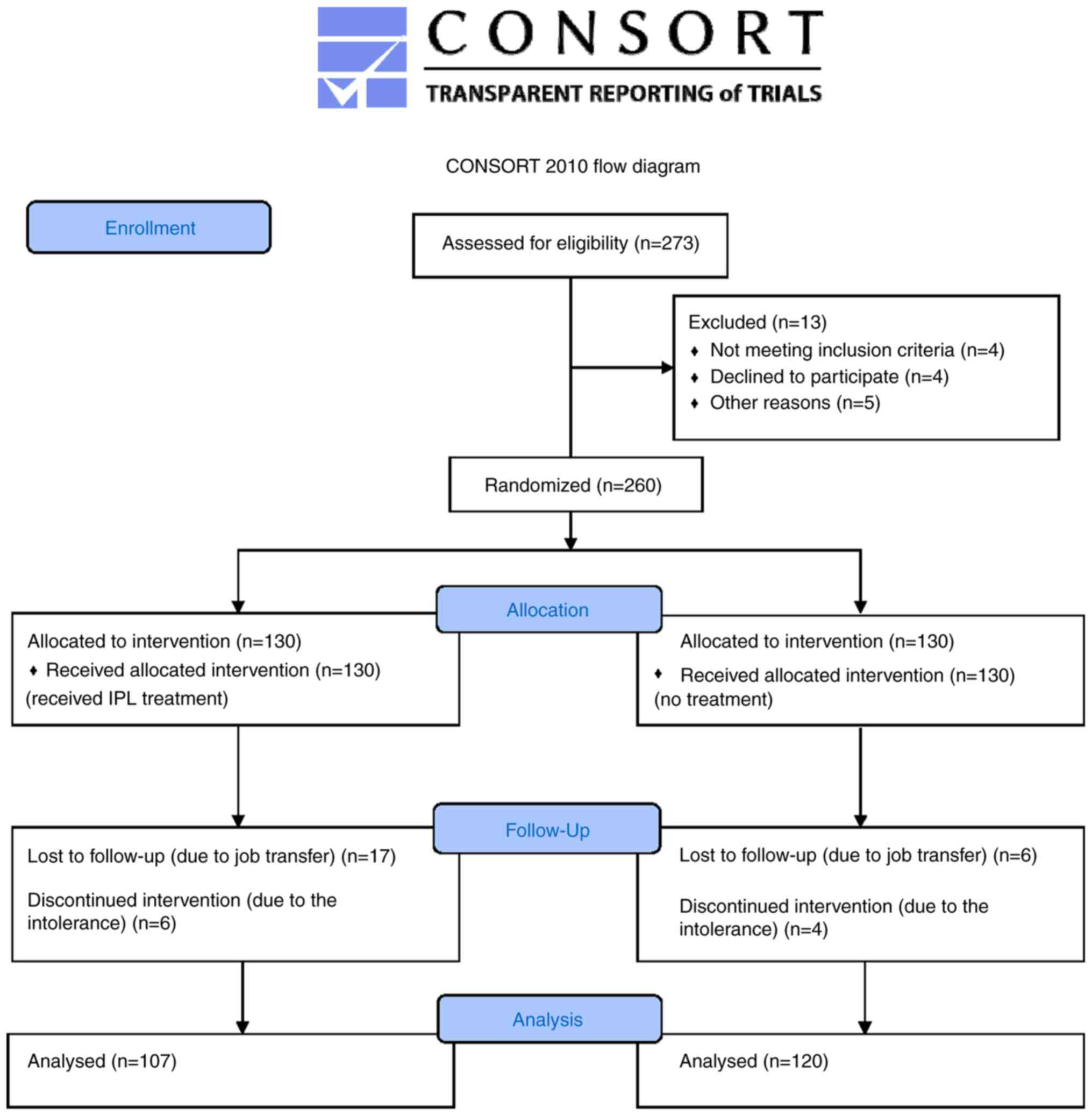
Of the 260 patients who were initially enrolled, 227
patients completed the study and were included in the final
analysis. In the IPL group, 23 patients were lost to follow-up,
specifically 17 due to job transfer and 6 due to intolerance to IPL
treatment, with symptoms including blisters, burning, redness, and
edema. In the control group, 10 patients were lost to follow-up,
specifically 6 due to job transfer and 4 due to the aggravation of
skin lesions. The 10 patients with aggravation in both groups were
withdrawn from clinical observation and were offered medication.
For the final analysis, there were 107 patients in the IPL
experimental group and 120 in the control group (Fig. 1).
Of the 227 patients included in the final analysis,
202 (88.99%) were women and 25 (11.01%) were men. The overall age
ranged from 18to 60 years (mean age, 40.20±10.76 years), and the
disease course ranged from 1 month to 20 years (mean disease
course, 2.28±3.28 years). Of the 107 patients in the IPL
experimental group, 97 (90.65%) were women and 10 (9.35%) were men,
with a mean age of 36.2±10.45 years and a mean disease course of
2.19±3.21 years. By contrast, of the 120 patients in the control
group, 105(87.50%) were women and 15 (12.50%) were men, with a mean
age of 38.5±10.51 years and a mean disease course of 2.27±3.20
years. There were no significant differences in age, sex and
disease course between the IPL and the control group (P>0.05),
making the two groups comparable in treatment efficacy
analyses.
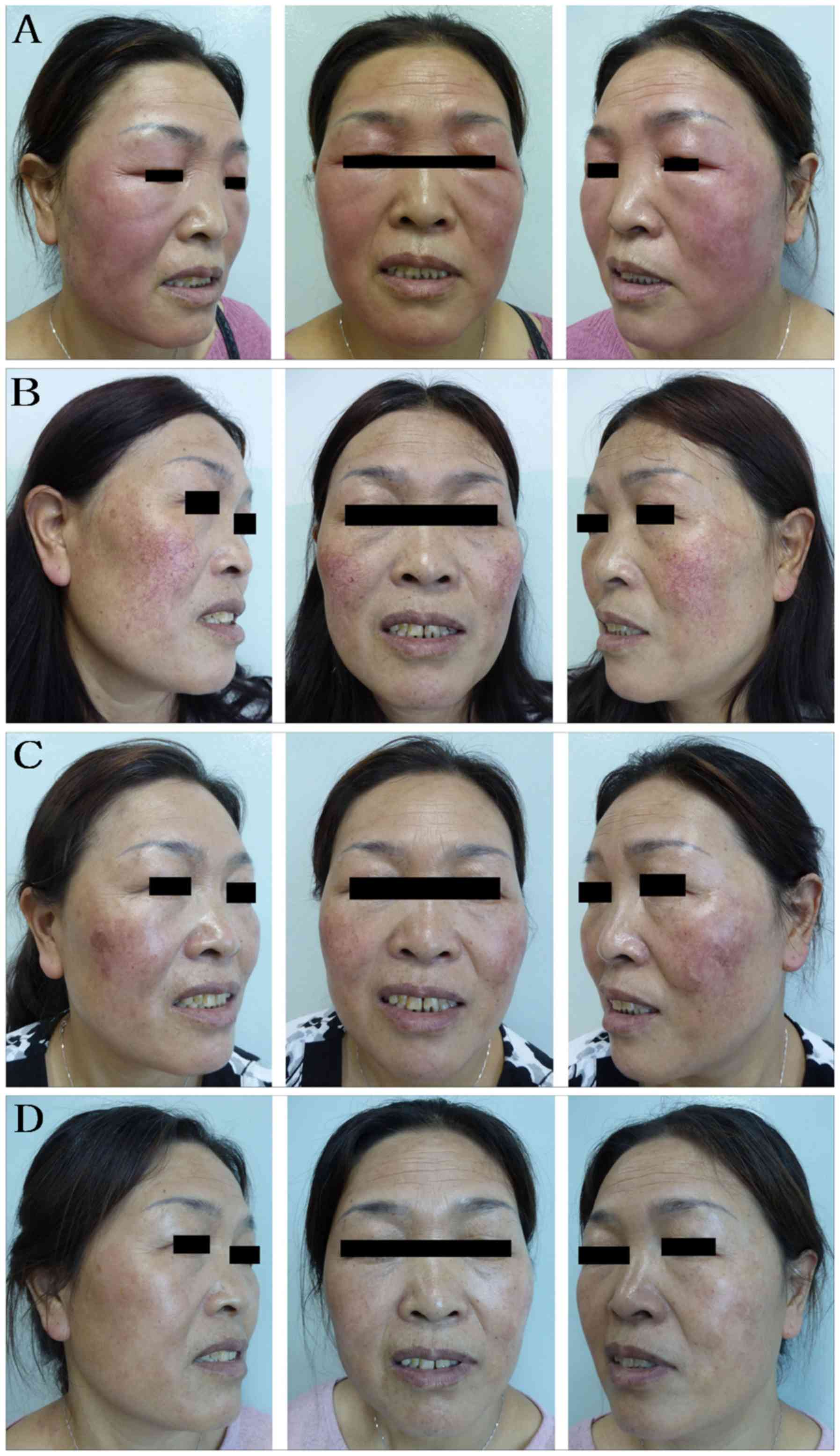
IPL efficacy
All patients were followed-up for ≥2 years. The
indications and disease course of representative patients are shown
in Fig. 2 for the experimental group
and Fig. 3 for the control group.
For the IPL group, 1 month after the initial IPL treatment, 19
patients (17.76%) with facial telangiectasia were characterized as
improved (Table I; Fig. 2C). At 3 months, the total efficacy
rate had increased to 39.25% (Table
I; Fig. 2D). At 4 months, 72
patients (67.29%) showed some degree of improvement, where 35
patients (32.71%) showed effective treatment. At 6 months, 102
patients (95.33%) showed improvement, where 71 patients (66.36%)
were characterized as effective (Table
I).
 | Table ITotal efficacy rates of the IPL
experimental group at 1-month follow-up intervals. |
Table I
Total efficacy rates of the IPL
experimental group at 1-month follow-up intervals.
| Month | Effective | Markedly
improved | Improved | Ineffective | Total (%) |
|---|
| 1 | 0 (0.00) | 0 (0.00) | 19 (17.76) | 88 (82.24) | 17.76 |
| 2 | 0 (0.00) | 12 (11.21) | 12 (11.21) | 83 (77.57) | 22.43 |
| 3 | 17 (15.89) | 13 (12.15) | 12 (11.21) | 65 (66.75) | 39.25 |
| 4 | 35 (32.71) | 21 (19.63) | 16 (14.95) | 35 (32.71) | 67.29 |
| 5 | 48 (44.86) | 27 (25.23) | 18 (16.82) | 14 (13.08) | 86.92 |
| 6 | 71 (66.36) | 22 (20.56) | 9 (8.41) | 5 (4.67) | 95.33 |
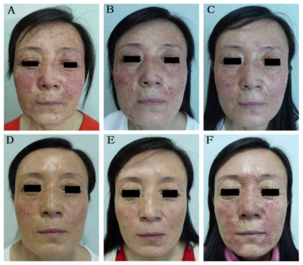
Patients with telangiectasia in the control group
demonstrated little or no recovery nor significant improvement
during the follow-up period (Fig. 3D
and E; Tables II and III). At 6 months after the baseline
assessments, the condition of 37 patients (30.83%) had improved,
whilst 83 patients (69.17%) were characterized as ‘ineffective’ in
terms of telangiectasia values from the baseline assessment. The
total efficacy rate of the control group was significantly lower
compared with that in the IPL group (χ2=99.113,
P<0.001).
| Table IIPatients showing improvement in the
IPL and control groups at one-month follow-up intervals. |
Table II
Patients showing improvement in the
IPL and control groups at one-month follow-up intervals.
| Month | IPL | Control | χ2 | P-value |
|---|
| 1 | 19 (17.76) | 0 (0.00) | N.A. | N.A. |
| 2 | 24 (22.43) | 5 (4.17) | 16.931 | P<0.001 |
| 3 | 42 (39.25) | 16 (13.33) | 19.976 | P<0.001 |
| 4 | 72 (67.29) | 25 (20.83) | 49.885 | P<0.001 |
| 5 | 93 (86.92) | 33 (27.50) | 80.854 | P<0.001 |
| 6 | 102 (95.33) | 37 (30.83) | 99.113 | P<0.001 |
| Table IIITotal efficacy rates of the IPL and
control groups at 6 months after baseline assessment. |
Table III
Total efficacy rates of the IPL and
control groups at 6 months after baseline assessment.
| Treatment
group | Effective | Markedly
improved | Improved | Ineffective | Total (%) |
|---|
| IPL | 71 (66.36) | 22 (20.56) | 9 (8.41) | 5 (4.67) | 95.33 |
| Control | 0 (0.00) | 12 (10.00) | 25 (20.83) | 83 (69.17) | 30.83 |
During the 2-year follow-up period, recurrence
occurred in 9 patients (8.41%) in the IPL group and 58 patients
(48.33%) in the control group (Fig.
3F; Table IV). Relapses were
observed mainly at 3-6 months after anti-mite treatment. The
recurrence rate of the IPL group was significantly lower compared
with that of the control group (χ2=43.333, P<0.001;
Table IV).
| Table IVRecurrence rates of the IPL and
control groups at 3-24 months after baseline assessment. |
Table IV
Recurrence rates of the IPL and
control groups at 3-24 months after baseline assessment.
| | Follow-up
month | |
|---|
| Treatment
group | 3 | 6 | 12 | 18 | 24 | Total |
|---|
| IPL | 5 (4.67) | 2 (1.87) | 1 (0.93) | 1 (0.93) | 0 (0) | 9 (8.41) |
| Control | 32 (26.67) | 18 (15.00) | 5 (4.17) | 2 (1.67) | 1 (0.83) | 58 (48.33) |
Adverse reactions
Regarding adverse reactions associated with IPL
treatment, within 30 min of the IPL treatment and the cold
compress, adverse reactions, including mild burning sensation,
temporary skin flushing and local skin edema, faded spontaneously.
In total, there were 11, 8, 3 and 2 cases of facial burning, facial
swelling, hyperpigmentation and facial blisters, respectively. In
the 2 patients with facial blisters, they were 0.1-0.3 cm in
diameter. All signs of redness, burns and blisters subsided without
further treatment within 1 week and hyperpigmentation vanished
spontaneously within 3 months of IPL treatment.
Discussion
In the present study, 540 nm-wavelength IPL was
administered on patients with late-stage rosacea following
sequential ornidazole-based anti-mite treatment. It was concluded
that 540 nm-IPL treatment was associated with a significant
improvement in telangiectasia and a reduced rate of recurrence by
comparing the rates of effective treatment (≥90% clearance of
telangiectasia), improvement (≥30% clearance) and recurrence
(original or neo-location) of the IPL and control groups. The 227
patients were followed-up for 2 years, where IPL treatment was
deemed safe and effective.
Rosacea is a chronic inflammatory cutaneous syndrome
characterized by transient facial flushing or persistent erythema,
papules, pustules and telangiectasia, with or without burning or
itching. According to The National Rosacea Society Expert
Committee, there are four subtypes of rosacea:
Erythematotelangiectatic, papulopustular (papulae and pustules),
phymatous (including enlargement of the nose) and ocular (eye
irritation) (9). Among these
subtypes, erythematotelangiectatic rosacea is the most prevalent,
with symptoms that include flushing, redness and telangiectasia
(1). Telangiectasia is a common
clinical symptom among a variety of facial skin diseases that is
characterized by dotted, patchy, linear or reticular distributions
of the capillaries, resulting in reddening of the facial skin. The
incidence of secondary telangiectasia has been increasing due to
irregular skin care, improper use of skin care products, abuse of
acid and alkali chemicals and long-term external use of hormone
preparations (19).
The causes of rosacea remain controversial, although
it has been previously hypothesized to involve the innate immune
defense system of the skin, microbial infections, genetic factors,
damage to barrier function, vasomotor dysfunction of the facial
vessels and dysfunction of neurovascular regulation (27,28).
Physiological and pathological expansion of the capillaries result
in increased blood flow (flushing) during early stage rosacea,
where pathological thickening and expansion of the blood vessels
occur over time (27). In addition,
Demodex folliculorum mite infestation is considered to be
closely associated with the etiology and pathogenesis of rosacea
(29,30).
Topical or internal drugs, or combinations, are used
routinely to alleviate the clinical symptoms of rosacea and avoid
scar formation. These include isotretinoin, antibiotics (mainly
tetracyclines), hydroxychloroquine and metronidazole (31). Although they are mostly
photosensitive for erythema, papules, and pustules, they remain
ineffective for the treatment of telangiectasia (31). In a previous clinical trial, 200
patients with mite-associated folliculitis were sequentially
treated with either an ornidazole- or metronidazole-based regimen
(13). Skin lesions in ~61%of the
patients were transiently aggravated during treatment. Compound
betamethasone injection (CBI) significantly alleviated the
inflammation and itching of facial lesions that occurred following
ornidazole treatment (13).
Application of a topical gel containing recombinant bovine basic
fibroblast growth factor(rbFGF) significantly improved the repair
of skin lesions during both the course of treatment and the
follow-up period, as compared with patients without rbFGF treatment
(14). Therefore, a treatment
regimen that combined ornidazole, CBI and rbFGF gel, which were
administered sequentially, effectively killed Demodex
folliculorum and alleviated facial inflammation. However, such
treatment had no effect on patients with telangiectasia (14).
In the present study, 540 nm-wavelength IPL was used
to treat telangiectasia in late-stage rosacea. The 540
nm-wavelength light, excited by the IPL, overlaps with the
absorption peak of oxygenated hemoglobin. Therefore, the strong
pulse light can penetrate the skin and is absorbed by hemoglobin.
The hemoglobin then coagulates, forming blood clots and cause the
blood vessels to close, resulting in the dilated capillaries
disappearing (32,33). Simultaneously, the thermal effect of
IPL can stimulate extracellular matrix protein secretion from
fibroblasts, whilst restricting the production of matrix
metalloproteinases, increasing the production of collagen
(including types I and III) in the dermis (34) and reducing the release of
inflammatory mediators (35). In
addition, IPL treatment modulates the local immune response, which
makes the skin shrink and thicken (32). Therefore, IPL treatment increases
skin elasticity, improves skin texture and removes fine wrinkles
(32). However, further research is
required to elucidate the mechanisms of action by which IPL
modulate the immune reactions that are associated with rosacea.
To achieve improved outcomes, the treatment area of
IPL was designed to overlap with the observed normal skin by 1-2
mm. Early-stage rosacea is characterized increased capillary
responsiveness and temporary expansion of blood vessels, leading to
symptoms including burning and tingling (36). The locations of continuously
expanding capillaries are difficult to determine precisely, which
requires the expansion of treatment coverage to avoid missing
telangiectasia that is invisible to the naked eye. To reduce
further adverse reactions, a post-treatment cold compress was
applied to reduce the skin temperature after IPL treatment. The
postoperative cold compress helped to relieve pain and burning
sensations, in addition to reducing the occurrence of adverse
reactions including redness, blisters and ecchymosis.
Telangiectasia is divided into primary and secondary
subtypes, with the latter sometimes attributed to systemic diseases
such as rosacea (27). Therefore,
the first step of treatment should be to identify and address the
underlying causes or contributing factors. It was previously found
that anti-mite treatment markedly improved the appearance of
papules and pustules caused by rosacea, which indicates the close
association between this disease and Demodex folliculorum
infestation (13). As the anti-mite
treatment dispelled the remote cause of telangiectasia associated
with rosacea, IPL was applied in the present study for the
coagulation of facial dilated blood vessels, thereby eliminating
dysfunctional blood vessels and effectively reducing the recurrence
rate of rosacea.
Compared with pulsed dye laser and pulsed laser in
rosacea-associated telangiectasia treatment, IPL is advantageous in
that it covers large areas of the skin, is more time efficient and
more likely to be accepted by patients. Although it may not be as
efficient as laser when used to treat certain types of skin lesions
including large dilated blood vessels (37). As a composite light with a long pulse
width, IPL has been shown to be an improvement in terms of reducing
pigmentation and telangiectasia, fine wrinkle elimination and the
smoothing and firming of the skin (38,39).
To conclude, the present study found that following
treatment with 540 nm IPL, the facial telangiectasia of patients
with rosacea was significantly improved, where the rates of
effectiveness and total efficacy were higher compared with those of
the untreated control group. In addition, IPL treatment was found
to be associated with a continuous reduction in the rate of rosacea
recurrence during the 2-year follow-up. However, since the present
study primarily investigated patients with telangiectasia in
late-stage rosacea, further investigation is required on the
effects of IPL on the recurrence of other manifestations of
rosacea.
Acknowledgements
Not applicable.
Funding
No funding was received.
Availability of data and materials
The datasets used and/or analyzed during the current
study are available from the corresponding author on reasonable
request.
Authors' contributions
YL designed the experiments. XLL collected and
interpreted the data. JHZ collected the follow-up data. LXW
analyzed the data. NZ collected and analyzed the data, and wrote
the manuscript. All authors read and approved the final version of
the manuscript.
Ethics approval and consent to
participate
The Ethics Committee of the 940th Hospital of the
Joint Logistics Support force of the Chinese People's Liberation
Army approved the study protocol (Lanzhou, China). Signed informed
consent was obtained from all patients included in the study.
Patient consent for publication
Not applicable.
Competing interests
The authors declare that they have no competing
interests.
References
|
1
|
Wilkin J, Dahl M, Detmar M, Drake L, Liang
MH, Odom R and Powell F: National Rosacea Society Expert C.
Standard grading system for rosacea: Report of the National Rosacea
Society Expert Committee on the classification and staging of
rosacea. J Am Acad Dermatol. 50:907–912. 2004.PubMed/NCBI View Article : Google Scholar
|
|
2
|
Crawford GH, Pelle MT and James WD:
Rosacea: I. Etiology, pathogenesis, and subtype classification. J
Am Acad Dermatol. 51:327–341; quiz 342-324. 2004.PubMed/NCBI View Article : Google Scholar
|
|
3
|
Wacker T and Lang GK: Demodex
folliculorum: Diagnosis and therapy today. Klin Monbl
Augenheilkd (German). 231:241–245. 2014.PubMed/NCBI View Article : Google Scholar
|
|
4
|
Alinia H, Tuchayi SM, James SM, Cardwell
LA, Nanda S, Bahrami N, Awosika O, Richardson I, Huang KE and
Feldman SR: Measurement of disease severity in a population of
rosacea patients. Dermatol Clin. 36:97–102. 2018.PubMed/NCBI View Article : Google Scholar
|
|
5
|
Oussedik E, Bourcier M and Tan J:
Psychosocial burden and other impacts of rosacea on patients'
quality of life. Dermatol Clin. 36:103–113. 2018.PubMed/NCBI View Article : Google Scholar
|
|
6
|
Egeberg A, Hansen PR, Gislason GH and
Thyssen JP: Patients with rosacea have increased risk of dementia.
Ann Neurol. 79:921–928. 2016.PubMed/NCBI View Article : Google Scholar
|
|
7
|
Faris C, Manjaly JG, Ismail-Koch H and
Caldera S: Rapid treatment of rhinophyma with powered
microdebrider. Case Rep Otolaryngol. 2013(621639)2013.PubMed/NCBI View Article : Google Scholar
|
|
8
|
Egeberg A, Hansen PR, Gislason GH and
Thyssen JP: Patients with rosacea have increased risk of depression
and anxiety disorders: A danish nationwide cohort study.
Dermatology. 232:208–213. 2016.PubMed/NCBI View Article : Google Scholar
|
|
9
|
Gallo RL, Granstein RD, Kang S, Mannis M,
Steinhoff M, Tan J and Thiboutot D: Standard classification and
pathophysiology of rosacea: The 2017 update by the National Rosacea
Society Expert Committee. J Am Acad Dermatol. 78:148–155.
2018.PubMed/NCBI View Article : Google Scholar
|
|
10
|
Micali G, Dall'Oglio F, Verzi AE, Luppino
I, Bhatt K and Lacarrubba F: Treatment of erythemato-telangiectatic
rosacea with brimonidine alone or combined with vascular laser
based on preliminary instrumental evaluation of the vascular
component. Lasers Med Sci. 33:1397–1400. 2018.PubMed/NCBI View Article : Google Scholar
|
|
11
|
Kennedy Carney C, Cantrell W and Elewski
BE: Rosacea: A review of current topical, systemic and light-based
therapies. G Ital Dermatol Venereol. 144:673–688. 2009.PubMed/NCBI
|
|
12
|
Del Rosso JQ, Tanghetti EA, Baldwin HE,
Rodriguez DA and Ferrusi IL: The burden of illness of
erythematotelangiectatic rosacea and papulopustular rosacea:
Findings from a web-based survey. J Clin Aesthet Dermatol.
10:17–31. 2017.PubMed/NCBI
|
|
13
|
Luo Y, Sun YJ, Zhang L and Luan XL:
Treatment of mites folliculitis with an ornidazole-based sequential
therapy: A randomized trial. Medicine (Baltimore).
95(e4173)2016.PubMed/NCBI View Article : Google Scholar
|
|
14
|
Luo Y, Luan XL, Sun YJ, Zhang L and Zhang
JH: Effect of recombinant bovine basic fibroblast growth factor gel
on repair of rosacea skin lesions: A randomized, single-blind and
vehicle-controlled study. Exp Ther Med. 17:2725–2733.
2019.PubMed/NCBI View Article : Google Scholar
|
|
15
|
Anzengruber F, Czernielewski J, Conrad C,
Feldmeyer L, Yawalkar N, Hausermann P, Cozzio A, Mainetti C,
Goldblum D, Goldblum D, et al: Swiss S1 guideline for the treatment
of rosacea. J Eur Acad Dermatol Venereol. 31:1775–1791.
2017.PubMed/NCBI View Article : Google Scholar
|
|
16
|
Lowe E and Lim S: Paradoxical erythema
reaction of long-term topical brimonidine gel for the treatment of
facial erythema of rosacea. J Drugs Dermatol. 15:763–765.
2016.PubMed/NCBI
|
|
17
|
Husain Z and Alster TS: The role of lasers
and intense pulsed light technology in dermatology. Clin Cosmetic
and Invest Dermatol. 9:29–40. 2016.PubMed/NCBI View Article : Google Scholar
|
|
18
|
Tanghetti EA, Jackson JM, Belasco KT,
Friedrichs A, Hougier F, Johnson SM, Kerdel FA, Palceski D, Hong
HC, Hinek A and Cadena MJ: Optimizing the use of topical
brimonidine in rosacea management: Panel recommendations. J Drugs
Dermatol. 14:33–40. 2015.PubMed/NCBI
|
|
19
|
Gao L, Gao N, Song W, Dang E, Yin R, Wang
L and Wang G: A retrospective study on efficacy of pulsed dye laser
and intense pulsed light for the treatment of facial
telangiectasia. J Drugs Dermatol. 16:1112–1116. 2017.PubMed/NCBI
|
|
20
|
Jansen T: Clinical presentations and
classification of rosacea. Ann Dermatol Venereol. 138:S192–S200.
2011.PubMed/NCBI View Article : Google Scholar
|
|
21
|
Jansen T: Clinical presentations and
classification of rosacea. Ann Dermatol Venereol. 138
(Suppl):S138–S147. 2011.PubMed/NCBI View Article : Google Scholar
|
|
22
|
Fitzpatrick TB: The validity and
practicality of sun-reactive skin types I through VI. Arch
Dermatol. 124:869–871. 1988.PubMed/NCBI View Article : Google Scholar
|
|
23
|
Goldberg DJ: Current trends in intense
pulsed light. J Clin Aesthet Dermatol. 5:45–53. 2012.PubMed/NCBI
|
|
24
|
Dummer R, Graf P, Greif C and Burg G:
Treatment of vascular lesions using the VersaPulse variable pulse
width frequency doubled neodymium: YAG laser. Dermatology.
197:158–161. 1998.PubMed/NCBI View Article : Google Scholar
|
|
25
|
Wilkin J, Dahl M, Detmar M, Drake L,
Feinstein A, Odom R and Powell F: Standard classification of
rosacea. National Rosacea Society Expert Committee: Report of the
National Rosacea Society Expert Committee on the classification and
staging of Rosacea. J Am Acad Dermatol. 46:584–587. 2002.PubMed/NCBI View Article : Google Scholar
|
|
26
|
Xu Z, Tan X, Ye J, Zhang C, Zhao M and
Zhan F: Evaluation of measles catch-up immunization campaign in
Hubei, 2010. Chin J Viral Dis. 1:208–211. 2011.
|
|
27
|
Cribier B: Pathophysiology of rosacea:
Redness, telangiectasia, and rosacea. Ann Dermatol Venereol. 138
(Suppl):S184–S191. 2011.PubMed/NCBI View Article : Google Scholar
|
|
28
|
Agnoletti AF, DEC E, Parodi A, Schiavetti
I, Savarino V, Rebora A, Paolino S, Cozzani E and Drago F:
Etiopathogenesis of rosacea: A prospective study with a three-year
follow-up. G Ital Dermatol Venereol. 152:418–423. 2017.PubMed/NCBI View Article : Google Scholar
|
|
29
|
Forton FM: Papulopustular rosacea, skin
immunity and Demodex: Pityriasis folliculorum as a missing
link. J Eur Acad Dermatol Venereol. 26:19–28. 2012.PubMed/NCBI View Article : Google Scholar
|
|
30
|
Turgut Erdemir A, Gurel MS, Koku Aksu AE,
Falay T, Inan Yuksel E and Sarikaya E: Demodex mites in acne
rosacea: Reflectance confocal microscopic study. Australas J
Dermatol. 58:e26–e30. 2017.PubMed/NCBI View Article : Google Scholar
|
|
31
|
Abokwidir M and Feldman SR: Rosacea
management. Skin Appendage Disord. 2:26–34. 2016.PubMed/NCBI View Article : Google Scholar
|
|
32
|
Campolmi P, Bonan P, Cannarozzo G,
Bruscino N, Troiano M, Prignano F and Lotti T: Intense pulsed light
in the treatment of non-aesthetic facial and neck vascular lesions:
Report of 85 cases. J Eur Acad Dermatol Venereol. 25:68–73.
2011.PubMed/NCBI View Article : Google Scholar
|
|
33
|
Liu J, Liu J, Ren Y, Li B and Lu S:
Comparative efficacy of intense pulsed light for different erythema
associated with rosacea. J Cosmet Laser Ther. 16:324–327.
2014.PubMed/NCBI View Article : Google Scholar
|
|
34
|
Wong WR, Shyu WL, Tsai JW, Hsu KH, Lee HY
and Pang JHS: Intense pulsed light modulates the expressions of
MMP-2, MMP-14 and TIMP-2 in skin dermal fibroblasts cultured within
contracted collagen lattices. J Dermatol Sci. 51:70–73.
2008.PubMed/NCBI View Article : Google Scholar
|
|
35
|
Vigo L, Giannaccare G, Sebastiani S,
Pellegrini M and Carones F: Intense pulsed light for the treatment
of dry eye owing to meibomian gland dysfunction. J Vis Exp.
146:2019.PubMed/NCBI View
Article : Google Scholar
|
|
36
|
Rainer BM, Kang S and Chien AL: Rosacea:
Epidemiology, pathogenesis, and treatment. Dermatoendocrinol.
9(e1361574)2017.PubMed/NCBI View Article : Google Scholar
|
|
37
|
Lim HS, Lee SC, Won YH and Lee JB: The
efficacy of intense pulsed light for treating
erythematotelangiectatic rosacea is related to severity and age.
Ann Dermatol. 26:491–495. 2014.PubMed/NCBI View Article : Google Scholar
|
|
38
|
Babilas P, Schreml S, Szeimies RM and
Landthaler M: Intense pulsed light (IPL): A review. Lasers Surg
Med. 42:93–104. 2010.PubMed/NCBI View Article : Google Scholar
|
|
39
|
Kawana S, Ochiai H and Tachihara R:
Objective evaluation of the effect of intense pulsed light on
rosacea and solar lentigines by spectrophotometric analysis of skin
color. Dermatol Surg. 33:449–454. 2007.PubMed/NCBI View Article : Google Scholar
|