Introduction
The femur is the longest bone and the major
weight-bearing bone of the lower limbs in the human body. Femoral
shaft fracture is one of the most common fractures in the clinic,
accounting for 18.5% of limb fractures and 6% of total bone
fractures (1). Femoral shaft
fracture refers to the fracture between the femoral condyle and
femur. Adult femoral shaft fractures are frequently caused by
high-energy injuries and post-fracture complications are common. If
it not treated properly, complications may seriously affect the
quality of life of patients and may even be life-threatening
(2).
With the continuous improvement of internal fixation
materials, surgical methods and changes in concepts of fracture
treatment methods, adult femoral shaft fractures tend to be treated
with surgical treatment combined with internal fixation. Since the
19th century, bone plate and intramedullary nail have been
gradually used in the treatment of femoral shaft fractures. Owing
to its compatibility with the bone mechanical properties and its
ability to align the bone fragments, intramedullary nail is the
first choice of treatment for femoral shaft fractures (3).
However, for complex femoral shaft fractures,
including Winquist III and IV femoral shaft fractures, traditional
intramedullary nails may still not provide sufficiently reliable
stability. Furthermore, non-surgical treatment is associated with a
long bed rest time and common complications thereof. In order to
solve this problem, a novel intramedullary nail system was
developed in the present study. The major characteristics of the
novel intramedullary nail are that the nail body are slotted and
the elastic modulus of the nail is reduced after rotation. The
novel intramedullary nail has two advantages when compared with
traditional nails: i) Fragments may be fixed by a Kirschner wire at
the grooved site and ii) elasticity may be reduced to accommodate
different bone fractures, particularly in elderly patients with
osteoporosis.
However, as the human body is a complex mechanical
structure, it is challenging to simulate human mechanics using
biomechanical experiments. The finite element method has been
mainly applied to biomechanical research in the medical field. As
the finite element method may simulate the biomechanical behavior
of the human body, it was used as an effective method to understand
its mechanical characteristics. Its effectiveness and superiority
have been fully demonstrated in basic experiments and clinical
applications (4-6).
The finite element method may divide the continuous elastic bodies
into a finite number of units, synthesize the research subject and
study the properties of each unit by using the finite element
software in the computer.
In the present study, the finite element method was
employed to analyze and evaluate the biomechanical status of novel
and traditional intramedullary nails prior to and after femoral
shaft fracture healing. The present study aimed to provide a
biomechanical basis for surgeons to select a more effective
fixation method with satisfactory therapeutic effects.
Materials and methods
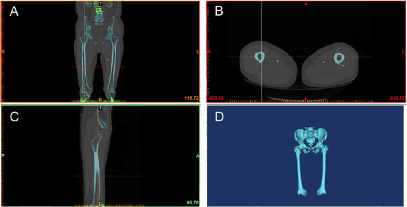
Volunteer and femoral scan
A 30-year-old healthy male with a body height of 172
cm and a body weight of 75 kg was selected as a volunteer. The
subject had no history of femoral fracture or trauma, and femoral
lesions and injuries were also ruled out by X-ray imaging. The
spiral axis scan of the bilateral femurs was performed by using a
CT scanner (Optima CT 660; GE Healthcare) from top to bottom using
the scanning condition of 120 KV, 125 mA and a layer thickness of 1
mm. The above machine had an advanced image post-processing and
camera function.
After the original data were interpolated and
enlarged on the machine, a continuous picture of the layer
thickness was obtained, and was burned to the optical disc in the
international standard dicom format. The study was approved by the
Ethics Committee of The Third Hospital of Hebei Medical University
(Shijiazhuang, China). The internal fixation model was built using
Solidwork 2016 software (Dassault); the novel intramedullary nail
system was designed by the Third Hospital of Hebei Medical
University (patent number, ZL201721394941.3).
Construction of normal femur 3D
model
The femur computed tomography (CT) image (dicom
format) was imported into the Mimics 17.0 software (Materialise) to
segment bonefrom the entire model bythresholding; the images were
subsequently selectively segmented using ‘split-mask’ according to
the anatomical structure. Finally, a geometric model of the femur
was established through regional expansion, smoothing all parts of
the femur, filling small gaps, and making the outer contour of the
femur smooth and continuous.
The constructed femur information was saved in an
STL format file, and the STL format file was repaired and optimized
by reverse engineering software Geomagic Studio 2012 (Raindrop). In
brief, the non-characteristic masses and indentations on the
surface of the model were removed and the surface was smoothed.
Furthermore, non-characteristic high curvature and the generation
of self-intersecting faces were prevented. The smooth surface of
the model was used to fit the triangular surface of the model
surface; the femoral cortical bone and cancellous bone were
distinguished by using the offset function. At last, a continuous
curved surface model was generated.
Boundary and load information
It was assumed that the fracture surface was
completely broken and in a contact state. The friction coefficient
was 0.2 according to a previous study (7). The femur was subjected to numerous
loads, including muscle force, joint force and dynamic impact
force. In view of numerous uncertainties, including the number and
direction of load during finite element simulation of muscle
loading, it was relatively complex to accurately simulate the
loading conditions in the dynamic state (8). The simple model method of Duda et
al (9) was used in the present
study. To simulate the load of the femur while standing and
walking, analysis ofthe static mechanics of the model was
performed. The condition was that the distal knee joint had a
degree of freedom of 0 and the distal nodes have a displacement of
0 on the X, Y and Z axes. Furthermore, a load of 700 N along the
direction of the force line at the outer surface of the contact
area between the femoral head and the acetabulum was applied.
According to Duda et al (9), the intact femur received a torque of
~0.04 body weight meters (BWM) during the gait cycle. However,
three weeks after the intramedullary nailing of the femoral shaft
fracture, the maximum torque of the femur was 0.01 BWM (10). Therefore, based on the load of 700 N,
the maximum torque of the femur was 7-28 Nm. Therefore, in the
torsion analysis under exclusion of the specific effects of each
muscle group, the boundary load condition was fixation on the
femoral condyles and a 7-Nm external rotation torque was applied at
the proximal end of the femur along the same axis as compressive
loading.
Results
Construction and properties of the
normal femur 3D model
In general, biological tissue belongs to an
anisotropic non-linear body. The application of bone finite element
analysis in the field of biomechanics is mostly based on the
isotropic, homogeneous continuous linear elastomer. Therefore, the
model utilized in the present study assumed that the femoral bone
is an isotropic, homogeneous linear elastic material, which
consists of cortical bone, cancellous bone and internal fixation
(Table I), and also referred to the
data reported by previous studies (11,12).
Only two materials of bone and internal fixation were considered in
the model, and muscle and tendon stress were simplified, regardless
of the friction between the joints and cartilage. A femoral
geometric model was established by using Mimics 17.0 (Materialise)
and Geomagic Studio 2012 (Raindrop) software based on the CT image
of the femur (Fig. 1).
 | Table IProperties of materials included in
the model. |
Table I
Properties of materials included in
the model.
| Material name | Modulus of elasticity
(MPa) | Poisson's ratio |
|---|
| Cortical bone | 16,800 | 0.29 |
| Cancellous bone | 260 | 0.20 |
| Traditional
intramedullary rod | 110,000 | 0.30 |
| Novel intramedullary
rod | 110,000 | 0.30 |
Construction of two different internal
fixation models for comminuted fracture of the femoral shaft
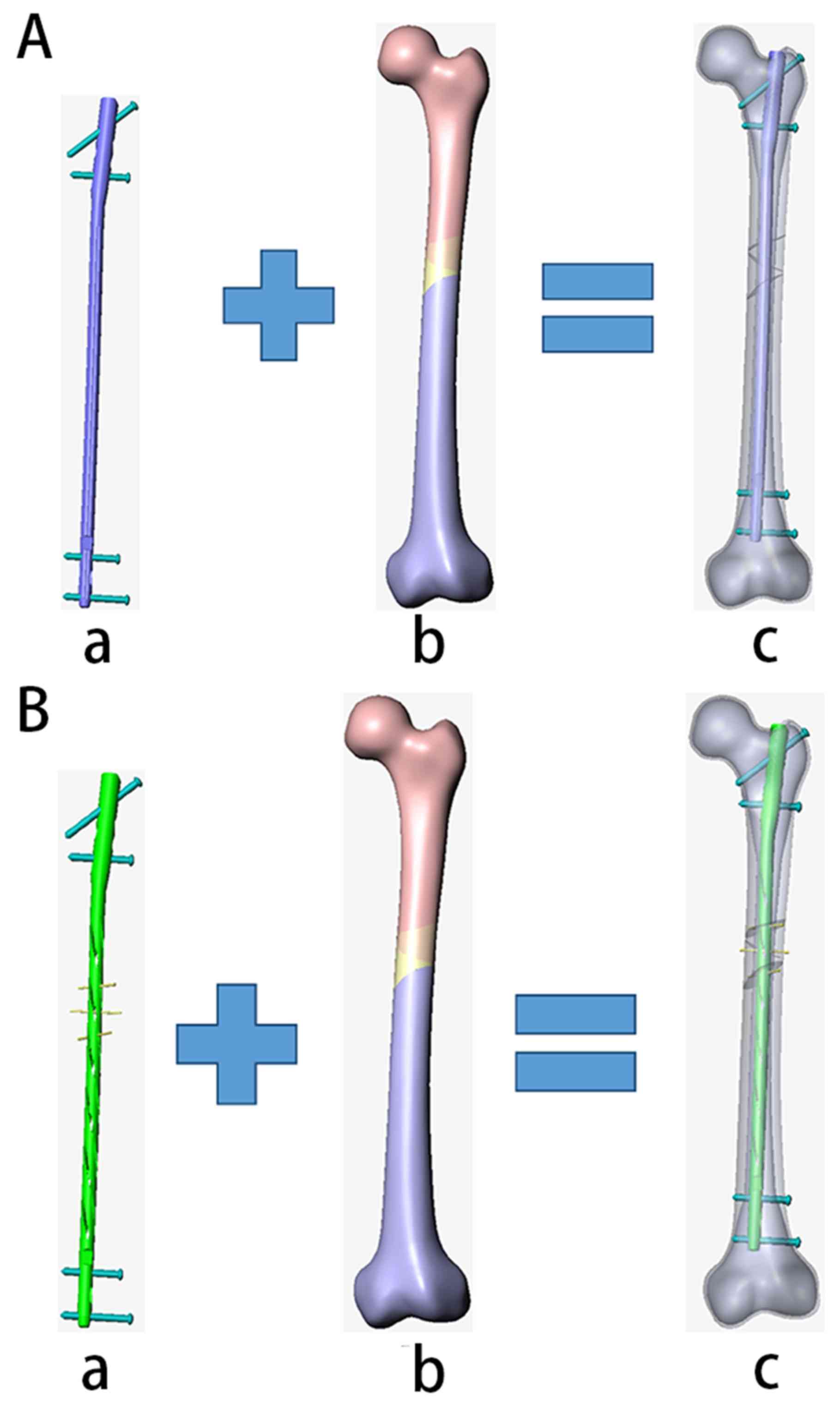
The traditional intramedullary nail system (Fig. 2) was based on the intramedullary nail
(diameter, 10 mm; length, 380 mm) produced by Double Medical
Technology Co., Ltd. (Fig. 2Aa).
Solidwork 2016 (Dassault) is a software for simulation design and
has been applied in numerous fields, including aviation and
machinery, where the modeling results were reliable (6). The internal fixation model was built
using the Solidwork 2016 (Dassault) software, a novelintramedullary
nail system designed in The Third Hospital of Hebei Medical
University in 2017 (patent no: ZL201721394941.3; Fig. 2Bb). The geometric structure and the
specific parameters of the novel intramedullary nailing system are
provided in Fig. S1. It is designed
as a hollow nail body, including the head, body and tail of the
nail. The nail body includes a slotted section and a corresponding
connecting section. The upper ring of the side wall of each grooved
section has several grooves connected to the mouth of the nail
body, which are spirally arranged along the extension direction of
the nail body.
The femoral solid model in IGES format generated by
Geomagic Studio 2012 (Raindrop) was imported into the design
software Solidwork 2016 (Dassault) to simulate femoral shaft
fractures (Fig. 2Ac and Bc). Combined with the above internal
fixation model, two different internal fixation femoral shaft
fractures in three-dimensional finite element models were
established (Fig. 2Ad and Be).
Finite element analysis of the
complete femur model and comminuted femoral shaft fracture
After verifying the validity of the above
traditional and novel femoral finite element models (Fig. 2A and B), the femoral comminuted fractures were
fixed with the traditional and novel intramedullary nail systems.
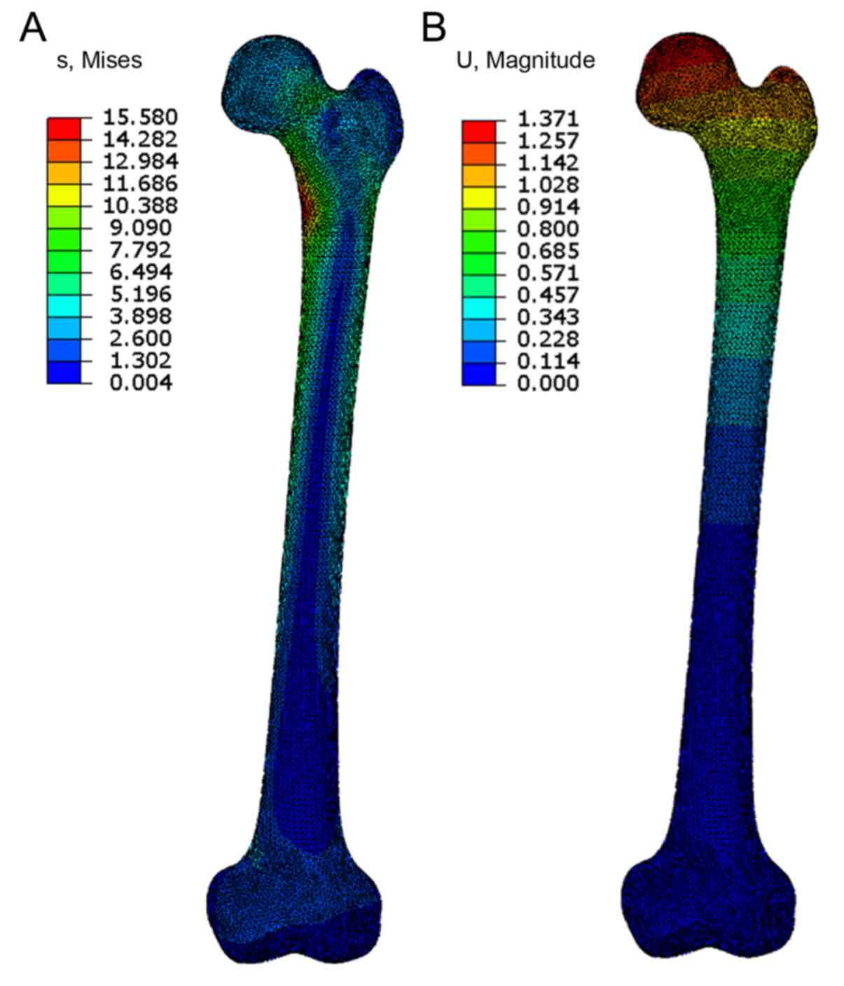
Subsequently, the finite element method was used to analyze a
complete femoral model under a 700-N load. The stress cloud map
(Fig. 3A) and the displacement cloud
map (Fig. 3B) were also provided.
The stress was mainly distributed in the middle and lower part of
the femur, around the femoral neck and the large and small
trochanter. There was also an obvious stress concentration at the
small trochanter of the femur (Figs.
4-6), prior to healing as well as after healing.
Under the compressive load, the maximum stresses,
the maximum displacements, broken bone stresses and broken bone
displacements among the traditional and the novel intramedullary
nail fixation prior to healing, the traditional and novel
intramedullary nailing after healing, and the complete femur were
also provided in Table II. Under
torsional load, the traditional and novel intramedullary nail
fixations prior to healing were also compared (Table III).
| Table IIComparison of bone stress and
displacement in different models of nail fixation under compressive
load prior to and after healing. |
Table II
Comparison of bone stress and
displacement in different models of nail fixation under compressive
load prior to and after healing.
| Item | Maximum stress
(MPa) | Maximum displacement
(mm) | Bone fragment stress
(MPa) | Bone fragment
displacement (mm) |
|---|
| Traditional
intramedullary nail fixation prior to healing | 73.1 | 3.84 | 5.30 | 0.691 |
| Novel intramedullary
nail fixation prior to healing | 330 | 2.49 | 17.5 | 0.343 |
| Traditional
intramedullary nail fixation after healing | 38.8 | 1.21 | | |
| Novel intramedullary
nail fixation after healing | 81.0 | 1.28 | | |
| Complete femur | 15.6 | 1.37 | | |
| Table IIIComparison of bone stress and
displacement in the traditional and novel intramedullary nail
fixation prior to healing under torsional load. |
Table III
Comparison of bone stress and
displacement in the traditional and novel intramedullary nail
fixation prior to healing under torsional load.
| Item | Maximum stress
(MPa) | Maximum displacement
(mm) | Bone fragment stress
(MPa) | Bone fragment
displacement (mm) |
|---|
| Traditional
intramedullary nail fixation prior to healing | 166 | 2.59 | 0.749 | 0.767 |
| Novel intramedullary
nail fixation prior to healing | 701 | 2.84 | 39.7 | 0.754 |
When subjected to compressive or torsional load
prior to complete healing, the stability of the novel IM nail
fixation was greater compared with that of the traditional IM nail
fixation, as indicated by the smaller resulting maximum fracture
site displacement (Table III). The
novel IM nail shares more loads as indicated by the higher maximum
stress in the novel intramedullary nail group compared with the
traditional IM nail group (Table
III). The fragment stress in the novel IM nail under both
loading conditions were higher because of the higher load
concentration around Kirschner wires in the fragment.
It was suggested that the novel intramedullary nail
fixation system was superior compared with traditional
intramedullary nail fixation systems with regards to maximum stress
and displacement, which stimulates bone growth.
Discussion
Femoral shaft fracture is a common type of fracture
encountered in the clinic. At present, the traditional
intramedullary nail may not provide sufficiently reliable results
for partial complex femoral shaft fractures. The present study
mainly discussed the partial stress distribution of the femur after
internal fixation with the novel or traditional nail under various
static loading conditions. The major difference between the novel
and the traditional intramedullary nails is that the body of the
novel nail includes a plurality of slots, and the slot is spirally
arranged along the extending direction of the body portion. It may
adjust the curvature of the local part of the nail body to match
the medullary cavity of different patients, fix the bone fragments
and shorten the healing time of the fracture.
Intramedullary nail acts as a load sharing device
with peri-fracture site compared to the load bearing metallic bone
plate. In the early post-fracture period, the novel intramedullary
nail facilitates greater force transmission through the fracture
site, which is beneficial for fracture healing. The stress of the
intramedullary nail after fracture healing is less than the stress
endured by the healthy femur, which indicates that force after
healing was mainly performed by the femur. There was also an
obvious stress concentration at the small trochanter of the femur
prior to and after healing. This was consistent with previous
studies (13,14), suggesting the successful
establishment of an effective and reliable model in the present
study. The displacement of the femur under load was small,
indicating that the femur has a large stiffness. In the early stage
of healing, the femur should not be subjected to a large stress,
and as the affected area heals, the femur also bears more force.
The novel intramedullary nail fixation system is able to eliminate
the stress shielding effect along the long axis of the femur and
allows for axial compression of the fracture site. The fretting
continuous axial load of the fracture site may promote fracture
healing (15).
In conclusion, the present study indicated that
appropriate pressure at the fracture end eliminates the gap at the
fracture end. This improves the static friction between the broken
femurs, which increases the fixing strength and mechanical
stability, and is also beneficial for bone growth and crawling
replacement. The novel intramedullary nail fixation reduced stress
shielding and promoted stress transmission. As further demonstrated
in another study, compressive stress-mediated changes may
effectively stimulate cell differentiation, which may promote
fracture healing and stimulate bone metabolism during the fixed
pressurization (16). Finally,
displacement of the femoral head and the broken end in the novel
intramedullary nail fixation system were far less than those in the
traditional intramedullary nail system, providing a significantly
improved fixation effect of the intramedullary nail. These
improvements in stability have a beneficial role in promoting
fracture healing.
Supplementary Material
(A) Geometric structure and (B)
specific parameters of the novel intramedullary nail.
Acknowledgements
Not applicable.
Funding
No funding was received.
Availability of data and materials
The datasets used and analyzed during the present
study are available from the corresponding author on reasonable
request.
Authors' contributions
YWC was responsible to the study concepts,
manuscript preparation and editing; WZX was responsible for the
clinical studies; ZHP was responsible for the definition of
intellectual content and literature research; ZGK was in charge of
the data acquisition; LiS was responsible for the statistical
analysis; LeS was responsible to the data analysis; XDC was
responsible to the experimental studies; CCL was the guarantor of
integrity of the entire study, study design and manuscript review.
All authors approved the final version of this manuscript.
Ethics approval and consent to
participate
The study was approved by the Third Hospital of
Hebei Medical University (Shijiazhuang, China). Informed consent of
images publication and data usage were also obtained (patent
number, ZL201721394941.3).
Patient consent for publication
Not applicable.
Competing interests
The authors declare that they have no competing
interests.
References
|
1
|
Fakhry SM, Rutledge R, Dahners LE and
Kessler D: Incidence, management and outcome of femoral shaft
fracture: A statewide population-based analysis of 2805 adult
patients in a rural state. J Trauma. 37:255–260; discussion
260-261. 1994.PubMed/NCBI
|
|
2
|
Morishige M, Muramatsu K, Tominaga Y,
Hashimoto T and Taguchi T: Surgical treatment of metastatic femoral
fractures: Achieving an improved quality of life for cancer
patients. Anticancer Res. 35:427–432. 2015.PubMed/NCBI
|
|
3
|
Cheung G, Zalzal P, Bhandari M, Spelt JK
and Papini M: Finite element analysis of a femoral retrograde
intramedullary nail subject to gait loading. Med Eng Phys.
26:93–108. 2004.PubMed/NCBI View Article : Google Scholar
|
|
4
|
Abueidda DW, Dalaq AS, Abu Al-Rub RK and
Younes HA: Finite element predictions of effective multifunctional
properties of interpenetrating phase composites with novel triply
periodic solid shell architectured reinforcements. Int J Mech Sci.
92:80–89. 2015. View Article : Google Scholar
|
|
5
|
Ramos Verri F, Santiago Junior JF, de
Faria Almeida DA, de Oliveira GB, de Souza Batista VE, Marques
Honório H, Noritomi PY and Pellizzer EP: Biomechanical influence of
crown-to-implant ratio on stress distribution over internal hexagon
short implant: 3-D finite element analysis with statistical test. J
Biomech. 48:138–145. 2015.PubMed/NCBI View Article : Google Scholar
|
|
6
|
Sternheim A, Giladi O, Gortzak Y, Drexler
M, Salai M, Trabelsi N, Milgrom C and Yosibash Z: Pathological
fracture risk assessment in patients with femoral metastases using
CT-based finite element methods. A retrospective clinical study.
Bone. 110:215–220. 2018.PubMed/NCBI View Article : Google Scholar
|
|
7
|
Robinson PS, Placide R, Soslowsky LJ and
Born CT: Mechanical strength of repairs of the hip piriformis
tendon. J Arthroplasty. 19:204–210. 2004.PubMed/NCBI View Article : Google Scholar
|
|
8
|
Paul JP: Stress and strain distribution
within the intact femur: Compression or bending? by Taylor et
al. Med Eng Phys. 19(97): 99–100. 1997.PubMed/NCBI View Article : Google Scholar
|
|
9
|
Duda GN, Schneider E and Chao EY: Internal
forces and moments in the femur during walking. J Biomech.
30:933–941. 1997.PubMed/NCBI View Article : Google Scholar
|
|
10
|
Schneider E, Michel MC, Genge M, Zuber K,
Ganz R and Perren SM: Loads acting in an intramedullary nail during
fracture healing in the human femur. J Biomech. 34:849–857.
2001.PubMed/NCBI View Article : Google Scholar
|
|
11
|
Perez JV, Warwick DJ, Case CP and
Bannister GC: Death after proximal femoral fracture-an autopsy
study. Injury. 26:237–240. 1995.PubMed/NCBI View Article : Google Scholar
|
|
12
|
Goosen JH, Mulder MC, Bongers KJ and
Verheyen CC: High revision rate after treatment of femoral neck
fractures with an optionally (un) cemented stem. Arch Orthop Trauma
Surg. 129:801–805. 2009.PubMed/NCBI View Article : Google Scholar
|
|
13
|
Valliappan S, Svensson NL and Wood RD:
Three dimensional stress analysis of the human femur. ComputBiol
Med. 7:253–264. 1977.PubMed/NCBI View Article : Google Scholar
|
|
14
|
Couteau B, Hobatho MC, Darmana R, Brignola
JC and Arlaud JY: Finite element modelling of the vibrational
behaviour of the human femur using CT-based individualized
geometrical and material properties. J Biomech. 31:383–386.
1998.PubMed/NCBI View Article : Google Scholar
|
|
15
|
Bottlang M, Tsai S, Bliven EK, von
Rechenberg B, Klein K, Augat P, Henschel J, Fitzpatrick DC and
Madey SM: Dynamic stabilization with active locking plates delivers
faster, stronger, and more symmetric fracture-healing. J Bone Joint
Surg Am. 98(466)2016.PubMed/NCBI View Article : Google Scholar
|
|
16
|
Cole JD, Blum DA and Ansel LJ: Outcome
after fixation of unstable posterior pelvic ring injuries. Clin
Orthop Relat Res. 160–179. 1996.PubMed/NCBI View Article : Google Scholar
|