Introduction
Epilepsy is a group of neurological disorders that
is characterized by epileptic seizures (1,2). As of
2015, ~39 million people were suffering from epilepsy (3), and it has been reported that ~80% of
cases occur in the developing world (4). Epilepsy resulted in 125,000 deaths in
2015, compared with 112,000 in 1990 (5,6).
Specifically, children in the 5-9 years age group are particularly
susceptible to morbidity associated with active epilepsy (7). Currently, seizures can be controlled
with medication in ~70% patients (8). However, for the remaining ~30%
patients with epilepsy, seizures cannot be controlled with drugs
due to adverse reactions (9,10).
Therefore, it remains essential to explore novel treatment
strategies for epilepsy.
A number of studies have previously demonstrated
that β-hydroxybutyrate (BHB) may serve an important role in
epilepsy progression. Suzuki et al (11) found that BHB induced by a ketogenic
diet (KD) may increase the concentration of γ-aminobutyric acid
(GABA) in the epileptic brain by inhibiting astrocytic GABA
degradation, which may account for its antiepileptic effects.
Samoilova et al (12) showed
that BHB is more suitable for treating epilepsy associated with
metabolic disorders compared with that caused by KD. Additionally,
BHB has been found to prevent neuronal injury induced by
glutamate-mediated lipid oxidation and glycolysis inhibition
(13). The administration of BHB
improved glutamate transport in the brain and conferred an
anticonvulsant effect (14).
Abdelmalik et al (15) found
that pretreatment with BHB reduced the frequency of seizures
induced by acute hypoglycemia. BHB has also been reported to
exhibit anticonvulsant effects on epileptic models induced by
pilocarpine, flurothyl and 4-aminopyridine (16-19).
A previous study demonstrated that exogenous BHB
administration at a dose of 4 mmol/kg served as an alternative to
KD in exerting protective effects in a kainic acid (KA)-induced the
epilepsy model (20). Therefore, in
the present study, the potential antiepileptic effects of exogenous
BHB on KA-induced epilepsy were explored further. The expression
levels of neuro-specific enolase (NSE) and glial fibrillary acidic
protein (GFAP) were evaluated using double immunofluorescence
labeling, whilst the contents of glutathione (GSH), GABA and ATP
were measured using ELISA.
Materials and methods
Animals
A total of 60 male Wistar rats (age, 3 weeks;
weight, 60±10 g) were obtained from The Shandong University Animal
Center. Rats had free access to food and tap water and were housed
at a standard temperature (22±1˚C) and humidity (50±5%) under a
12-h light/dark cycle. The rats were maintained under standard
housing conditions until the time of the experiment. The present
study was approved by the Ethics Committee of Shanghai Jiao Tong
University School of Medicine (Shanghai, China). All experimental
procedures were conducted according to the National Institute of
Health Guidelines (21).
Establishment of the KA-induced rat
epilepsy model
On postnatal day 21, 60 Wistar rats were randomly
assigned into the following four groups (n=15 rats in each group):
i) normal saline (NS); ii) NS + KA; iii) BHB + KA; and iv) BHB
groups. Rats in the BHB and NS groups were injected with 4 mmol/kg
BHB (1 mmol/ml, cat. no. H6501; Sigma-Aldrich, Merck KGaA) or 4
ml/kg NS, respectively. Rats in the BHB + KA group were pretreated
with 4 mmol/kg BHB (1 mmol/ml) that was administered
intraperitoneally 30 min prior to KA (10 mg/kg; cat. no. K0250;
Sigma-Aldrich, Merck KGaA) injection intraperitoneally. Rats in the
NS + KA group were administered NS intraperitoneally 30 min prior
to KA injection. Selection of the BHB dose was based upon a
previous study (20). Seizure
behavior of rats was analyzed for 2 h, 1 h after KA administration
according to the scale previously devised by Racine (22): i) stage I, facial clonus; ii) stage
II, head nodding or wet dog shaking; iii) stage III, forelimbs
clonus; iv) stage IV, rearing forelimbs; and v) stage V, rearing,
jumping or falling. Rats which presented with seizure behaviors of
stages ≥IV were considered to be epileptic. If the status
epilepticus continued for >90 min, 10% chloral hydrate (400
mg/kg; Sigma-Aldrich; Merck KGaA) was injected intraperitoneally to
stop seizure behavior. No rat exhibited any sign of peritonitis
after chloral hydrate injection.
NSE and GFAP expression
At 1, 3 and 7 days after KA administration (n=5 rats
at each time point), rats were anesthetized with 10% chloral
hydrate (400 mg/kg, intraperitoneal injection, n=5 rats at each
time point in each group) and decapitated before their skulls were
immediately cut open. No rats exhibited signs of pain after the
administration of chloral hydrate. The left hemisphere of the brain
was then obtained and immediately fixed in 4% paraformaldehyde for
24 h at 4˚C, which was embedded in paraffin and 4-µm thick coronal
paraffin sections were prepared for staining. The expression levels
of NSE and GFAP in the hippocampal tissues were assessed using a
double immunofluorescence labeling method. Coronal paraffin
sections were dewaxed successively in xylene for 10 min twice and
then rehydrated using a descending ethanol gradient before 0.3%
Triton X-100 was added for 15 min at 37˚C. After blocking with 5%
bovine serum albumin (Beijing Zhongshan Jinqiao Biotechnology Co.
Ltd; OriGene Technologies, Inc.) for 1 h at 37˚C, the coronal
paraffin sections were incubated with a rabbit NSE antibody (1:100
dilution; cat. no. ab79757; Abcam) and goat GFAP antibody (1:100
dilution; cat. no. ab53554; Abcam) overnight at 4˚C. After washing
three times, the sections were incubated with DyLight®
488-conjugated AffiniPure donkey anti-rabbit IgG H + L (1:1,000
dilution; cat. no. ab96919; Abcam) and Alexa Fluor 647-conjugated
AffiniPure donkey anti-goat IgG H + L (1:1,000 dilution; cat. no.
A21447; Life Technologies; Thermo Fisher Scientific, Inc.)
secondary antibodies for 2 h at room temperature. DAPI (100 ng/ml;
Beijing Solarbio Science & Technology Co., Ltd.) was used to
stain the nucleus for 15 min at room temperature. After washing for
a further three times, the sections were observed under a
fluorescence microscope at x400 magnification (Olympus
Corporation), with three view fields of view taken per section.
From the images, the mean optical density of NSE- and GFAP-positive
fibers was measured using the ImageJ (version 1.49; National
Institute of health) program to assess changes in neuron and
astrocyte content in the rat brains, respectively.
GSH and GABA content
Hippocampal tissues were removed from the right
hemisphere 1, 3 and 7 days after KA administration and immediately
stored at -80˚C. The frozen hippocampal tissues were defrosted to
room temperature and 9X weight of cold NS was added to the tissues
and grind was done in ice-cold NS. After the cells were fragmented,
10% homogenized hippocampal tissue (the ratio of tissue:NS was 1:9)
was centrifuged for 15 min at 510 x g at 4˚C. The supernatant was
then obtained for subsequent experimentation. Using a Bio-Rad Model
450 microplate reader (Bio-Rad Laboratories, Inc.), GSH (cat. no.
CEA294Ge) and GABA (cat. no. CEA900Ge) contents were measured using
the corresponding ELISA kits (Cloud-Clone Corp.) according to the
manufacturer's protocols.
Statistical analysis
Statistical analyses were performed using the SPSS
software 20.0 (IBM Corp.). All data are presented as the mean ±
standard error of the mean. Two-way analysis of variance was used
to analyze the main effect of treatment, the main effect of time,
and the interaction between treatment and time. Significant
differences between specific groups were analyzed using Bonferroni
corrections. In the present study, P<0.05 was considered to
indicate a statistically significant difference. All experiments
were performed in triplicate.
Results
NSE and GFAP expression
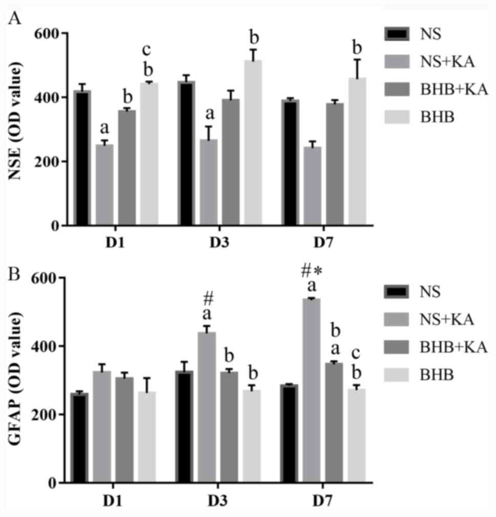
The expression levels of NSE and GFAP were evaluated
using double immunofluorescence (Figs.
1-3). For NSE, the interaction between time and treatment
revealed no statistically significant difference, whilst the main
effect of the treatment factor was statistically significant
(P<0.01). After KA administration, NSE expression was found to
be significantly lower in the NS + KA group compared with that in
the NS group (D1, P<0.01; D3, P<0.05) and the BHB group (D1,
D3, P<0.01; D7, P<0.05; Fig.
4A and Table SI). By contrast,
the expression of NSE was revealed to be significantly higher in
the BHB + KA group compared with that in the NS + KA group
(P<0.05) after 1 day of KA administration (Fig. 4A and Table SI). No significant differences in
NSE expression among different time points were observed,
suggesting that time exerted little influence on NSE
expression.
The interaction between treatment and time on GFAP
expression showed significant differences (P<0.01), whilst the
main effect of treatment on GFAP expression was also found to be
significant (P<0.01). After 3 and 7 days of KA administration,
GFAP expression was significantly higher in the NS + KA group
compared with that in the NS (D3, P<0.05; D7, P<0.01) and BHB
groups (both P<0.01, Fig. 4B),
whilst the expression of GFAP was significantly decreased in the
BHB + KA group compared with that in the NS + KA group (D3,
P<0.05; D7, P<0.01; Fig. 4B
and Table SII). However, after 7
days of KA administration, GFAP expression was significantly higher
in the BHB + KA compared with that in BHB group (P<0.01;
Fig. 4B and Table SII). In addition, time was also
found to significantly exert influence on GFAP expression
(P<0.01). Within the NS + KA groups, GFAP expression increased
along with time (P<0.05, D1 vs. D3, D3 vs. D7; P<0.01, D1 vs.
D7). By contrast, there was no significant difference among the
different time points in NS, BHB+KA and BHB groups.
These results suggested that KA can cause neuron
damage and compensatory astrocyte hyperplasia, which can be
reversed by BHB treatment. There were no differences in NSE and
GFAP expression between the BHB and NS groups at any point in time,
indicating that BHB did not exert toxic effects on the brain
tissues.
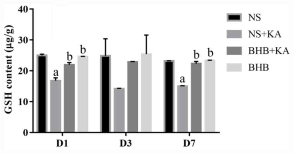
GSH content
There was no difference in the interaction between
time and treatment on GSH contents, where the main effect of time
also did not reveal significant influence. At 1 and 7 days after KA
administration, GSH content was found to be significantly lower in
the NS + KA groups compared with that in the NS groups (both
P<0.01) and the BHB groups (both P<0.01; Fig. 5 and Table SIII). GSH levels were also revealed
to be significantly higher in the BHB + KA group compared with
those in the NS + KA groups after 1 and 7 days (both P<0.01;
Fig. 5 and Table SIII). These results suggested that
BHB can alleviate the reduction in GSH caused by KA administration
in rats. In addition, no differences in the GSH contents were
observed between the BHB and NS groups, implicating the safety of
BHB.
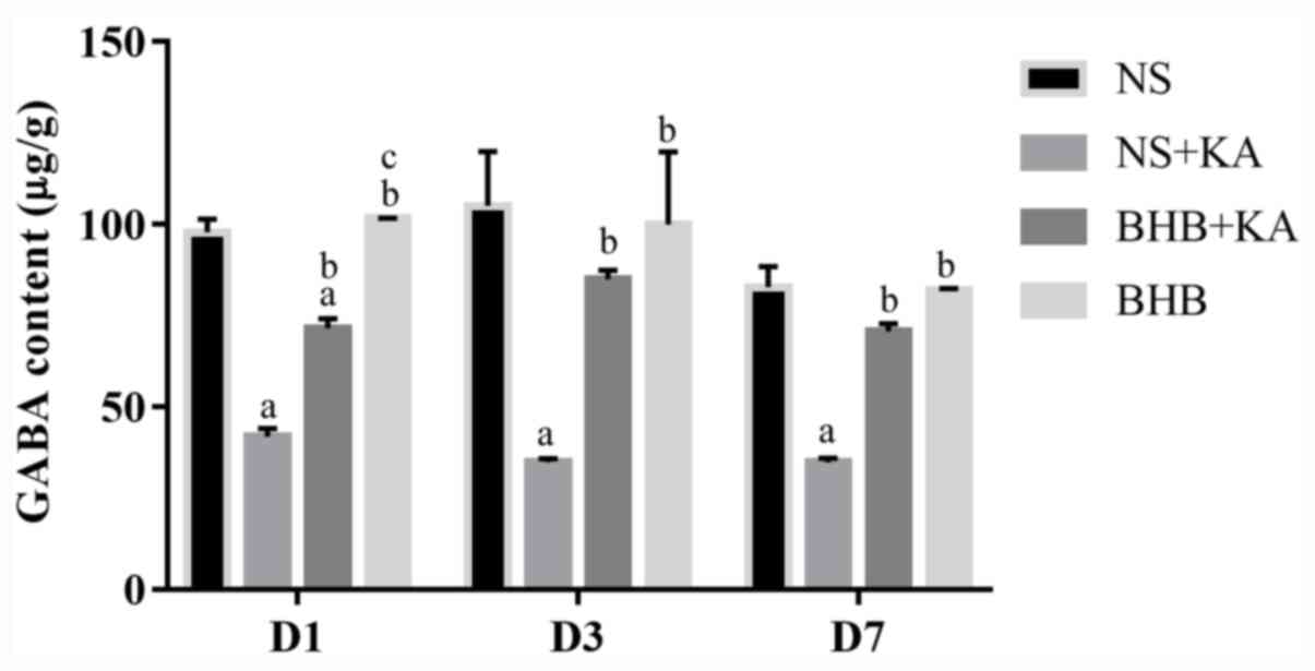
GABA contents
The interaction between time and treatment showed no
significant influence on GABA levels, where the main effect of time
also did not reveal statistical influence. After 1, 3 and 7 days of
KA administration, GABA levels were significantly reduced in the NS
+ KA groups compared with those in the NS groups (all P<0.01)
and the BHB groups (all P<0.01; Fig.
6 and Table SIV). At all three
time points, following pretreatment with BHB, the GABA contents
were found to be significantly higher in the BHB + KA group
compared with those in the NS + KA group (D1 and D7, P<0.01; D3,
P<0.05; Fig. 6 and Table SIV). However, in the BHB + KA
group, GABA contents remained significantly decreased compared with
those in NS (P<0.01) and BHB groups (P<0.01) 1 day after KA
administration (Fig. 6 and Table SIV). These results demonstrated
that KA administration reduced GABA levels whilst BHB alleviated
this decrease in GABA caused by KA treatment in rats. No
differences were found between BHB and NS groups in terms of GABA
levels.
Discussion
Epilepsy is one of the most prevalent serious
neurological disorders, for which it is important to develop novel
effective therapies. Previous studies have documented exogenous BHB
to be an anticonvulsant that exerts neuroprotective effects both
in vitro and in vivo (12,23).
In the present study, the antiepileptic effects of BHB in a
KA-induced epilepsy rat model were explored. Neuronal damage in the
hippocampus was demonstrated to be alleviated after rats were
pretreated with BHB. Additionally, the present study revealed that
BHB was capable of blocking the activation of astrocytes whilst
preserving the expression of GSH and GABA after KA
administration.
NSE levels have been previously reported to be
applicable for determining seizure durations and to estimate the
prognosis of brain injuries (24).
The NSE contents were found to be significantly higher in the serum
of children with epilepsy compared with those in unaffected
children, suggesting that elevated serum NSE after epileptic
seizures may be associated with brain damage (25,26).
GFAP is a glial cell marker in the development of the central
nervous system that is mainly expressed in activated astrocytes
(27,28). After epileptic seizure attacks, GFAP
expression was previously revealed to be significantly elevated in
astrocytes (29). In the present
study, NSE and GFAP were used to stain neurons and activated
astrocytes respectively, where KA injection resulted in an
inflammatory environment in rat brains, as indicated by the
extensive activation of glial cells. In addition, the number of
neurons was found to be increased in the BHB + KA group compared
with that in the NS + KA group whilst the degree of astrocyte
activation was reduced. These results indicated that neuronal
damage induced by KA was alleviated after the rats were pretreated
with BHB, which may be due in part to its ability to inhibit the
activation of glial cells.
It has been previously shown that oxidative stress
is one of the main pathological mechanisms of epilepsy (30,31).
During the progression of epilepsy, reactive oxygen species (ROS)
can damage the cell membrane, proteins, enzymes and DNA components
within the nucleus and the mitochondria (32). GSH is a part of the main antioxidant
system that neutralizes the excessive ROS. GSH is capable of
preventing damage to important cellular components caused by ROS,
including free radicals, peroxides, lipid peroxides and heavy
metals (33). Previous studies have
demonstrated that the elimination of GSH is closely associated with
a number of human diseases, including neurodegenerative diseases,
diabetes and acquired immune deficiency syndrome (34,35).
The present study showed that GSH levels in the hippocampal tissues
were significantly reduced after rats were treated with KA,
suggesting that the ability to eliminate free radicals is reduced
in epilepsy. Results from the present study also revealed that
administration of BHB reversed the reduction in GSH caused by KA
administration in the rat hippocampus. Therefore, it can be
potentially concluded that BHB can diminish ROS damage caused by KA
by preserving GSH levels. This is in accordance with a previous
study that also showed that BHB treatment can reduce the
overproduction of ROS and activate GSH further in the epileptic
hippocampus (36).
GABA is the main inhibitory neurotransmitter in the
central nervous system that serves a critical role in the
development of epilepsy (37,38).
Increased GABA synaptic activity can reduce the excitability of
neurons (39), whilst a reduced
GABA level can enhance the excitability of neurons (37). GABAA receptors are
ligand-gated ion channels that hyperpolarize neurons by increasing
inward chloride conductance (38).
Since the activation of these receptors results in a rapid
inhibitory effect, they serve a principal role in nerve
transmission processes in the central nervous system (38). GABAB receptors can reduce
calcium entry and mainly mediate slow synaptic inhibition, which is
involved with numerous types of epilepsy and cognitive impairment
(37,38). It was demonstrated that ketones can
alter glutamate metabolism by increasing GABA synthesis, which
would in turn dampen seizure activity (40). It has also been previously
demonstrated that BHB can reduce the incidence of seizure-like
activity in a GABAB-dependent manner (41). In the present study, GABA levels in
the hippocampal tissues were markedly reduced after rats were
treated with KA. This reduction in GABA can increase the
excitability of the neurons, thereby reducing the threshold of
epileptic seizures. GABA levels in the hippocampus tissue were
significantly increased after the rats were pretreated with BHB.
These results suggest that elevations in the levels of GABA
following the application of BHB can dampen seizure activity. In
the present study, a correlation analysis between the GABA content
and the number of neurons and astrocytes was not performed, which
would be of significance for understanding the mechanism of BHB
further. This is a limitation of the present study.
It has been previously demonstrated that BHB is a
more efficient energy source compared with glucose and that the
presence of BHB can reduce ATP production from glycolysis (42,43).
Glycolytic ATP is the primary source of energy that supports plasma
membrane functions, including ATP-sensitive potassium
(KATP) channels. Lower glycolytic ATP levels would lead
to higher KATP channel opening probability, which would
cause membrane hyperpolarization and reduce the influx of calcium
via voltage-gated calcium channels. This would in turn reduce the
release of excitatory amino acids and decreased neuron excitability
(44-46).
KATP channels, which are widely distributed in the
hippocampus, would open with higher probability in the presence of
BHB, which may underlie the anticonvulsive effects of ketone
bodies.
In the present study, only the ATP contents in the
BHB group were found to be greater compared with that of the
detection threshold on day 7 (46.26±0.81 ng/g). This experiment
could not detect ATP in other experimental groups. Considering the
rapid degradation of ATP during the tissue preparation process, the
frozen hippocampus tissues might have been the main cause of this.
Due to the significant elevations in ATP production, ATP could
still be detected in the BHB group despite its rapid
degradation.
Recently, several studies have demonstrated that BHB
confers neuroprotective effects on the central nervous system
against oxygen toxicity, Alzheimer's and Parkinson's disease
(47-49).
Although a series of studies have demonstrated that BHB has
protective effects in various epileptic models (9,20,36,50),
it remains necessary to verify the effects of BHB in other
epileptic models. Additionally, it is difficult to maintain stable
BHB concentrations in the blood, which limits the efficacy of BHB
administration for clinical application (45). Therefore, further studies focused on
BHB treatment for antiepileptic therapy are required to confirm its
efficacy and explore the underlying mechanisms.
Taken together, the similarity between the results
mediated by BHB and KD in epileptic models suggest that exogenous
BHB could replace KD as an anticonvulsant treatment for epilepsy.
In particular, there are some limitations of KD applications,
including nausea, constipation and abdominal pain (51). By contrast, BHB administration has
not been reported to cause adverse effects, which may improve the
patients' quality of life. Therefore, the application of exogenous
BHB may serve as a novel therapeutic technique in treating
epilepsy. However, it is essential to explore the therapeutic
effect of exogenous BHB further in the future.
Supplementary Material
P-values for the comparison of
neuron-specific enolase between the two indicated groups at each
time point.
P-values for the comparison of glial
fibrillary acidic protein between the two indicated groups at each
time point.
P-values for the comparison of
glutathione between the two indicated groups at each time
point.
P-values for the comparison of
γ-aminobutyric acid between the two indicated groups at each time
point.
Acknowledgements
Not applicable.
Funding
The present study was supported by a project of the
Shandong Province Science and Technology Program (grant no.
2014GSF118179) and the Special Foundation for Taishan Scholars
(grant no. ts20110814).
Availability of data and materials
The datasets used and/or analyzed during the present
study are available from the corresponding author on reasonable
request.
Authors' contributions
JW designed the study and supervised the project. JS
performed the experiments and completed the manuscript. YW
contributed to the acquisition, analysis and interpretation of data
for the study. JX performed the statistical analyses, helped with
supervising the whole project and was accountable for revision of
the manuscript. All authors read and approved the final
manuscript.
Ethics approval and consent to
participate
The present study was approved by the Ethics
Committee of Shanghai Jiao Tong University School of Medicine
(Shanghai, China).
Patient consent for publication
Not applicable.
Competing interests
The authors declare that they have no competing
interests.
References
|
1
|
Chang BS and Lowenstein DH: Epilepsy. N
Engl J Med. 349:1257–1266. 2003.PubMed/NCBI View Article : Google Scholar
|
|
2
|
Fisher RS, Acevedo C, Arzimanoglou A,
Bogacz A, Cross JH, Elger CE, Engel J Jr, Forsgren L, French JA,
Glynn M, et al: ILAE official report: A practical clinical
definition of epilepsy. Epilepsia. 55:475–482. 2014.PubMed/NCBI View Article : Google Scholar
|
|
3
|
GBD 2015 Disease and Injury Incidence and
Prevalence Collaborators. Global, regional, and national incidence,
prevalence, and years lived with disability for 310 diseases and
injuries, 1990-2015: A systematic analysis for the Global Burden of
Disease Study 2015. Lancet. 388:1545–1602. 2016.PubMed/NCBI View Article : Google Scholar
|
|
4
|
Kernich CA: Patient and family fact sheet
Epilepsy. Neurologist. 9:265–266. 2003.PubMed/NCBI View Article : Google Scholar
|
|
5
|
GBD 2015 Mortality and Causes of Death
Collaborators. Global, regional, and national life expectancy,
all-cause mortality, and cause-specific mortality for 249 causes of
death, 1980-2015: A systematic analysis for the Global Burden of
Disease Study 2015. Lancet. 388:1459–1544. 2016.PubMed/NCBI View Article : Google Scholar
|
|
6
|
GBD 2013 Mortality and Causes of Death
Collaborators. Global, regional, and national age-sex specific
all-cause and cause-specific mortality for 240 causes of death,
1990-2013: A systematic analysis for the Global Burden of Disease
Study 2013. Lancet. 385:117–171. 2015.PubMed/NCBI View Article : Google Scholar
|
|
7
|
Brodie MJ, Elder AT and Kwan P: Epilepsy
in later life. Lancet Neurol. 8:1019–1030. 2009.PubMed/NCBI View Article : Google Scholar
|
|
8
|
Eadie MJ: Shortcomings in the current
treatment of epilepsy. Expert Rev Neurother. 12:1419–1427.
2012.PubMed/NCBI View Article : Google Scholar
|
|
9
|
Kwan P and Brodie MJ: Early identification
of refractory epilepsy. N Engl J Med. 342:314–319. 2000.PubMed/NCBI View Article : Google Scholar
|
|
10
|
Wei CX, Bian M and Gong GH: Current
research on antiepileptic compounds. Molecules. 20:20741–20776.
2015.PubMed/NCBI View Article : Google Scholar
|
|
11
|
Suzuki Y, Takahashi H, Fukuda M, Hino H,
Kobayashi K, Tanaka J and Ishii E: β-Hydroxybutyrate alters
GABA-transaminase activity in cultured astrocytes. Brain Res.
1268:17–23. 2009.PubMed/NCBI View Article : Google Scholar
|
|
12
|
Samoilova M, Weisspapir M, Abdelmalik P,
Velumian AA and Carlen PL: Chronic in vitro ketosis is
neuroprotective but not anti-convulsant. J Neurochem. 113:826–835.
2010.PubMed/NCBI View Article : Google Scholar
|
|
13
|
Maalouf M and Rho JM: Oxidative impairment
of hippocampal long-term potentiation involves activation of
protein phosphatase 2A and is prevented by ketone bodies. J
Neurosci Res. 86:3322–3330. 2008.PubMed/NCBI View Article : Google Scholar
|
|
14
|
Likhodii SS and Burnham WM: Ketogenic
diet: Does acetone stop seizures? Med Sci Monit. 8:HY19–HY24.
2002.PubMed/NCBI
|
|
15
|
Abdelmalik PA, Shannon P, Yiu A, Liang P,
Adamchik Y, Weisspapir M, Samoilova M, Burnham WM and Carlen PL:
Hypoglycemic seizures during transient hypoglycemia exacerbate
hippocampal dysfunction. Neurobiol Dis. 26:646–660. 2007.PubMed/NCBI View Article : Google Scholar
|
|
16
|
Yum MS, Ko TS and Dong WK: Anticonvulsant
effects of β-hydroxybutyrate in mice. J Epilepsy Res. 2:29–32.
2012.PubMed/NCBI View Article : Google Scholar
|
|
17
|
Yum MS, Ko TS and Kim DW:
β-Hydroxybutyrate increases the pilocarpine-induced seizure
threshold in young mice. Brain Dev. 34:181–184. 2012.PubMed/NCBI View Article : Google Scholar
|
|
18
|
Minlebaev M and Khazipov R: Antiepileptic
effects of endogenous beta-hydroxybutyrate in suckling infant rats.
Epilepsy Res. 95:100–109. 2011.PubMed/NCBI View Article : Google Scholar
|
|
19
|
Thio LL, Wong M and Yamada KA: Ketone
bodies do not directly alter excitatory or inhibitory hippocampal
synaptic transmission. Neurology. 54:325–331. 2000.PubMed/NCBI View Article : Google Scholar
|
|
20
|
Si J, Wang S, Liu N, Yang X, Wang Y, Li L,
Wang J and Lv X: Anticonvulsant effect of exogenous
β-hydroxybutyrate on kainic acid-induced epilepsy. Exp Ther Med.
14:765–770. 2017.PubMed/NCBI View Article : Google Scholar
|
|
21
|
National Research Council: Guide for the
Care and Use of Laboratory Animals. 8th edition. Washington (DC):
National Academies Press (US); 2011. Available from: urihttps://www.ncbi.nlm.nih.gov/books/NBK54050/simplehttps://www.ncbi.nlm.nih.gov/books/NBK54050/
doi: 10.17226/12910ß.
|
|
22
|
Racine RJ: Modification of seizure
activity by electrical stimulation II Motor seizure.
Electroencephalogr Clin Neurophysiol. 32:281–294. 1972.PubMed/NCBI View Article : Google Scholar
|
|
23
|
Xie G, Tian W, Wei T and Liu F: The
neuroprotective effects of β-hydroxybutyrate on Aβ-injected rat
hippocampus in vivo and in Aβ-treated PC-12 cells in vitro. Free
Radic Res. 49:139–150. 2015.PubMed/NCBI View Article : Google Scholar
|
|
24
|
Bindra A, Kaushal A, Prabhakar H,
Chaturvedi A, Chandra PS, Tripathi M, Subbiah V, Sathianathan S,
Banerjee J and Prakash C: Neuroprotective role of dexmedetomidine
in epilepsy surgery: A preliminary study. Neurol India. 67:163–168.
2019.PubMed/NCBI View Article : Google Scholar
|
|
25
|
Maiti R, Mishra BR, Sanyal S, Mohapatra D,
Parida S and Mishra A: Effect of carbamazepine and oxcarbazepine on
serum neuron-specific enolase in focal seizures: A randomized
controlled trial. Epilepsy Res. 138:5–10. 2017.PubMed/NCBI View Article : Google Scholar
|
|
26
|
Yardimoğlu M, Ilbay G, Dalcik C, Dalcik H,
Sahin D and Ates N: Immunocytochemistry of neuron specific enolase
(NSE) in the rat brain after single and repeated epileptic
seizures. Int J Neurosci. 118:981–993. 2008.PubMed/NCBI View Article : Google Scholar
|
|
27
|
Jacque CM, Vinner C, Kujas M, Raoul M,
Racadot J and Baumann NA: Determination of glial fibrillary acidic
protein (GFAP) in human brain tumors. J Neurol Sci. 35:147–155.
1978.PubMed/NCBI View Article : Google Scholar
|
|
28
|
Venkatesh K, Srikanth L, Vengamma B,
Chandrasekhar C, Sanjeevkumar A, Mouleshwara Prasad BC and Sarma
PV: In vitro differentiation of cultured human CD34+ cells into
astrocytes. Neurol India. 61:383–388. 2013.PubMed/NCBI View Article : Google Scholar
|
|
29
|
Alese OO and Mabandla MV: Upregulation of
hippocampal synaptophysin, GFAP and mGluR3 in a pilocarpine rat
model of epilepsy with history of prolonged febrile seizure. J Chem
Neuroanat. 100(101659)2019.PubMed/NCBI View Article : Google Scholar
|
|
30
|
Puttachary S, Sharma S, Stark S and
Thippeswamy T: Seizure-induced oxidative stress in temporal lobe
epilepsy. Biomed Res Int. 2015(745613)2015.PubMed/NCBI View Article : Google Scholar
|
|
31
|
Yuen AWC, Keezer MR and Sander JW:
Epilepsy is a neurological and a systemic disorder. Epilepsy Behav.
78:57–61. 2018.PubMed/NCBI View Article : Google Scholar
|
|
32
|
Pearson-Smith JN and Patel M: Metabolic
dysfunction and oxidative stress in epilepsy. Int J Mol Sci.
18(2365)2017.PubMed/NCBI View Article : Google Scholar
|
|
33
|
Pompella A, Visvikis A, Paolicchi A, De
Tata V and Casini AF: The changing faces of glutathione, a cellular
protagonist. Biochem Pharmacol. 66:1499–1503. 2003.PubMed/NCBI View Article : Google Scholar
|
|
34
|
Lutchmansingh FK, Hsu JW, Bennett FI,
Badaloo AV, McFarlane-Anderson N, Gordon-Strachan GM, Wright-Pascoe
RA, Jahoor F and Boyne MS: Glutathione metabolism in type 2
diabetes and its relationship with microvascular complications and
glycemia. PLoS One. 13(e0198626)2018.PubMed/NCBI View Article : Google Scholar
|
|
35
|
Sprietsma JE: Cysteine, glutathione (GSH)
and zinc and copper ions together are effective, natural,
intracellular inhibitors of (AIDS) viruses. Med Hypotheses.
52:529–538. 1999.PubMed/NCBI View Article : Google Scholar
|
|
36
|
Guo Q, Liu S, Wang S, Wu M, Li Z and Wang
Y: Beta-hydroxybutyric acid attenuates neuronal damage in
epilepticmice. Acta Histochem. 121:455–459. 2019.PubMed/NCBI View Article : Google Scholar
|
|
37
|
Bhagat K, Singh JV, Pagare PP, Kumar N,
Sharma A, Kaur G, Kinarivala N, Gandu S, Singh H, Sharma S and Bedi
PMS: Rational approaches for the design of various GABA modulators
and their clinical progression. Mol Divers 2020 (Epub ahead of
print).
|
|
38
|
Treiman DM: GABAergic mechanisms in
epilepsy. Epilepsia. 42 (Suppl 3):S8–S12. 2001.PubMed/NCBI View Article : Google Scholar
|
|
39
|
Cepeda C, Levinson S, Nariai H, Yazon VW,
Tran C, Barry J, Oikonomou KD, Vinters HV, Fallah A, Mathern GW and
Wu JY: Pathological high frequency oscillations associate with
increased GABA synaptic activity in pediatric epilepsy surgery
patients. Neurobiol Dis. 134(104618)2020.PubMed/NCBI View Article : Google Scholar
|
|
40
|
Simeone TA, Simeone KA and Rho JM: Ketone
bodies as anti-seizure agents. Neurochem Res. 42:2011–2018.
2017.PubMed/NCBI View Article : Google Scholar
|
|
41
|
Li J, O'Leary EI and Tanner GR: The
ketogenic diet metabolite beta-hydroxybutyrate (β-HB) reduces
incidence of seizure-like activity (SLA) in a Katp- and
GABAb-dependent manner in a whole-animal Drosophila melanogaster
model. Epilepsy Res. 133:6–9. 2017.PubMed/NCBI View Article : Google Scholar
|
|
42
|
Mejía-Toiber J, Montiel T and Massieu L:
D-beta-hydroxybutyrate prevents glutamate-mediated lipoperoxidation
and neuronal damage elicited during glycolysis inhibition in vivo.
Neurochem Res. 31:1399–1408. 2006.PubMed/NCBI View Article : Google Scholar
|
|
43
|
Mudaliar S, Alloju S and Henry RR: Can a
shift in fuel energetics explain the beneficial cardiorenal
out-comes in the EMPA-REG OUTCOME study? A unifying hypothesis.
Diabetes Care. 39:1115–1122. 2016.PubMed/NCBI View Article : Google Scholar
|
|
44
|
Lund TM, Ploug KB, Iversen A, Jensen AA
and Jansen-Olesen I: The metabolic impact of β-hydroxybutyrate on
neurotransmission: Reduced glycolysis mediates changes in calcium
responses and KATP channel receptor sensitivity. J Neurochem.
132:520–531. 2015.PubMed/NCBI View Article : Google Scholar
|
|
45
|
Tanner G, Lutas A, Martínez-François JR
and Yellen G: Single K ATP channel opening in response to action
potential firing in mouse dentate granule neurons. J Neurosci.
31:8689–8696. 2011.PubMed/NCBI View Article : Google Scholar
|
|
46
|
Giménez-Cassina A, Martínez-François JR,
Fisher JK, Szlyk B, Polak K, Wiwczar J, Tanner GR, Lutas A, Yellen
G and Danial NN: BAD-dependent regulation of fuel metabolism and
K(ATP) channel activity confers resistance to epileptic seizures.
Neuron. 74:719–730. 2012.PubMed/NCBI View Article : Google Scholar
|
|
47
|
Soto-Mota A, Norwitz NG and Clarke K: Why
a d-β-hydroxybutyrate monoester? Biochem Soc Trans. 48:51–59.
2020.PubMed/NCBI View Article : Google Scholar
|
|
48
|
Krishnan M, Hwang JS, Kim M, Kim YJ, Seo
JH, Jung J and Ha E: β-hydroxybutyrate impedes the progression of
Alzheimer's disease and atherosclerosis in ApoE-deficient mice.
Nutrients. 12(471)2020.PubMed/NCBI View Article : Google Scholar
|
|
49
|
Norwitz NG, Hu MT and Clarke K: The
mechanisms by which the ketone body D-β-hydroxybutyrate may improve
the multiple cellular pathologies of Parkinson's disease. Front
Nutr. 6(63)2019.PubMed/NCBI View Article : Google Scholar
|
|
50
|
Yum MS, Lee M, Woo DC, Kim DW, Ko TS and
Velíšek L: β-Hydroxybutyrate attenuates NMDA-induced spasms in rats
with evidence of neuronal stabilization on MR spectroscopy.
Epilepsy Res. 117:125–132. 2015.PubMed/NCBI View Article : Google Scholar
|
|
51
|
Giordano C, Marchiò M, Timofeeva E and
Biagini G: Neuroactive peptides as putative mediators of
antiepileptic ketogenic diets. Front Neurol. 5(63)2014.PubMed/NCBI View Article : Google Scholar
|