|
1
|
Cassidy J, Carroll L, Peloso P, Borg J,
von Holst H, Holm L, Kraus J and Coronado V: WHO Collaborating
Centre Task Force on Mild Traumatic Brain Injury: Incidence, risk
factors and prevention of mild traumatic brain injury: Results of
the WHO collaborating Centre task force on mild traumatic brain
injury. J Rehabil Med. 36 (Suppl 43):S28–S60. 2004.PubMed/NCBI View Article : Google Scholar
|
|
2
|
Smith DH, Meaney DF and Shull WH: Diffuse
axonal injury in head trauma. J Head Trauma Rehabil. 18:307–316.
2003.PubMed/NCBI View Article : Google Scholar
|
|
3
|
Kraus MF, Susmaras T, Caughlin BP, Walker
CJ, Sweeney JA and Little DM: White matter integrity and cognition
in chronic traumatic brain injury: A diffusion tensor imaging
study. Brain. 130:2508–2519. 2007.PubMed/NCBI View Article : Google Scholar
|
|
4
|
McKee AC, Stern RA, Nowinski CJ, Stein TD,
Alvarez VE, Daneshvar DH, Lee HS, Wojtowicz SM, Hall G, Baugh CM,
et al: The spectrum of disease in chronic traumatic encephalopathy.
Brain. 136:43–64. 2013.PubMed/NCBI View Article : Google Scholar
|
|
5
|
Xiong Y, Mahmood A and Chopp M: Animal
models of traumatic brain injury. Nat Rev Neurosci. 14:128–142.
2013.PubMed/NCBI View
Article : Google Scholar
|
|
6
|
Namjoshi DR, Cheng WH, McInnes KA, Martens
KM, Carr M, Wilkinson A, Fan J, Robert J, Hayat A, Cripton PA and
Wellington CL: Merging pathology with biomechanics using chimera
(closed-head impact model of engineered rotational acceleration): A
novel, Surgery-free model of traumatic brain injury. Mol
Neurodegen. 9(55)2014.PubMed/NCBI View Article : Google Scholar
|
|
7
|
Li XY, Dai GH, Feng DF, Li J and Gu L:
Experimental device for inducing diffuse brain injury in small
animals. BME Clin Med. 13:489–493. 2009.
|
|
8
|
Xiang L, Zhang YT, Liang P, Wei H, Peng LL
and Li LS: Study on the establishment of rat model of different
degree of diffuse axonal injury. Chongqing Med. 45:2881–2884.
2016.
|
|
9
|
Mayer AR, Ling J, Mannell MV, Gasparovic
C, Phillips JP, Doezema D, Reichard R and Yeo RA: A prospective
diffusion tensor imaging study in mild traumatic brain injury.
Neurology. 74:643–650. 2010.PubMed/NCBI View Article : Google Scholar
|
|
10
|
McAllister TW, Ford JC, Ji S, Beckwith JG,
Flashman LA, Paulsen K and Greenwald RM: Maximum principal strain
and strain rate associated with concussion diagnosis correlates
with changes in corpus callosum white matter indices. Ann Biomed
Eng. 40:127–140. 2012.PubMed/NCBI View Article : Google Scholar
|
|
11
|
Eierud C, Craddock RC, Fletcher S, Aulakh
M, King-Casas B, Kuehl D and LaConte SM: Neuroimaging after mild
traumatic brain injury: Review and meta-analysis. Neuroimage Clin.
4:283–294. 2014.PubMed/NCBI View Article : Google Scholar
|
|
12
|
Bramlett HM, Kraydieh S, Green EJ and
Dietrich WD: Temporal and regional patterns of axonal damage
following traumatic brain injury: A beta-amyloid precursor protein
immunocyto-chemical study in rats. J Neuropathol Exp Neurol.
56:1132–1141. 1997.PubMed/NCBI View Article : Google Scholar
|
|
13
|
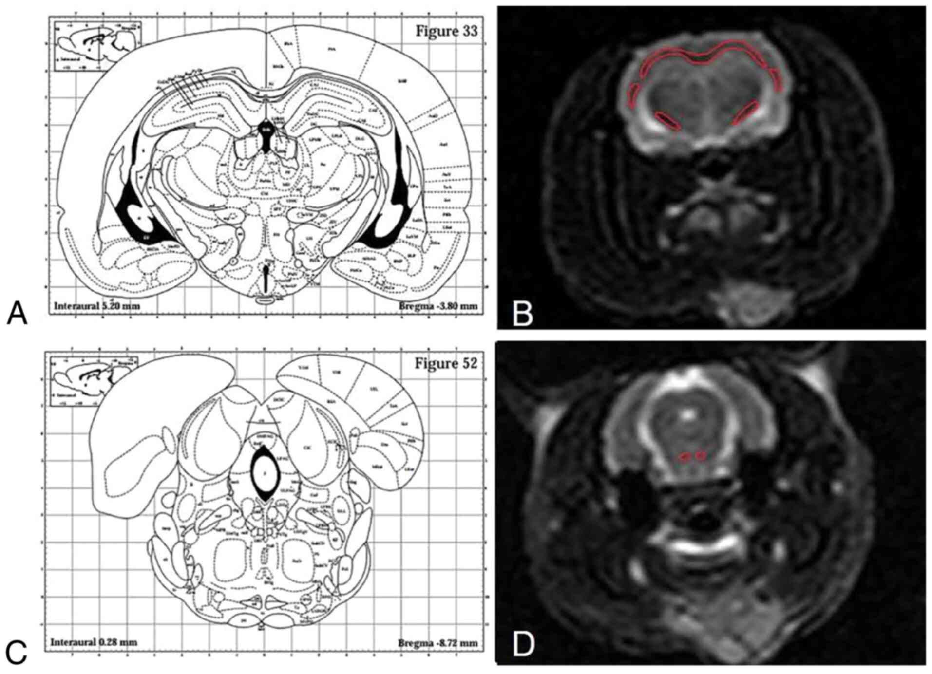
Paxinos G and Watson C: The Rat Brain in
Stereotaxic Coordinates. Elsevier Publishing, Amsterdam, pp82-100,
1997.
|
|
14
|
Kilbourne M, Kuehn R, Tosun C, Caridi J,
Keledjian K, Bochicchio G, Scalea T, Gerzanich V and Simard JM:
Novel model of frontal impact closed head injury in the rat. J
Neurotrauma. 26:2233–2243. 2009.PubMed/NCBI View Article : Google Scholar
|
|
15
|
Fehily B and Fitzgerald M: Repeated mild
traumatic brain injury: Potential mechanisms of damage. Cell
Transplant. 26:1131–1155. 2017.PubMed/NCBI View Article : Google Scholar
|
|
16
|
Xiang L, Zhang YT, Liang P, Wei H, Peng LL
and Li LS: Value of β-APP and NF-L as markers in evaluation of rat
diffuse axonal injury. J Third Military Med Univ. 37:2255–2260.
2015.
|
|
17
|
Browne KD, Chen XH, Meaney DF and Smith
DH: Mild traumatic brain injury and diffuse axonal injury in swine.
J Neurotrauma. 28:1747–1755. 2011.PubMed/NCBI View Article : Google Scholar
|
|
18
|
Gasparetto EL, Rueda Lopes FC and
Domingues RC and Domingues RC: Diffusion imaging in traumatic brain
injury. Neuroimaging Clin N Am. 21:115–125. 2011.PubMed/NCBI View Article : Google Scholar
|
|
19
|
McCrory P, Meeuwisse WH, Aubry M, Cantu B,
Dvorák J, Echemendia RJ, Engebretsen L, Johnston K, Kutcher JS,
Raftery M, et al: Consensus statement on concussion in sport: The
4th International Conference on Concussion in Sport held in Zurich,
November 2012. Br J Sports Med. 47:250–258. 2013.PubMed/NCBI View Article : Google Scholar
|
|
20
|
Ham TE and Sharp DJ: How can investigation
of network function inform rehabilitation after traumatic brain
injury. Curr Opin Neurol. 25:662–669. 2012.PubMed/NCBI View Article : Google Scholar
|
|
21
|
Lipton ML, Gellella E, Lo C, Gold T,
Ardekani BA, Shifteh K, Bello JA and Branch CA: Multifocal white
matter ultrastructural abnormalities in mild traumatic brain injury
with cognitive disability: A voxel-wise analysis of diffusion
tensor imaging. J Neurotrauma. 25:1335–1342. 2008.PubMed/NCBI View Article : Google Scholar
|
|
22
|
Wang Y, Wang Q, Haldar JP, Yeh FC, Xie M,
Sun P, Tu TW, Trinkaus K, Klein RS, Cross AH and Song SK:
Quantification of increased cellularity during inflammatory
demyelination. Brain. 134:3590–601. 2011.PubMed/NCBI View Article : Google Scholar
|