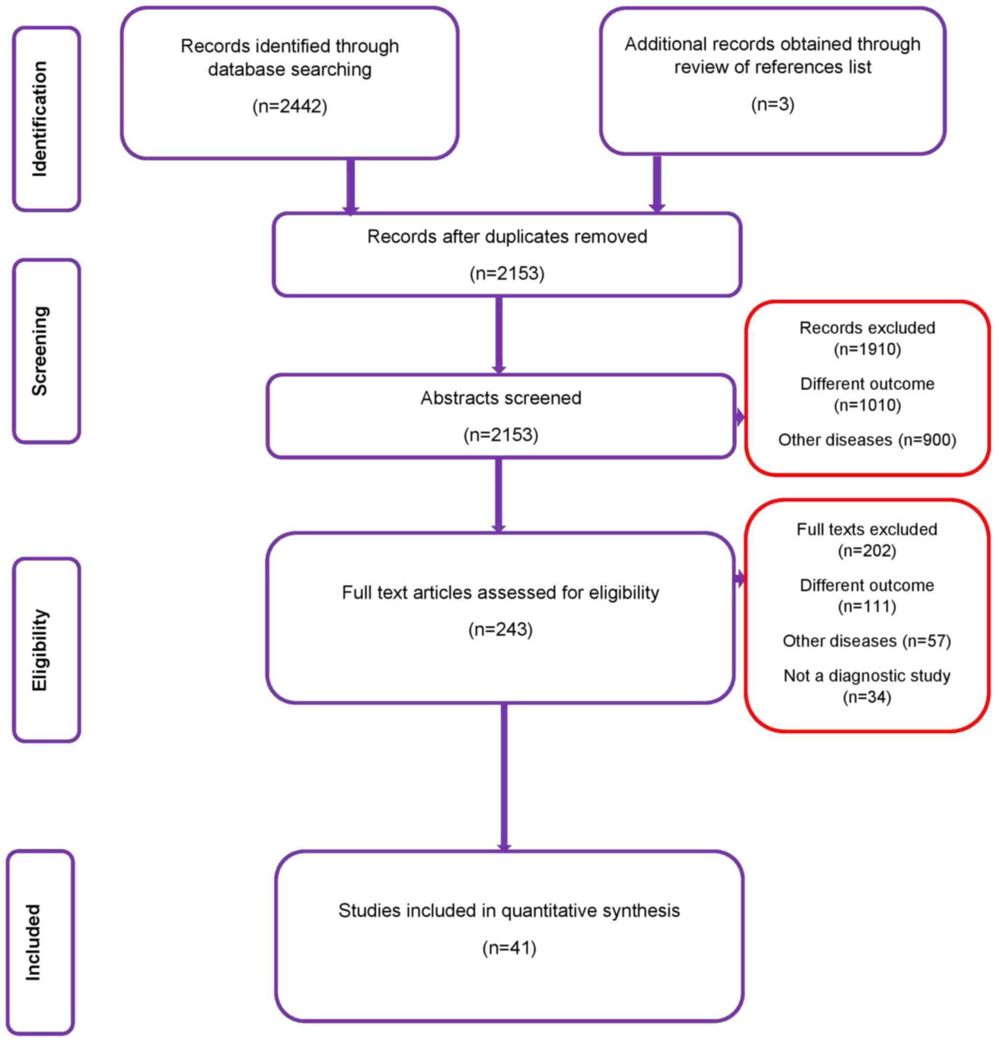
|
1
|
Griffin N, Grant LA and Sala E: Adnexal
masses: Characterization and imaging strategies. Semin Ultrasound
CT MR. 31:330–346. 2010.PubMed/NCBI View Article : Google Scholar
|
|
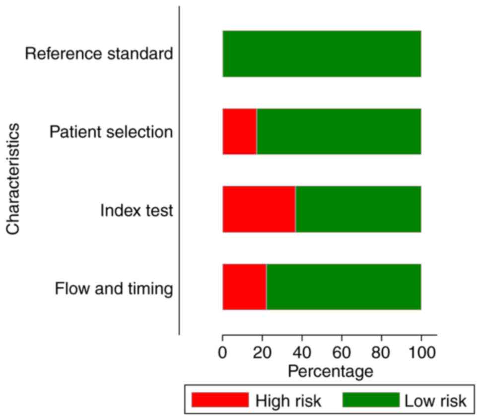
2
|
Givens V, Mitchell GE, Harraway-Smith C,
Reddy A and Maness DL: Diagnosis and management of adnexal masses.
Am Fam Physician. 80:815–820. 2009.PubMed/NCBI
|
|
3
|
Hakoun AM, AbouAl-Shaar I, Zaza KJ,
Abou-Al-Shaar H and A Salloum MN: Adnexal masses in pregnancy: An
updated review. Avicenna J Med. 7:153–157. 2017.PubMed/NCBI View Article : Google Scholar
|
|
4
|
Fischerova D, Zikan M, Dundr P and Cibula
D: Diagnosis, treatment, and follow-up of borderline ovarian
tumors. Oncologist. 17:1515–1533. 2012.PubMed/NCBI View Article : Google Scholar
|
|
5
|
Curtin JP: Management of the adnexal mass.
Gynecol Oncol. 55:S42–S46. 1994.PubMed/NCBI View Article : Google Scholar
|
|
6
|
American College of Obstetricians and
Gynecologists: ACOG practice bulletin. Management of adnexal
masses. Obstet Gynecol 110: 201-214, 2007.
|
|
7
|
Coleman BG: Transvaginal sonography of
adnexal masses. Radiol Clin North Am. 30:677–691. 1992.PubMed/NCBI
|
|
8
|
Whiting PF, Rutjes AWS, Westwood ME,
Mallett S, Deeks JJ, Reitsma JB, Leeflang MM, Sterne JA and Bossuyt
PM: QUADAS-2 Group: QUADAS-2: A revised tool for the quality
assessment of diagnostic accuracy studies. Ann Intern Med.
155:529–536. 2011.PubMed/NCBI View Article : Google Scholar
|
|
9
|
Alcázar JL, Pascual MÁ, Olartecoechea B,
Graupera B, Aubá M, Ajossa S, Hereter L, Julve R, Gastón B, Peddes
C, et al: IOTA simple rules for discriminating between benign and
malignant adnexal masses: Prospective external validation.
Ultrasound Obstetr Gynecol. 42:467–471. 2013.PubMed/NCBI View Article : Google Scholar
|
|
10
|
Daemen A, Valentin L, Fruscio R, Van
Holsbeke C, Melis GB, Guerriero S, Czekierdowski A, Jurkovic D,
Ombelet W, Rossi A, et al: Improving the preoperative
classification of adnexal masses as benign or malignant by
second-stage tests. Ultrasound Obstet Gynecol. 37:100–106.
2011.PubMed/NCBI View
Article : Google Scholar
|
|
11
|
Ruiz de Gauna B, Rodriguez D,
Olartecoechea B, Aubá M, Jurado M, Gómez Roig MD and Alcázar JL:
Diagnostic performance of IOTA simple rules for adnexal masses
classification: A comparison between two centers with different
ovarian cancer prevalence. Eur J Obstet Gynecol Reprod Biol.
191:10–14. 2015.PubMed/NCBI View Article : Google Scholar
|
|
12
|
Fathallah K, Huchon C, Bats AS, Metzger U,
Lefrère-Belda MA, Bensaid C and Lécuru F: External validation of
simple ultrasound rules of Timmerman on 122 ovarian tumors. Gynecol
Obstet Fertil. 39:477–481. 2011.(In French). PubMed/NCBI View Article : Google Scholar
|
|
13
|
Granberg S, Norström A and Wikland M:
Tumors in the lower pelvis as imaged by vaginal sonography. Gynecol
Oncol. 37:224–229. 1990.PubMed/NCBI View Article : Google Scholar
|
|
14
|
Guerriero S, Alcazar JL, Ajossa S, Galvan
R, Laparte C, García-Manero M, Lopez-Garcia G and Melis GB:
Transvaginal color Doppler imaging in the detection of ovarian
cancer in a large study population. Int J Gynecol Cancer.
20:781–786. 2010.PubMed/NCBI View Article : Google Scholar
|
|
15
|
Hartman CA, Juliato CRT, Sarian LO, Toledo
MC, Jales RM, Morais SS, Pitta DD, Marussi EF and Derchain S:
Ultrasound criteria and CA 125 as predictive variables of ovarian
cancer in women with adnexal tumors. Ultrasound Obstet Gynecol.
40:360–366. 2012.PubMed/NCBI View Article : Google Scholar
|
|
16
|
Jain KA, Friedman DL, Pettinger TW,
Alagappan R, Jeffrey RB Jr and Sommer FG: Adnexal masses:
Comparison of specificity of endovaginal US and pelvic MR imaging.
Radiology. 186:697–704. 1993.PubMed/NCBI View Article : Google Scholar
|
|
17
|
Jain KA: Prospective evaluation of adnexal
masses with endovaginal gray-scale and duplex and color Doppler US:
Correlation with pathologic findings. Radiology. 191:63–67.
1994.PubMed/NCBI View Article : Google Scholar
|
|
18
|
Knafel A, Nocun A, Banas T, Wiechec M,
Jach R, Pietrus M and Pitynski K: IOTA simple ultrasound-based
rules; why do we inconclusive results? Int J Gynecol Cancer.
2013(8)2013.
|
|
19
|
Komatsu T, Konishi I, Mandai M, Togashi K,
Kawakami S, Konishi J and Mori T: Adnexal masses: Transvaginal US
and gadolinium-enhanced MR imaging assessment of intratumoral
structure. Radiology. 198:109–115. 1996.PubMed/NCBI View Article : Google Scholar
|
|
20
|
Lucidarme O, Akakpo JP, Granberg S, Sideri
M, Levavi H, Schneider A, Autier P, Nir D and Bleiberg H: Ovarian
HistoScanning Clinical Study Group: A new computer-aided diagnostic
tool for non-invasive characterisation of malignant ovarian masses:
Results of a multicentre validation study. Eur Radiol.
20:1822–1830. 2010.PubMed/NCBI View Article : Google Scholar
|
|
21
|
Mancuso A, De Vivo A, Triolo O and Irato
S: The role of transvaginal ultrasonography and serum CA 125 assay
combined with age and hormonal state in the differential diagnosis
of pelvic masses. Eur J Gynaecol Oncol. 25:207–210. 2004.PubMed/NCBI
|
|
22
|
Moszynski R, Szpurek D, Szubert S and
Sajdak S: Analysis of false negative results of subjective
ultrasonography assessment of adnexal masses. Ginekol Pol.
84:102–107. 2013.PubMed/NCBI View
Article : Google Scholar
|
|
23
|
Nunes N, Yazbek J, Ambler G, Hoo W,
Naftalin J and Jurkovic D: Prospective evaluation of the IOTA
logistic regression model LR2 for the diagnosis of ovarian cancer.
Ultrasound Obstet Gynecol. 40:355–359. 2012.PubMed/NCBI View Article : Google Scholar
|
|
24
|
Nunes N, Ambler G, Hoo WL, Naftalin J, Foo
X, Widschwendter M and Jurkovic D: A prospective validation of the
IOTA logistic regression models (LR1 and LR2) in comparison to
subjective pattern recognition for the diagnosis of ovarian cancer.
Int J Gynecol Cancer. 23:1583–1589. 2013.PubMed/NCBI View Article : Google Scholar
|
|
25
|
Radosa MP, Vorwergk J, Fitzgerald J,
Kaehler C, Schneider U, Camara O, Runnebaum IB and Schleußner E:
Sonographic discrimination between benign and malignant adnexal
masses in premenopause. Ultraschall Med. 35:339–344.
2014.PubMed/NCBI View Article : Google Scholar
|
|
26
|
Romagnolo C, Trivella G, Bonacina M,
Fornalè M, Maggino T and Ferrazzi E: Preoperative diagnosis of 221
consecutive ovarian masses: Scoring system and expert evaluation.
Eur J Gynaecol Oncol. 27:487–489. 2006.PubMed/NCBI
|
|
27
|
Roman LD, Muderspach LI, Stein SM,
Laifer-Narin S, Groshen S and Morrow CP: Pelvic examination, tumor
marker level, and gray-scale and Doppler sonography in the
prediction of pelvic cancer. Obstet Gynecol. 89:493–500.
1997.PubMed/NCBI View Article : Google Scholar
|
|
28
|
Salle B, Gaucherand P, Ecochard R and
Rudigoz RC: Role of pulsed color Doppler in the presurgical
evaluation of pelvic masses. J Gynecol Obstet Biol Reprod (Paris).
24:234–240. 1995.(In French). PubMed/NCBI
|
|
29
|
Sayasneh A, Wynants L, Preisler J, Kaijser
J, Johnson S, Stalder C, Husicka R, Abdallah Y, Raslan F, Drought
A, et al: Multicentre external validation of IOTA prediction models
and RMI by operators with varied training. Br J Cancer.
108:2448–2454. 2013.PubMed/NCBI View Article : Google Scholar
|
|
30
|
Sayasneh A, Kaijser J, Preisler J, Smith
AA, Raslan F, Johnson S, Husicka R, Ferrara L, Stalder C,
Ghaem-Maghami S, et al: Accuracy of ultrasonography performed by
examiners with varied training and experience in predicting
specific pathology of adnexal masses. Ultrasound Obstet Gynecol.
45:605–612. 2015.PubMed/NCBI View Article : Google Scholar
|
|
31
|
Shetty J, Reddy G and Pandey D: Role of
sonographic gray-scale pattern recognition in the diagnosis of
adnexal masses. J Clin Diagn Res. 11:QC12–QC15. 2017.PubMed/NCBI View Article : Google Scholar
|
|
32
|
Shetty J, Saradha A, Pandey D, Bhat R,
Pratap K and Bharatnur S: IOTA simple ultrasound rules for triage
of adnexal mass: Experience from South India. J Obstet Gynecol
India. 69:356–362. 2019.PubMed/NCBI View Article : Google Scholar
|
|
33
|
Silvestre L, Martins WP and
Candido-dos-Reis FJ: Limitations of three-dimensional power Doppler
angiography in preoperative evaluation of ovarian tumors. J Ovarian
Res. 8(47)2015.PubMed/NCBI View Article : Google Scholar
|
|
34
|
Sohaib SA, Mills TD, Sahdev A, Webb JA,
Vantrappen PO, Jacobs IJ and Reznek RH: The role of magnetic
resonance imaging and ultrasound in patients with adnexal masses.
Clin Radiol. 60:340–348. 2005.PubMed/NCBI View Article : Google Scholar
|
|
35
|
Sokalska A, Timmerman D, Testa AC, Van
Holsbeke C, Lissoni AA, Leone FP, Jurkovic D and Valentin L:
Diagnostic accuracy of transvaginal ultrasound examination for
assigning a specific diagnosis to adnexal masses. Ultrasound Obstet
Gynecol. 34:462–470. 2009.PubMed/NCBI View
Article : Google Scholar
|
|
36
|
Stein SM, Laifer-Narin S, Johnson MB,
Roman LD, Muderspach LI, Tyszka JM and Ralls PW: Differentiation of
benign and malignant adnexal masses: Relative value of gray-scale,
color Doppler, and spectral Doppler sonography. AJR Am J
Roentgenol. 164:381–386. 1995.PubMed/NCBI View Article : Google Scholar
|
|
37
|
Strigini FA, Gadducci A, Del Bravo B,
Ferdeghini M and Genazzani AR: Differential diagnosis of adnexal
masses with transvaginal sonography, color flow imaging, and serum
CA 125 assay in pre- and postmenopausal women. Gynecol Oncol.
61:68–72. 1996.PubMed/NCBI View Article : Google Scholar
|
|
38
|
Tantipalakorn C, Wanapirak C,
Khunamornpong S, Sukpan K and Tongsong T: IOTA simple rules in
differentiating between benign and malignant ovarian tumors. Asian
Pac J Cancer Prev. 15:5123–5126. 2014.PubMed/NCBI View Article : Google Scholar
|
|
39
|
Testa A, Kaijser J, Wynants L, Fischerova
D, Van Holsbeke C, Franchi D, Savelli L, Epstein E, Czekierdowski
A, Guerriero S, et al: Strategies to diagnose ovarian cancer: New
evidence from phase 3 of the multicentre international IOTA study.
Br J Cancer. 111:680–688. 2014.PubMed/NCBI View Article : Google Scholar
|
|
40
|
Timmerman D, Verrelst H, Bourne TH, De
Moor B, Collins WP, Vergote I and Vandewalle J: Artificial neural
network models for the preoperative discrimination between
malignant and benign adnexal masses. Ultrasound Obstet Gynecol.
13:17–25. 1999.PubMed/NCBI View Article : Google Scholar
|
|
41
|
Timmerman D, Ameye L, Fischerova D,
Epstein E, Melis GB, Guerriero S, Van Holsbeke C, Savelli L,
Fruscio R, Lissoni AA, et al: Simple ultrasound rules to
distinguish between benign and malignant adnexal masses before
surgery: Prospective validation by IOTA group. BMJ.
341(c6839)2010.PubMed/NCBI View Article : Google Scholar
|
|
42
|
Utrilla-Layna J, Alcázar JL, Aubá M,
Laparte C, Olartecoechea B, Errasti T, Juez L, Mínguez JÁ,
Guerriero S and Jurado M: Performance of three-dimensional power
Doppler angiography as third-step assessment in differential
diagnosis of adnexal masses. Ultrasound Obstet Gynecol. 45:613–617.
2015.PubMed/NCBI View Article : Google Scholar
|
|
43
|
Valentin L: Prospective cross-validation
of Doppler ultrasound examination and gray-scale ultrasound imaging
for discrimination of benign and malignant pelvic masses.
Ultrasound Obstet Gynecol. 14:273–283. 1999.PubMed/NCBI View Article : Google Scholar
|
|
44
|
Valentin L, Jurkovic D, Van Calster B,
Testa A, Van Holsbeke C, Bourne T, Vergote I, Van Huffel S and
Timmerman D: Adding a single CA 125 measurement to ultrasound
imaging performed by an experienced examiner does not improve
preoperative discrimination between benign and malignant adnexal
masses. Ultrasound Obstet Gynecol. 34:345–354. 2009.PubMed/NCBI View
Article : Google Scholar
|
|
45
|
Van Calster B, Timmerman D, Bourne T,
Testa AC, Van Holsbeke C, Domali E, Jurkovic D, Neven P, Van Huffel
S and Valentin L: Discrimination between benign and malignant
adnexal masses by specialist ultrasound examination versus serum
CA-125. J Natl Cancer Inst. 99:1706–1714. 2007.PubMed/NCBI View Article : Google Scholar
|
|
46
|
Van Gorp T, Veldman J, Van Calster B,
Cadron I, Leunen K, Amant F, Timmerman D and Vergote I: Subjective
assessment by ultrasound is superior to the risk of malignancy
index (RMI) or the risk of ovarian malignancy algorithm (ROMA) in
discriminating benign from malignant adnexal masses. Eur J Cancer.
48:1649–1656. 2012.PubMed/NCBI View Article : Google Scholar
|
|
47
|
Van Holsbeke C, Van Calster B, Testa AC,
Testa AC, Domali E, Lu C, Van Huffel S, Valentin L and Timmerman D:
Prospective internal validation of mathematical models to predict
malignancy in adnexal masses: Results from the international
ovarian tumor analysis study. Clin Cancer Res. 15:684–691.
2009.PubMed/NCBI View Article : Google Scholar
|
|
48
|
van Trappen PO, Rufford BD, Mills TD,
Sohaib SA, Webb JA, Sahdev A, Carroll MJ, Britton KE, Reznek RH and
Jacobs IJ: Differential diagnosis of adnexal masses: Risk of
malignancy index, ultrasonography, magnetic resonance imaging, and
radioimmunoscintigraphy. Int J Gynecol Cancer. 17:61–67.
2007.PubMed/NCBI View Article : Google Scholar
|
|
49
|
Yamashita Y, Torashima M, Hatanaka Y,
Harada M, Higashida Y, Takahashi M, Mizutani H, Tashiro H, Iwamasa
J, Miyazaki K, et al: Adnexal masses: Accuracy of characterization
with transvaginal US and precontrast and postcontrast MR imaging.
Radiology. 194:557–565. 1995.PubMed/NCBI View Article : Google Scholar
|
|
50
|
Karnik A, Tembey RA and Mani S: Value of
MRI in characterizing adnexal masses. J Obstet Gynaecol India.
65:259–266. 2015.PubMed/NCBI View Article : Google Scholar
|
|
51
|
Kaijser J, Vandecaveye V, Deroose CM,
Rockall A, Thomassin-Naggara I, Bourne T and Timmerman D: Imaging
techniques for the pre-surgical diagnosis of adnexal tumours. Best
Pract Res Clin Obstet Gynaecol. 28:683–695. 2014.PubMed/NCBI View Article : Google Scholar
|
|
52
|
Dochez V, Caillon H, Vaucel E, Dimet J,
Winer N and Ducarme G: Biomarkers and algorithms for diagnosis of
ovarian cancer: CA125, HE4, RMI and ROMA, a review. J Ovarian Res.
12(28)2019.PubMed/NCBI View Article : Google Scholar
|
|
53
|
Ferraro S, Braga F, Lanzoni M, Boracchi P,
Biganzoli EM and Panteghini M: Serum human epididymis protein 4 vs
carbohydrate antigen 125 for ovarian cancer diagnosis: A systematic
review. J Clin Pathol. 66:273–281. 2013.PubMed/NCBI View Article : Google Scholar
|
|
54
|
Wang J, Gao J, Yao H, Wu Z, Wang M and Qi
J: Diagnostic accuracy of serum HE4, CA125 and ROMA in patients
with ovarian cancer: A meta-analysis. Tumour Biol. 35:6127–6138.
2014.PubMed/NCBI View Article : Google Scholar
|
|
55
|
Zhen S, Bian LH, Chang LL and Gao X:
Comparison of serum human epididymis protein 4 and carbohydrate
antigen 125 as markers in ovarian cancer: A meta-analysis. Mol Clin
Oncol. 2:559–566. 2014.PubMed/NCBI View Article : Google Scholar
|
|
56
|
Li F, Tie R, Chang K, Wang F, Deng S, Lu
W, Yu L and Chen M: Does risk for ovarian malignancy algorithm
excel human epididymis protein 4 and CA125 in predicting epithelial
ovarian cancer: A meta-analysis. BMC Cancer. 12(258)2012.PubMed/NCBI View Article : Google Scholar
|
|
57
|
Pewsner D, Battaglia M, Minder C, Marx A,
Bucher HC and Egger M: Ruling a diagnosis in or out with ‘SpPIn’
and ‘SnNOut’: A note of caution. BMJ. 329:209–213. 2004.PubMed/NCBI View Article : Google Scholar
|