Introduction
Natural killer (NK)/T-cell lymphomas (NKTCLs) are
non-Hodgkin lymphomas mostly extranodal, which are frequently of NK
and of (rare) T-cell origin, being closely related to the
Epstein-Barr virus (EBV). The World Health Organization (WHO)
classifies NKTCL into nodal, extranodal (sometimes referred to as
non-nasal)-cutaneous and other extranodal types, and disseminated
(leukemic).
Nasal type NKTCL, previously referred to as lethal
midline granuloma or angiocentric T-cell lymphoma, involves the
upper aero-digestive tract with the nasal cavity, nasopharynx,
paranasal sinuses, tonsils or palate. Non-nasal NKTCLs frequently
involve the skin, testis or the gastrointestinal tract, but it can
affect many other sites. The disseminated type, as the name
implies, has the capacity to involve many organs, all the while
having a leukemic phase (1,2).
The most frequent malignant intra-orbital tumor in
adults is represented by lymphoma which can unilaterally determine
eyeball protrusion, eyelid ptosis, diplopia, pupil anomalies and
ocular discomfort.
The cutaneous manifestations of NKTCL are highly
variable and can change as the illness progresses, varying from
erythema, itching, eczematous eruption, pruritic plaques,
lichenoid, to even nodular lesions.
Both intra-orbital and cutaneous manifestations were
present in our case. Following careful and thorough investigations,
our patient was diagnosed with NKTCL.
Case report
A 31-year-old patient presented at the Emergency
Unit of Saint Andrew Emergency Clinical Hospital of Galati in March
2020 being transferred from the Infectious Diseases Hospital where
he was assessed for respiratory failure, fever and acute
pneumonitis. The patient presented with a poor health status with
fever, bilateral pleurisy and marked ascites.
The patient provided informed consent before he was
taken into consideration as a case study for possible journal
publication. The study was conducted in accordance with the
Declaration of Helsinki, and the protocol was approved by the
Ethics Committee of Saint Andrew Emergency Clinical Hospital of
Galati (approval no. 9436 from 28.04.2020).
The clinical examination revealed partial eyelid
ptosis of the right eye and multiple cutaneous eczematous-type
lesions localized on the thorax, abdomen, limbs and face. The
lesions evolved into plaques and were found in different stages of
evolution on different body sites, having a more or less
erythematous aspect. Some of the lesions acquired nodular
features.
The patient's medical history revealed that the
onset of the skin lesions was 3 months prior to this hospital
admission, along with a progressive onset of partial right eyelid
ptosis. The patient was underweight, had multiple tattoos on the
arms and chest, denied alcohol and illicit substance consumption
and was a non-smoker. He suffered from marked ascites, bilateral
pleurisy and the inferior pole of the spleen could not be
palpable.
Due to the patient's poor general health status, he
was admitted to the Intensive Care Unit. The laboratory
investigations revealed significant aspects.
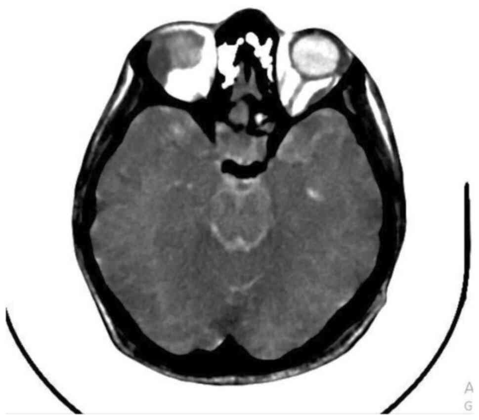
The cranial computed tomography (CT) found a
heterogeneous 20/11-mm mass located on the lateral wall of the
right orbit, bilateral circumferential polypoid mucosal thickening
of the maxillary sinuses and posterior nasal concha hypertrophy; no
brain lesions were identified (Fig.
1).
The abdominal ultrasound measured a 139-mm spleen in
its longitudinal axis and revealed large amounts of ascites. By
analyzing the pleural liquid collected by aspiration, high values
of lactic dehydrogenase (3,107 IU/l) were found and the smear
identified frequent lymphocytes without the presence of any
microorganisms (18,000 leukocytes/mm3, 100%
lymphocytes). Blood cultures returned positive for
methicillin-resistant Staphylococcus aureus and the blood
procalcitonin level was 30.65 ng/ml.
The complete blood count results revealed
9.46x10³ white blood cells with 7.27x10³
neutrophils, 0.95x10³ lymphocytes, 1.18x10³
monocytes, 0.01x10³ eosinophils and 0.05x10³
basophils; the hemoglobin level was low (11.9 g/l) and the
platelets numbered 179x103/µl. Peripheral blood smear
revealed 5% unsegmented and 72% segmented neutrophils with 10%
lymphocytes, 13% monocytes and no eosinophils, basophils or
atypical lymphocytes.
Pleural puncture was performed. Microscopic
examination of the pleural fluid revealed frequent leukocytes with
a predominance of lymphocytes (18,000/mm3), of which
over 90% were small lymphocytes. This result raised the suspicion
of non-Hodgkin's malignant lymphoma.
The bone marrow biopsy and aspiration revealed
granulocyte suppression, plasmacytosis and eosinophilia, raising
the suspicion of a T-cell non-Hodgkin lymphoma without medullar
infiltration or any discharge into the peripheral bloodstream.
A skin biopsy was made from a tumor nodule and the
pathology report revealed, on hematoxylin and eosin-stained slides,
a focal hyperkeratotic epidermis with uneven granular layer, mild
irregular acanthosis with discrete spongiosis, lymphocyte
exocytosis and disproportionate epidermotropism, and interface
vacuolar change with atypical lymphocytes along the dermo-epidermal
junction. The superficial and deep dermis was characterized by a
heavy infiltrate of atypical lymphocytes having irregular,
cerebriform nuclei, with perivascular, perifollicular and
intrafollicular location; the malignant cells also infiltrated the
arrectorpili muscle. The histopathological diagnosis was peripheral
NK/T cell lymphoma, no special type.
Immunohistochemistry studies were conducted as the
differential diagnosis with mycosis fungoides was suspected. After
careful investigation, the malignant proliferation was revealed to
be made up of medium sized CD3+ T cells with CD56 (NK
marker) and isolated CD30 expression; the tumor cells were
CD4- and CD8-, negative for the blastic
marker TdT (terminal deoxynucleotidyltransferase) and also for the
B cell marker CD20. Isolated cells (under 5%) expressed CD30, an
activated lymphocyte marker, while the cytotoxic activation marker,
granzyme B, had no specific immunophenotyping expression.
Following results of the histopathology and
immunohistochemistry, the final conclusion entailed the diagnosis
of medium T/NK cell non-Hodgkin lymphoma, possibly nasal type. Its
clinical and microscopic features are highlighted in
Figs. 2-5.
The patient died 72 h after admission and was not
tested for EBV; he proved to be HIV-negative. The lymphoma-specific
treatment was not initiated.
Discussion
Natural killer (NK)/T-cell lymphomas (NKTCLs) are a
rare and distinct type of malignant non-Hodgkin lymphoma
(frequently extranodal lymphomas, most of them of the nasal type).
They are identified in 12% of lymphoma patients, 68% of them having
the nasal type, 26% extranasal type and 6% aggressive or
unclassifiable type (3,4). A higher incidence is observed in Asia
when compared with Europe (22 vs. 5%) with an average life
expectancy of 8 months (5,6).
Nasal type lymphoma is frequently located in the
upper aero-digestive tract: Nasal cavity, nasopharynx, paranasal
sinuses, tonsils, hypopharynx and larynx, being frequently
associated with the Epstein-Barr virus (EBV) (7,8). Nasal
type NKTCLs are, in almost 95% of cases, associated with EBV; the
exact EBV pathway mechanism involved in the malignant
transformation is not yet fully understood (6). The immunohistochemistry profile
features CD3+, CD43+, CD45RO+,
CD56+, EBV-, CD8-, Epstein-Barr
virus-encoded small RNA-positive (EBER+), with
positivity for cytotoxic granular proteins (7-11).
The extranodal pattern of involvement seems to be
connected with the CD56 marker, which is the neural cell adhesion
molecule (NCAM) possessing hemophilic connection properties. The
neoplastic cells are thus redistributed to other sites and evolve
as new malignancy sites. The skin is the most common site for NKTCL
dissemination (12).
NKTCL is rarely found in Caucasians or in the
Western population, having a prevalence estimated at around
0.17-1.15% of all non-Hodgkin lymphomas, with 45% of these having
NKTCL origin (5).
NKTCL appears most frequently in patients over the
age of 60 years, although studies have shown that it can be found
in both geriatric and pediatric patients. The mortality rate is
higher than in other lymphomas and has poorer response rates to
radiation and chemotherapy (3).
The general manifestations of NKTCLs include signs
and symptoms located mainly in the face and neck regions: Facial
pain, diplopia, visual impairment, eyeball protrusion, eyelid
ptosis, pupil anomalies, nasal obstruction, refractory sinusitis,
velo-palatal motor disturbances, cranial nerve neuropathies,
intra-orbital and intrasinus masses. Other associations consist of
respiratory failure and liver and spleen enlargement (7).
In our patient's case, the intra-orbital tumor mass
was altering the ocular axis and was responsible for partial right
eyelid ptosis, being considered an intra-orbital lymphoma
metastasis. Unfortunately, biopsy was not performed in order to
confirm this theory. Nevertheless, considering the previously
described clinical aspects which were correlated with the
laboratory investigations, we consider that the diagnosis of
intra-orbital metastatic lymphoma can be supported.
In adults, lymphomas are the most frequent malignant
intra-orbital tumors. They manifest as swellings with increase in
intra-orbital volume causing mass-effect. Diplopia, eye motility
disturbances and eyelid dysfunction appear due to the invasion or
compression of orbital contents (13).
In regards to skin lesions, the differential
diagnosis with mycosis fungoides must be made, being the most
common cutaneous T-cell lymphoma. This type of lesion is more
common in individuals younger than 20 years of age, with a sex
ratio that favors males. It is an unusual manifestation of
skin-associated CD4+ T cells, which, in early stages,
can resemble eczema or psoriasis (14,15).
In our case, some of the skin biopsy aspects that
were found resembled mycosis fungoides: Atypical cerebriform
lymphocytes in the dermis and epidermis, some of them in and around
hair follicles (resembling folliculotropic mycosis fungoides) and
epidermotropism with lymphocytes at the dermo-epidermal junction
(sentinel sign). The differential diagnosis between NKTCL and
mycosis fungoides was made with the help of immunohistochemistry
studies.
Extranodal nasal-type NKTCLs are rare malignancies
for which standardized therapy has not yet been established. To
date, there are no randomized clinical trials which can clearly
determine such a scheme. For these patients it is recommended that
they enroll in clinical trials and that they receive treatment in
highly specialized clinics (16).
The therapeutic strategy in this type of lymphoma
requires collaboration between hematologists, oncologists,
radiotherapists, neurologists, pathologists, and treatment needs to
be individualized according to the site of the lymphoma (nasal or
extra-nasal) and disease stage (17).
Patients in the first stage of disease evolution are
stratified by age groups, by performance status and dependency
level, by regional lymph node involvement and by the increased
level of lactate dehydrogenase (1)
(a situation which was present also in our patient's case).
Concerning nasal-type NKTCLs, one can use
radiotherapy at a dose of at least 50 Gy, as single therapy, but
this is associated with a high rate of local and distant
recurrence. As such, combined therapy is recommended, using the
CHOP treatment scheme (cyclophosphamide, doxorubicin, vincristine,
prednisone); thus, the 5-year survival rate is increased from 20 to
80% (18,19).
For patients in the second stage of disease, in
addition to radiotherapy and cysplatin, 3 cycles of DeVIC
(dexamethasone, etoposide, ifosfamide, and carboplatin) or
sequential SMILE (etoposide, ifosfamide, methotrexate and
dexamethasone) chemo-radiotherapy is suggested (20).
For stage four nasal-type lymphoma, and also any
stage extranasal types, poly-chemotherapy with pegaspargase (with
or without radiotherapy) can be administered (18).
Permanent re-staging is conducted according to
imaging studies including CT, magnetic resonance imaging (MRI),
positron emission tomography-computed tomography(PET-CT scan),
endoscopy or repeated biopsies (5,21).
Unfortunately, progression is registered even though treatment is
applied (18).
Studies are being carried out which evaluate the
beneficial role of stem cell transplantation in NKTCLs, but
definitive conclusions cannot be yet drawn concerning this
therapeutic alternative (22).
The case presented here describes the steps carried
out in order to establish the diagnosis of nasal-type NKTCL,
starting from the evaluation of neurological lesions (eye and
eyelid mobility dysfunction) associated with suggestive skin
lesions; all of these correlated with the cranial CT scan results
which were suggestive for intra-orbital lymphoma dissemination.
The case was unusual as it presented with
intra-orbital secondary dissemination of the primary NKTCL and as
it evolved quickly, with an exitus of approximately 3 months-a life
expectancy much lower than that cited in other research (3), with positive blood cultures in the
context of acute pneumonitis set in a background of immune
suppression.
The intra-orbital mass could not be confirmed
through biopsy as a lymphoma, but by correlating the clinical and
laboratory data mentioned earlier, the orbital mass was suspected
as being a lymphoma; this is also valid for the sinus
involvement.
The cutaneous lesions which clinically and
morphologically resembled mycosis fungoides, were confirmed to be
NKTCL. An essential tool for diagnosis is immunohistochemistry
which revealed the medium T cell tumor proliferation to be positive
for CD3 and CD30 and negative for CD4, CD8 and CD20.
The diagnosis of lymphoma was also supported by the
pleural liquid fine needle aspiration appearance.
Acknowledgements
Not applicable.
Funding
No funding was received.
Availability of data and materials
Any further information concerning the case study is
available from the corresponding author upon reasonable
request.
Author's contributions
ML, AT, DCV and EN contributed to the study design,
analyzed and interpreted the patient's clinical data regarding the
hematological disease, and prepared the manuscript. EN performed
the histological examination of the cutaneous biopsy. VS, AT, AF,
VZC, GS, MP and EME contributed to collecting the relevant
literature, data analysis, reviewed and critically interpreted the
information. All authors read and approved the final version of the
manuscript.
Ethics approval and consent to
participate
Approval from ‘Saint Andrew’ Emergency Clinical
Hospital Ethics Committee (Decision no. 9436 from 28.04.2020) was
obtained. Informed consent for participation in the study or use of
the patient tissue was obtained from our participant.
Patient consent for publication
Informed consent for publication was obtained from
our patient.
Competing interests
The authors declare that they have no competing
interests.
References
|
1
|
Tse E and Kwong YL: NK/T-cell lymphomas.
Best Pract Res Clin Haematol. 32:253–261. 2019.PubMed/NCBI View Article : Google Scholar
|
|
2
|
Gutiérrez A, Caballero MD, Pérez-Manga G
and Rodriguez J: Hematopoietic SCT for peripheral T-cell lymphoma.
Bone Marrow Transplant. 42:773–781. 2008.PubMed/NCBI View Article : Google Scholar
|
|
3
|
Su YJ, Wang PN, Chang H, Shih LY, Lin TL,
Kuo MC, Chuang WY, Wu JH, Tang TC, Hung YS, et al: Extranodal
NK/T-cell lymphoma, nasal type: Clinical features, outcome, and
prognostic factors in 101 cases. Eur J Haematol. 101:379–388.
2018.PubMed/NCBI View Article : Google Scholar
|
|
4
|
Jaffe ES, Nicolae A and Pittaluga S:
Peripheral T-cell and NK-cell lymphomas in the WHO clasification:
Pearls and pitfalls. Mod Pathol. 26 (Suppl 1):S71–S87.
2013.PubMed/NCBI View Article : Google Scholar
|
|
5
|
Aozasa K and Zaki MA: Epidemiology and
pathogenesis of nasal NK/T-cell lymphoma: A mini-review.
ScientificWorldJournal. 11:422–428. 2011.PubMed/NCBI View Article : Google Scholar
|
|
6
|
Suzuki R, Suzumia J, Yamaguchi M, Nakamura
S, Kameoka J, Kojima H, Abe M, Kinoshita T, Yoshino T, Iwatsuki K,
et al: Prognostic factors for mature natural killer (NK) cell
neoplasms: Aggressive NK cell leukemia and extranodal NK cell
lymphoma, nasal type. Ann Oncol. 21:1032–1040. 2010.PubMed/NCBI View Article : Google Scholar
|
|
7
|
Kimura H and Fujiwara S: Overview of
EBV-associated T/NK-cell lymphoproliferative diseases. Front
Pediatr. 6(417)2018.PubMed/NCBI View Article : Google Scholar
|
|
8
|
Park S and Ko YH: Epstein-Barr
virus-associated T/natural killer-cell lymphoproliferative
disorders. J Dermatol. 41:29–39. 2014.PubMed/NCBI View Article : Google Scholar
|
|
9
|
Chen Z, Guan P, Shan T, Ye Y, Gao L, Wang
Z, Zhao S, Zhang W, Zhang L, Pan L, et al: CD30 expression and
survival in extranodal NK/T-cell lymphoma: A systematic review and
meta-analysis. Oncotarget. 9:16547–16556. 2018.PubMed/NCBI View Article : Google Scholar
|
|
10
|
Boda D: Cellomics as integrative omics for
cancer. Curr Proteomics. 10:237–245. 2013.
|
|
11
|
Ion A, Popa IM, Papagheorghe LM, Lisievici
C, Lupu M, Voiculescu V, Caruntu C and Boda D: Proteomic approaches
to biomarker discovery in cutaneous T-cell lymphoma. Dis Markers.
2016(9602472)2016.PubMed/NCBI View Article : Google Scholar
|
|
12
|
Shimizu I, Hamano Y, Sato S, Takeda W,
Kirihara T, Sato K, Ueki T, Hiroshima Y, Sumi M, Ueno M, et al:
Neurolymphomatosis in a patient with extranodal NK/T-cell lymphoma,
nasal-type: A case report and literature review. Intern Med.
53:471–475. 2014.PubMed/NCBI View Article : Google Scholar
|
|
13
|
Tailor TD, Gupta D, Dalley RW, Keene CD
and Anzai Y: Orbital neoplasms in adults: Clinical, radiologic, and
pathologic review. Radiographics. 33:1739–1758. 2013.PubMed/NCBI View Article : Google Scholar
|
|
14
|
Hwang ST, Janik JE, Jaffe ES and Wilson
WH: Mycosis fungoides and Sézary syndrome. Lancet. 371:945–957.
2018.
|
|
15
|
Cioplea M, Caruntu C, Zurac S, Bastian A,
Sticlaru L, Cioroianu A, Boda D, Jugulete G, Nichita L and Popp C:
Dendritic cell distribution in mycosis fungoides vs. inflammatory
dermatosis and other T-cell skin lymphoma. Oncol Lett.
17:4055–4059. 2019.PubMed/NCBI View Article : Google Scholar
|
|
16
|
Khariwala SS, Litman DA, McQuone SJ, Hess
JL and Thaler ER: Quiz case 2. Natural killer (NK) cell/peripheral
T-cell lymphoma. Arch Otolaryngol Head Neck Surg. 126:1391–1393.
2000.PubMed/NCBI
|
|
17
|
Kommalapati A, Tella SH, Ganti AK and
Armitage JO: Natural Killer/T-cell neoplasms: Analysis of
incidence, patient characteristics, and survival outcomes in the
United States. Clin Lymphoma Myeloma Leuk. 18:475–479.
2018.PubMed/NCBI View Article : Google Scholar
|
|
18
|
Kim BS, Kim TY, Kim CW, Kim JY, Heo DS,
Bang YJ and Kim NK: Therapeutic outcome of extranodal NK/T-cell
lymphoma initially treated with chemotherapy-result of chemotherapy
in NK/T-cell lymphoma. Acta Oncol. 42:779–783. 2003.PubMed/NCBI View Article : Google Scholar
|
|
19
|
Ciobotaru OR, Lupu MN, Rebegea L,
Ciobotaru OC, Miulescu M and Kamel E: Dexamethasone-chemical
structure and mechanisms of action in prophylaxis of postoperative
side effects. Rev Chim (Bucharest). 70:843–847. 2019.
|
|
20
|
Tse E and Kwong YL: Practical management
of natural killer/T-cell lymphoma. Curr Opin Oncol. 24:480–486.
2012.PubMed/NCBI View Article : Google Scholar
|
|
21
|
Chim CS, Ma SY, Au WY, Choy C, Lie AKW,
Liang R, Yau CC and Kwong YL: Primary nasal natural killer cell
lymphoma: Long-term treatment outcome and relationship with the
International Prognostic Index. Blood. 103:216–221. 2004.PubMed/NCBI View Article : Google Scholar
|
|
22
|
Lin HN, Liu CY, Pai JT, Chang FP, Yang CF,
Yu YB, Hsiao LT, Chiou TJ, Liu JH, Gau JP, et al: How to predict
the outcome in mature T and NK cell lymphoma by currently used
prognostic models? Blood Cancer J. 2(e93)2012.PubMed/NCBI View Article : Google Scholar
|