Introduction
Osteosarcoma (OS) is the most frequent primary solid
malignancy of the bone characterized by the presence of malignant
mesenchymal cells producing osteoid or immature bone (1). The incidence of OS is estimated at 5
per million persons, accounting for 45.3% of bone tumors worldwide
(2,3). The alterations in the expression of
the corresponding genes that encode tumor suppressors and promotors
are closely associated with the incidence of OS. In the last
decades, the therapeutic strategies for OS have rapidly improved,
including T-cell related, occupational and physical therapy
(4,5); however, the prognosis of patients with
OS remains poor due to the high rates of tumor metastasis and
recurrence (6-9).
At present, the identification of new therapeutic targets that are
associated with the occurrence, development and metastasis of
cancers has drawn special attention, which may provide novel
approaches for the clinical management and treatment of OS
(10).
MicroRNAs (miRNAs) are a class of non-coding RNAs
that have been reported to regulate the expression of target genes
by binding to the 3'-untranslated region of target mRNAs (11). Furthermore, miRNAs have been
demonstrated to serve crucial roles in tumor cell development,
differentiation, proliferation and apoptosis. Subsequently, various
miRNAs have been identified as regulators in the biogenesis and
progression of certain types of cancer, such as miR-154 in gastric
cancer, miR-133a in non-small cell lung cancer, miR-19b in breast
cancer and miR-769 in colorectal cancer (12-15).
Numerous aberrantly expressed miRNAs were reported to participate
in the development and prognosis of OS. For example, it was
demonstrated that downregulated miR-223-3p can predict the poor
prognosis of patients with OS and that miR-223-3p can inhibit OS
cell proliferation, invasion and migration by regulating the
expression of cadherin 6(16).
Furthermore, miR-552-5p was reported to serve as a tumor promoter
that could facilitate the development and progression of OS
(17). A previous study
demonstrated that miR-588 is downregulated in breast cancer and
that it can inhibit the proliferation of breast cancer cells
(18). The dysregulation and
functional role of miR-588 in tumor cell development was observed
in prostate cancer, gastric cancer and lung squamous cell carcinoma
(19-21).
A previous study reported that miR-588 is downregulated in OS
(22); however, its function and
specific role in the progression of OS and the prognosis of
patients remain unclear.
The present study aimed to evaluate the expression
and biological function of miR-588 in OS tissues and cells and to
determine the prognostic value of miR-588 in patients with OS.
Materials and methods
Tissues and cell lines
This study was approved by the Ethics Committee of
The Second Affiliated Hospital of Mudanjiang University (approval
no. 200934; Mudanjiang, China). A total of 104 patients diagnosed
with OS at The Second Affiliated Hospital of Mudanjiang University
were recruited between October 2010 and November 2014. The
inclusion criteria for patients with OS were as follows: i)
Patients were diagnosed with OS as a primary malignancy; ii) none
of the patients had received any anti-tumor therapy, radiotherapy
or chemotherapy; iii) all patients signed written informed consent
and participated in a 5-year follow-up survey; and iv) the
clinicopathological characteristics of patients with OS was
provided. Patients with a history of other cancers were excluded
from this study. Paired OS tissues and adjacent normal tissues were
collected during resection surgery and confirmed by at least two
pathologists. Tissues were immediately frozen in liquid nitrogen
and stored at -80˚C prior to following experiments.
The OS cell lines MG63, U2OS, KHOS-240S and SAOS-2,
and the human osteoblast cell line hFOB 1.19 were purchased from
the American Type Culture Collection. All cells were cultured in
DMEM medium (Thermo Fisher Scientific, Inc.) supplemented with 10%
FBS (Thermo Fisher Scientific, Inc.) and placed at 37˚C in a
humidified incubator containing 5% CO2.
Cell transfection
In order to regulate the expression of miR-588 in OS
cells, miR-588 mimic (50 nM; 5'-UUG GCCACAAUGGGUUAGAAC-3'), miR-588
inhibitor (50 nM; 5'-GUUCUAACCCAUUGUGGCCAA-3'), mimic negative
control (mimic NC; 50 nM; 5'-UUCUCCGAACGUGUCACG UU-3') and
inhibitor negative control (inhibitor NC; 50 nM,
5'-UUGCUAGUGCGACACUACUUCTT-3') (all from Shanghai GenePharma Co.,
Ltd.) were transfected into OS cells using Lipofectamine 2000™
reagent (Invitrogen; Thermo Fisher Scientific, Inc.) according to
the manufacturer's instructions. The transfection mix was prepared
at room temperature and cells were incubated with the transfection
complexes at 37˚C for 4 h. Transfected cells were subjected to
subsequent experiments after 48 h of transfection. Non-transfected
cells were used as the mock group.
RNA extraction and reverse
transcription quantitative (RT-q) PCR
Total RNA from tissues and cells was extracted using
TRIzol® reagent (Invitrogen; Thermo Fisher Scientific,
Inc.). Following reverse transcription to cDNA using the TaqMan
MicroRNA Reverse Transcription kit (Invitrogen; Thermo Fisher
Scientific, Inc.), RT-qPCR was used to determine the expression of
miR-588 with the SYBR Green I Master Mix kit (Invitrogen; Thermo
Fisher Scientific, Inc.). The sequences of the primers were as
follows: miR-588, forward 5'-GGGUUG GCCACAAUGGGU-3', reverse
5'-GTCGTATCCAGTGCG TGT-3'; and U6, forward 5'-CTCGCTTCGGCAGCACA-3'
and reverse 5'-AACGCTTCACGAATTTGCGT-3'. The thermocycling
conditions were as follows: 95˚C for 30 sec, followed by 40 cycles
of annealing at 95˚C for 10 sec and extension at 60˚C for 30 sec.
The relative expression levels of miR-588 were normalized to
endogenous control U6 and were expressed as
2-ΔΔCq (23).
Cell proliferation assay
OS cell proliferation was evaluated with the Cell
Counting Kit-8 (CCK-8; Dojindo Molecular Technologies, Inc.) assay.
Briefly, 5x10³ cells were seeded per well into a 96-well
plate for 0, 24, 48 and 72 h. A total of 10 µl CCK-8 was added to
each well and cells were incubated for 4 h at 37˚C with 5%
CO2. The absorbance was read at 450 nm using a
microplate reader (Beckman Coulter, Inc.). Experiments were
performed in triplicate.
Cell migration and invasion assay
Transwell assays were used to detect the migratory
and invasive abilities of OS cells. Briefly, 1x10⁴ cells
were seeded into the upper chamber of a 24-well Transwell chamber
(8-µm pore size; Corning, Inc.) and cultured with serum-free
medium. The bottom chamber was filled with medium supplemented with
10% FBS used as a chemoattractant. For the invasion assay, the
upper chamber was pre-coated with Matrigel solution (BD
Biosciences) at 37˚C for 1 h prior to cell seeding. Following 48 h
incubation at 37˚C, the number of cells that have migrated to the
bottom chamber were stained with 1% crystal violet at room
temperature for 30 min and counted using a light microscope
(magnification, x200). Experiments were performed in
triplicate.
Statistical analysis
Data were expressed as the means ± standard
deviation obtained from at least three independent experiments and
were analyzed by SPSS 20.0 software (IBM Corp.) and GraphPad Prism
7.0 software (GraphPad Software, Inc.). The difference between
groups was evaluated by paired Student's t-test or one-way ANOVA
followed by Tukey's post hoc test. The association between miR-588
expression and the clinicopathological characteristics of patients
with OS was analyzed using χ2 test. The overall survival
of patients with OS according to their miR-588 expression level was
estimated by Kaplan-Meier analysis and log-rank test. The
prognostic value of miR-588 in OS was further evaluated by Cox
regression analysis. P<0.05 was considered to indicate a
statistically significant difference.
Results
miR-588 is significantly downregulated
in OS tissues and cells
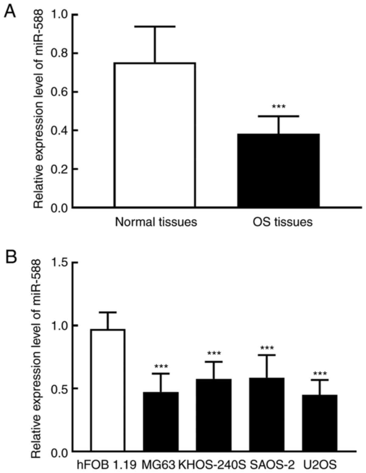
The results from RT-qPCR demonstrated that miR-588
expression was significantly decreased in OS tumor tissues compared
with adjacent normal tissues (P<0.001; Fig. 1A). Similarly, miR-588 expression was
significantly downregulated in OS cells (MG63, KHOS-240S, SAOS-2,
and U2OS) compared with hFOB1.19 cells (P<0.001; Fig. 1B). miR-588 downregulation in OS
tissues and cells suggested that it may have a role in the
development of OS.
miR-588 downregulation is associated
with Musculoskeletal Tumor Society (MSTS) staging of patients with
OS
According to the mean expression level of miR-588 in
OS tumor tissues (0.384), 104 patients with OS were divided into a
low miR-588 expression group (n=63) and a high miR-588 expression
group (n=41). The results demonstrated that miR-588 expression was
associated with MSTS staging (P=0.033) of patients with OS
(Table I). No significant
association was found between miR-588 expression and the other
clinicopathological characteristics of patients (age, sex, tumor
size and location; P<0.05; Table
I).
 | Table IAssociation between miR-588 expression
and the clinicopathological characteristics of patients with
osteosarcoma. |
Table I
Association between miR-588 expression
and the clinicopathological characteristics of patients with
osteosarcoma.
| | miR-588 | |
|---|
| Characteristics | Cases (n=104) | Low expression
(n=63) | High expression
(n=41) | P-value |
|---|
| Age, years | | | | 0.633 |
|
<20 | 55 | 31 | 24 | |
|
≥20 | 49 | 32 | 17 | |
| Sex | | | | 0.731 |
|
Male | 60 | 35 | 25 | |
|
Female | 44 | 28 | 16 | |
| Tumor size, cm | | | | 0.194 |
|
<8 | 57 | 30 | 27 | |
|
≥8 | 47 | 33 | 14 | |
| MSTS staging | | | | 0.033 |
|
I-II | 74 | 40 | 34 | |
|
III | 30 | 23 | 7 | |
| Location | | | | 0.374 |
|
Tibia/femur | 68 | 37 | 31 | |
|
Elsewhere | 36 | 26 | 10 | |
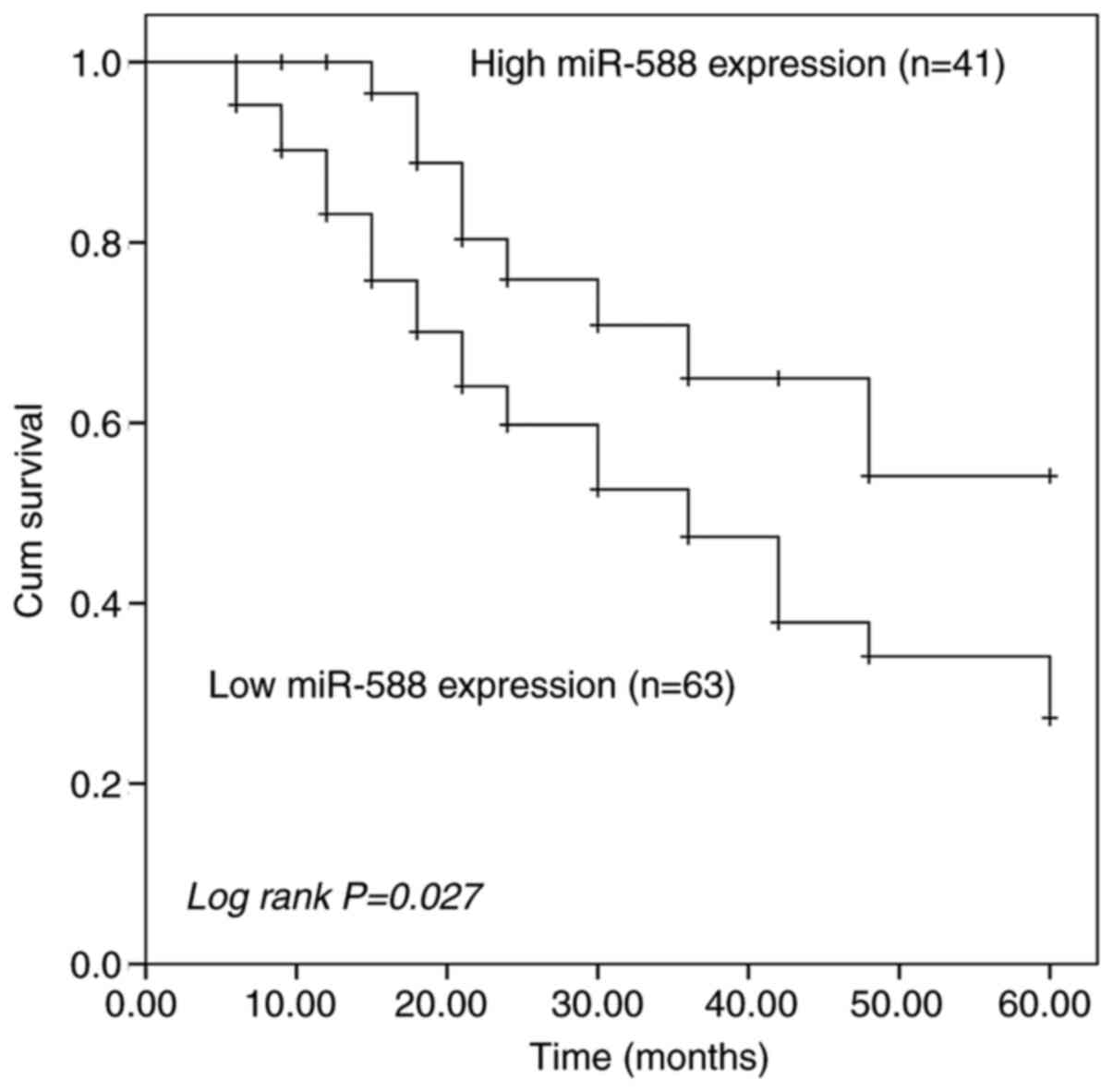
Prognostic value of miR-588 in OS
The Kaplan-Meier curve of patients with OS
demonstrated that patients with high miR-588 expression levels had
a higher 5-year survival rate than patients with low miR-588
expression levels (log-rank P=0.027; Fig. 2). The prognostic value of miR-588
was further assessed by Cox regression analysis. The results showed
that miR-588 expression [hazard ratio (HR) =2.533; 95% confidence
interval (CI) =1.187-5.407; P=0.016] and MSTS staging (HR=2.517;
95% CI=1.088-5.824; P=0.031) were independent predictors for the
prognosis of patients with OS (Table
II).
| Table IIImpacts of clinicopathological
characteristics on prognosis in patients with osteosarcoma. |
Table II
Impacts of clinicopathological
characteristics on prognosis in patients with osteosarcoma.
|
Characteristics | HR factor | 95% CI | P-value |
|---|
| miR-588
expression | 2.533 | 1.187-5.407 | 0.016 |
| Age | 1.195 | 0.625-2.287 | 0.590 |
| Sex | 1.163 | 0.599-2.258 | 0.656 |
| Tumor size | 1.353 | 0.686-2.671 | 0.383 |
| MSTS staging | 2.517 | 1.088-5.824 | 0.031 |
| Tumor location | 1.745 | 0.853-3.569 | 0.127 |
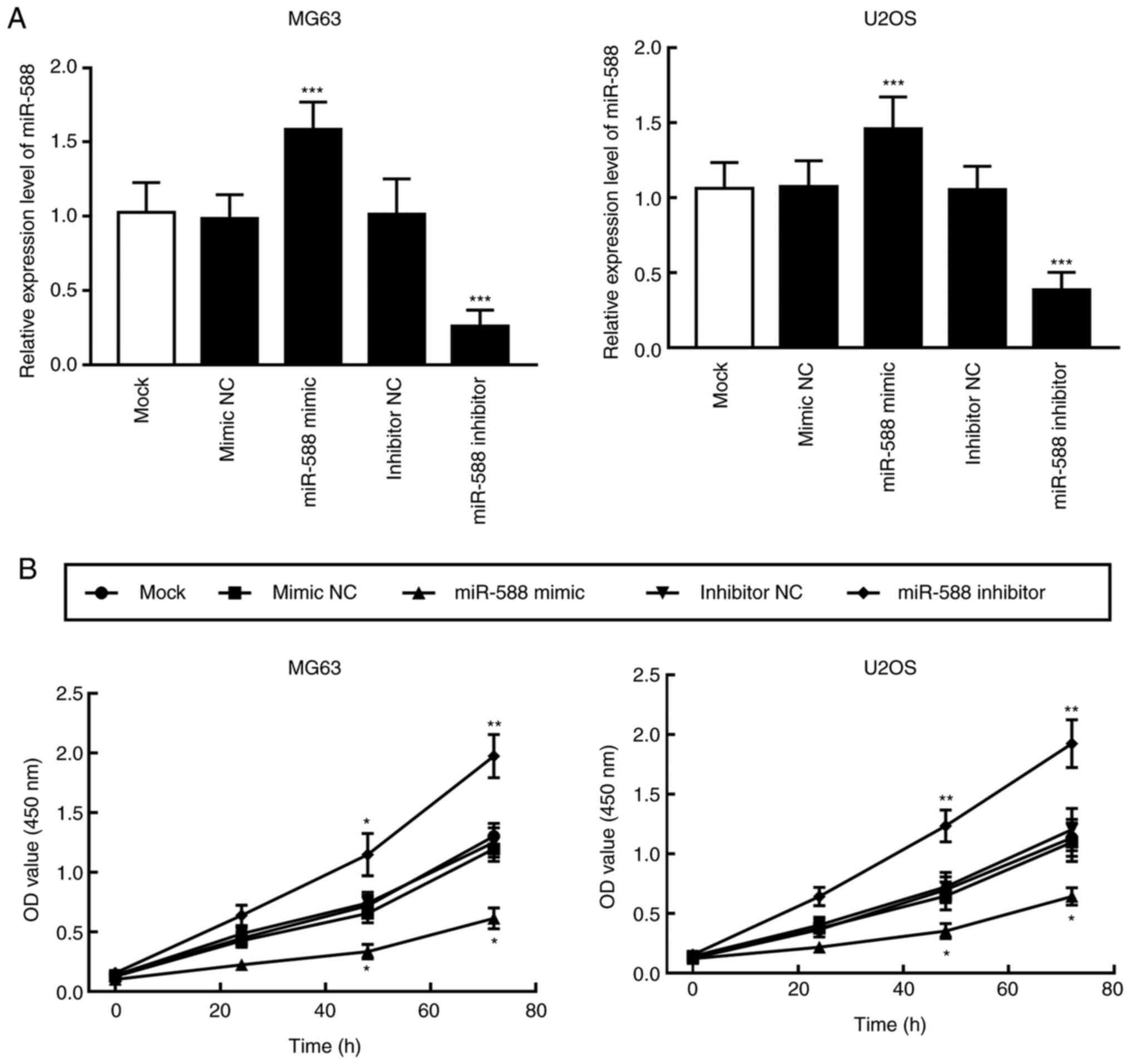
miR-588 inhibits the proliferation of
OS cells
Cell transfections with miR-588 mimic or miR-588
inhibitor were performed to regulate the expression of miR-588 in
MG63 and U2OS cells, which presented the lowest miR-588 expression
(Fig. 1B). Compared with the Mock
and NC groups, miR-588 expression in MG63 and U2OS cells
transfected with miR-588 mimic was significantly increased, whereas
its expression was significantly decreased in miR-588 inhibitor
transfected cells (P<0.001; Fig.
3A). Furthermore, the effect of transfections with miR-588
mimic and miR-588 inhibitor on OS cell proliferation was determined
with the CCK-8 assay. The results demonstrated that miR-588
overexpression significantly inhibited the proliferation of MG63
and U2OS cells, whereas miR-588 knockdown significantly stimulated
the proliferation of OS cells (P<0.01 and P<0.05; Fig. 3B).
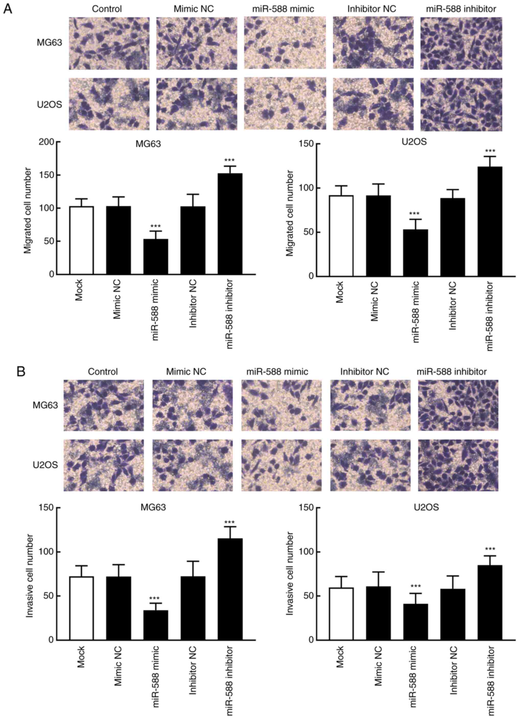
miR-588 inhibits the migratory and
invasive abilities of OS cells
The migratory and invasive abilities of OS
transfected cells were evaluated by Transwell assays. The results
demonstrated that the number of migrated and invasive cells in the
miR-588 mimic group was significantly decreased compared with that
in the Mock and NC groups. Conversely, miR-588 knockdown
significantly increased the number of migrated and invasive MG63
and U2OS cells (P<0.001; Fig. 4A
and B).
Discussion
OS is a common bone tumor characterized by rapid
development, high metastatic potential and poor prognosis (24,25).
Novel efficient biomarkers are therefore required to improve the
survival rate of patients with OS. Previous studies reported that
miRNAs are involved in various cellular functions and biological
processes, including cell proliferation, differentiation, apoptosis
and invasion (26,27). Therefore, determining the miRNAs
involved in OS tumor formation might help the development of novel
therapeutic strategies for the treatment and management of OS
(28). It has been demonstrated
that several miRNAs are dysregulated in OS and serve crucial roles
in OS tumor progression. For example, it was reported that
miR-139-5p expression is downregulated in OS tissues and cells and
that miR-139-5p overexpression can significantly inhibit the
progression of OS (29). It is
therefore necessary to determine the function of dysregulated
miRNAs in OS progression.
In prostate cancer, miR-588 was demonstrated to be
upregulated, and its expression is associated with the poor
clinical prognosis and survival rate of patients (19). However, the expression of miR-588
was different in OS. In the present study, the downregulation of
miR-588 in OS was demonstrated to be associated with MSTS staging
of patients with OS, which was consistent with the previously
reported miRNA expression profile of OS (22). Similar findings in breast cancer
showed that significant miR-588 downregulation could promote cancer
cell proliferation and decrease cisplatin chemosensitivity
(18). Furthermore, miR-588
downregulation also predicted the poor prognosis of patients with
OS. In the present study, miR-588 expression and MSTS staging were
demonstrated to act as independent prognostic factors for OS. The
prognostic value of miR-588 was also previously demonstrated in
breast and prostate cancers (18,19).
The prognostic value of miRNAs has drawn special attention in
previous studies, and several miRNAs have been identified as
independent prognostic indicators for various types of cancer
(30). For instance, miR-1294 is
downregulated in gastric cancer and its expression is associated
with shorter disease-free survival and overall survival of
patients. miR-1294 was also identified as an independent risk
factor for gastric cancer prognosis by multivariate Cox analysis
(31). The relatively small
clinical sample size of the present study was a limitation, which
might also limit the interpretation of the results. It is therefore
necessary to conduct further investigation using a larger sample
size.
Regarding the biological function of miR-588 in the
development of OS, the present study demonstrated that miR-588
downregulation significantly stimulated OS cell proliferation and
migratory and invasive abilities, whereas miR-588 overexpression
had the opposite effect. Previously, miR-588 was also found to
inhibit gastric tumor progression to its inhibitory effect on
gastric cancer cell invasion and migration (20). Furthermore, miR-588 was reported to
be downregulated in lung squamous cell carcinoma and to act as a
tumor suppressor that could inhibit tumor cell migration and
invasion by targeting progranulin (21). These results indicated that miR-588
may be involved in OS tumor development and that it may serve a
tumor suppressor role in OS.
Although the expression and functional role of
miR-588 in OS were confirmed in the present study, the underlying
mechanism by which miR-588 regulates the progression and
development of OS was not determined and requires therefore further
investigation. In addition, in vivo studies are also necessary to
confirm the results from in vitro studies (32). The present study provided in vitro
evidence demonstrating the potential tumor suppressor role of
miR-588 in the development of OS, which needs to be validated by in
vivo experiments.
In conclusion, the present study demonstrated that
miR-588 was downregulated in OS tissues and cells. In addition,
miR-588 expression was associated with MSTS staging of patients
with OS and miR-588 downregulation indicated a poor prognosis for
these patients. Furthermore, the proliferation and migratory and
invasive abilities of OS cells were stimulated following miR-588
downregulation and inhibited by miR-588 overexpression. Taken
together, these findings suggested that miR-588 may be considered
as a biomarker for the prognosis of OS and a tumor suppressor in
the development of OS.
Acknowledgements
Not applicable.
Funding
Funding: No funding was received.
Availability of data and materials
The datasets used and/or analyzed during the current
study are available from the corresponding author on reasonable
request.
Authors' contributions
TY and SL analyzed and interpreted the data from
patients. TY, SL, TM and WS performed the experiments and wrote
manuscript. TY and WS confirm the authenticity of all the raw data.
All authors have read and approved the final manuscript.
Ethics approval and consent to
participate
This study was approved by the Ethics Committee of
The Second Affiliated Hospital of Mudanjiang University (approval
no. 200934; Mudanjiang, China). All patients signed written
informed consent.
Patient consent for publication
Not applicable.
Competing interests
The authors declare that they have no competing
interests.
References
|
1
|
Picci P: Osteosarcoma (osteogenic
sarcoma). Orphanet J Rare Dis. 2(6)2007.PubMed/NCBI View Article : Google Scholar
|
|
2
|
Ottaviani G and Jaffe N: The epidemiology
of osteosarcoma. Cancer Treat Res. 152:3–13. 2009.PubMed/NCBI View Article : Google Scholar
|
|
3
|
Liu W, Zhang X, Liu P, Shen X, Lan T, Li
W, Jiang Q, Xie X and Huang H: Effects of berberine on matrix
accumulation and NF-kappa B signal pathway in alloxan-induced
diabetic mice with renal injury. Eur J Pharmacol. 638:150–155.
2010.PubMed/NCBI View Article : Google Scholar
|
|
4
|
Yoshida K, Okamoto M, Aoki K, Takahashi J
and Saito N: A Review of T-Cell Related Therapy for Osteosarcoma.
Int J Mol Sci. 21(4877)2020.PubMed/NCBI View Article : Google Scholar
|
|
5
|
Punzalan M and Hyden G: The role of
physical therapy and occupational therapy in the rehabilitation of
pediatric and adolescent patients with osteosarcoma. Cancer Treat
Res. 152:367–384. 2009.PubMed/NCBI View Article : Google Scholar
|
|
6
|
Daw NC, Chou AJ, Jaffe N, Rao BN, Billups
CA, Rodriguez-Galindo C, Meyers PA and Huh WW: Recurrent
osteosarcoma with a single pulmonary metastasis: A
multi-institutional review. Br J Cancer. 112:278–282.
2015.PubMed/NCBI View Article : Google Scholar
|
|
7
|
Meazza C and Scanagatta P: Metastatic
osteosarcoma: A challenging multidisciplinary treatment. Expert Rev
Anticancer Ther. 16:543–556. 2016.PubMed/NCBI View Article : Google Scholar
|
|
8
|
Sun HH, Chen XY, Cui JQ, Zhou ZM and Guo
KJ: Prognostic factors to survival of patients with chondroblastic
osteosarcoma. Medicine (Baltimore). 97(e12636)2018.PubMed/NCBI View Article : Google Scholar
|
|
9
|
Imura Y, Takenaka S, Kakunaga S, Nakai T,
Wakamatsu T, Outani H, Tanaka T, Tamiya H, Oshima K, Hamada K, et
al: Survival analysis of elderly patients with osteosarcoma. Int
Orthop. 43:1741–1747. 2019.PubMed/NCBI View Article : Google Scholar
|
|
10
|
Liao YX, Yu HY, Lv JY, Cai YR, Liu F, He
ZM and He SS: Targeting autophagy is a promising therapeutic
strategy to overcome chemoresistance and reduce metastasis in
osteosarcoma. Int J Oncol. 55:1213–1222. 2019.PubMed/NCBI View Article : Google Scholar
|
|
11
|
Lagos-Quintana M, Rauhut R, Lendeckel W
and Tuschl T: Identification of novel genes coding for small
expressed RNAs. Science. 294:853–858. 2001.PubMed/NCBI View Article : Google Scholar
|
|
12
|
Qiao W, Cao N and Yang L: MicroRNA-154
inhibits the growth and metastasis of gastric cancer cells by
directly targeting MTDH. Oncol Lett. 14:3268–3274. 2017.PubMed/NCBI View Article : Google Scholar
|
|
13
|
Yang ZQ, Wu CA and Cheng YX: Prognostic
value of microRNA-133a expression and its clinicopathologic
significance in non-small cell lung cancer: a comprehensive study
based on meta-analysis and the TCGA database. Oncol Res Treat.
41:762–768. 2018.PubMed/NCBI View Article : Google Scholar
|
|
14
|
Li C, Zhang J, Ma Z, Zhang F and Yu W:
miR-19b serves as a prognostic biomarker of breast cancer and
promotes tumor progression through PI3K/AKT signaling pathway.
OncoTargets Ther. 11:4087–4095. 2018.PubMed/NCBI View Article : Google Scholar
|
|
15
|
Wang L, Xu M, Lu P and Zhou F:
microRNA-769 is downregulated in colorectal cancer and inhibits
cancer progression by directly targeting cyclin-dependent kinase 1.
OncoTargets Ther. 11:9013–9025. 2018.PubMed/NCBI View Article : Google Scholar
|
|
16
|
Ji Q, Xu X, Song Q, Xu Y, Tai Y, Goodman
SB, Bi W, Xu M, Jiao S, Maloney WJ, et al: miR-223-3p inhibits
human osteosarcoma metastasis and progression by directly targeting
CDH6. Mol Ther. 26:1299–1312. 2018.PubMed/NCBI View Article : Google Scholar
|
|
17
|
Cai W, Xu Y, Yin J, Zuo W and Su Z:
miR-552-5p facilitates osteosarcoma cell proliferation and
metastasis by targeting WIF1. Exp Ther Med. 17:3781–3788.
2019.PubMed/NCBI View Article : Google Scholar
|
|
18
|
Yu M, Zhang X, Li H, Zhang P and Dong W:
MicroRNA-588 is downregulated and may have prognostic and
functional roles in human breast cancer. Med Sci Monit.
23:5690–5696. 2017.PubMed/NCBI View Article : Google Scholar
|
|
19
|
Zhao N, Lin T, Zhao C, Zhao S, Zhou S and
Li Y: MicroRNA-588 is upregulated in human prostate cancer with
prognostic and functional implications. J Cell Biochem: Oct 5, 2017
(Epub ahead of print).
|
|
20
|
Zhou X and Xu M, Guo Y, Ye L, Long L, Wang
H, Tan P and Xu M: MicroRNA-588 regulates invasion, migration and
epithelial-mesenchymal transition via targeting EIF5A2 pathway in
gastric cancer. Cancer Manag Res. 10:5187–5197. 2018.PubMed/NCBI View Article : Google Scholar
|
|
21
|
Qian L, Lin L, Du Y, Hao X, Zhao Y and Liu
X: MicroRNA-588 suppresses tumor cell migration and invasion by
targeting GRN in lung squamous cell carcinoma. Mol Med Rep.
14:3021–3028. 2016.PubMed/NCBI View Article : Google Scholar
|
|
22
|
Chen G, Yu W, Li Z, Wang Q, Yang Q, Du Z,
Zhang G and Song Y: Potential regulatory effects of miR-182-3p in
osteosarcoma via targeting EBF2. BioMed Res Int.
2019(4897905)2019.PubMed/NCBI View Article : Google Scholar
|
|
23
|
Livak KJ and Schmittgen TD: Analysis of
relative gene expression data using real-time quantitative PCR and
the 2(-Delta Delta C(T)) Method. Methods. 25:402–408.
2001.PubMed/NCBI View Article : Google Scholar
|
|
24
|
Wang SY, Hu HZ, Qing XC, Zhang ZC and Shao
ZW: Recent advances of drug delivery nanocarriers in osteosarcoma
treatment. J Cancer. 11:69–82. 2020.PubMed/NCBI View Article : Google Scholar
|
|
25
|
Qiu S, Tao L and Zhu Y: Marital Status and
Survival in Osteosarcoma Patients: An Analysis of the Surveillance,
Epidemiology, and End Results (SEER) Database. Med Sci Monit.
25:8190–8203. 2019.PubMed/NCBI View Article : Google Scholar
|
|
26
|
Bushati N and Cohen SM: microRNA
functions. Annu Rev Cell Dev Biol. 23:175–205. 2007.PubMed/NCBI View Article : Google Scholar
|
|
27
|
Mohr AM and Mott JL: Overview of microRNA
biology. Semin Liver Dis. 35:3–11. 2015.PubMed/NCBI View Article : Google Scholar
|
|
28
|
Wang J, Liu S, Shi J, Li J, Wang S, Liu H,
Zhao S, Duan K, Pan X and Yi Z: The Role of miRNA in the Diagnosis,
Prognosis, and Treatment of Osteosarcoma. Cancer Biother
Radiopharm. 34:605–613. 2019.PubMed/NCBI View Article : Google Scholar
|
|
29
|
Shi YK and Guo YH: MiR-139-5p suppresses
osteosarcoma cell growth and invasion through regulating DNMT1.
Biochem Biophys Res Commun. 503:459–466. 2018.PubMed/NCBI View Article : Google Scholar
|
|
30
|
Iorio MV and Croce CM: MicroRNA
dysregulation in cancer: Diagnostics, monitoring and therapeutics.
A comprehensive review. EMBO Mol Med. 4:143–159. 2012.PubMed/NCBI View Article : Google Scholar
|
|
31
|
Shi YX, Ye BL, Hu BR and Ruan XJ:
Expression of miR-1294 is downregulated and predicts a poor
prognosis in gastric cancer. Eur Rev Med Pharmacol Sci.
22:5525–5530. 2018.PubMed/NCBI View Article : Google Scholar
|
|
32
|
Chen Y, Gao DY and Huang L: In vivo
delivery of miRNAs for cancer therapy: Challenges and strategies.
Adv Drug Deliv Rev. 81:128–141. 2015.PubMed/NCBI View Article : Google Scholar
|