Wound healing is a complex pathophysiological
process involving different types of cells, cytokines and the
extracellular matrix (1). Wound
healing mainly consists of four highly integrated and overlapping
phases, haemostasis, inflammation, proliferation and tissue
remodelling (1,2). Several endogenous and exogenous
factors, such as growth factors, cytokines and chemokines, play
important regulatory roles in each phase of wound healing (2-4).
Increasing evidence suggest that neurotrophins, particularly nerve
growth factor (NGF) and its receptors, play critical roles in the
regulation of wound healing (5-8).
In addition to its crucial role in the nervous system, NGF also
exerts a wide range of effects in wound healing. For example, NGF
promotes fibroblast and keratinocyte proliferation, extracellular
matrix component expression and secretion, angiogenesis and
myofibroblast differentiation (9-11).
The topical application of NGF is considered a potential treatment
strategy for promoting wound healing under pathological conditions,
without obvious side effects (12-14).
In addition, the Food and Drug Administration (FDA) has approved
NGF eye drops for the treatment of rare neurotrophic keratitis
(15). The present review discusses
the role of NGF and its potential molecular mechanisms in wound
healing in laboratory animal and clinical research.
NGF is synthesized in the endoplasmic reticulum in
its prosomal form (proNGF) and is subsequently folded and
transferred to the Golgi apparatus via the persistent secretory
pathway or the regulated secretory pathway (24). proNGF is subsequently cleaved by the
calcium-dependent serine protease, furin convertase, to expose the
biologically active carboxyl terminus, thereby forming the β-NGF
protein (24). In addition, some
proNGF is directly released from cells, and mature β-NGF proteins
are formed under the action of extracellular proteases (23,25,26).
NGF is predominantly secreted by nerve cells (23); however, some studies have reported
that NGF can also be synthesized and secreted by non-neuronal
cells, such as immune inflammatory cells, epithelial cells,
keratinocytes, smooth muscle cells and fibroblasts (27-29).
NGF and proNGF are essential neurotrophic factors
for the development and maintenance of the central and peripheral
nervous systems (30,31). In addition, NGF and proNGF play
critical roles in the degeneration and repair events of
neurological disorders, with different underlying aetiologies
(32-34).
An imbalance of NGF/proNGF is associated with early inflammation
and neurodegeneration (35).
Increasing evidence suggest that in addition to their neurotrophic
functions, proNGF and NGF participate in several biological
processes, including the immune response, inflammatory response,
pulmonary hypertension, wound healing and cancer metastasis
(23,33,36-42).
Studies on NGF and its receptors have identified novel therapeutic
approaches for malignant tumors, diabetes, chronic wounds and
cardiovascular diseases (13,40).
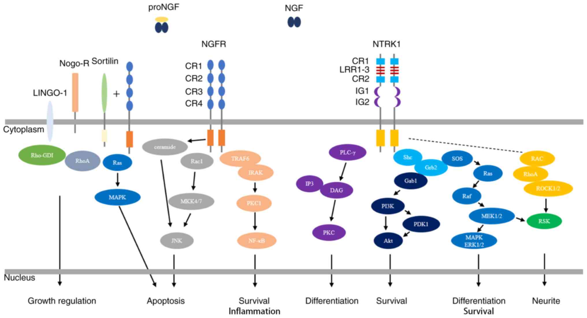
NGF exerts its biological functions through the
activation of the high-affinity tropomyosin-related kinase receptor
1 (NTRK1) and the low-affinity nerve growth factor receptor (NGFR),
which collaborate in mediating signaling (43-45)
(Fig. 1).
NTRK1, also known as Trk-A, TRKA, MTC, TRK, TRK1 and
p140-TrkA, is a 140-kDa transmembrane glycoprotein that belong to
the receptor tyrosine kinase family and is encoded by a gene
located on chromosome 1p23.1 (21,46).
NTRK1 consists of three domains, an extracellular receptor domain,
a single transmembrane helix and an intracellular tyrosine kinase
domain (47). The NTRK1
extracellular sequence, which exhibits intrinsic tyrosine kinase
activity, is composed of five distinct domains (D1-D5) (48). D1 and D3 contain cysteine-rich
repetitive motifs, D2 contains three leucine-rich repeat domains,
D4 and D5 are immunoglobulin-like domains, and D5 is responsible
for NGF binding (48,49). In the nervous system, NTRK1 is
primarily expressed in cholinergic neurons that are implicated in
spatial learning and memory, such as the central cortical pyramid,
basal forebrain, striatum, compartment and lateral geniculate body,
as well as in peripheral sensory neurons and sympathetic nerves
(50). However, NTRK1 is also
extensively expressed in non-neuronal tissues, including immune
cells, tumor cells and stem cells, suggesting pleiotropic functions
outside the nervous system (51,52).
NGF binds to the D5 extracellular domain of NTRK1,
resulting in dimerization of NTRK1 and activation of its
intracellular kinase (48). A total
of six tyrosine residues in the intracellular tyrosine kinase
domain, namely, Tyr670, Tyr674, Tyr675, Tyr490, Tyr751 and Tyr785,
are phosphorylated (53).
Autophosphorylation of Tyr490, Tyr751 and Tyr785 results in
activation of the three main downstream signal transduction
pathways, the mitogen-activated protein kinase/extracellular
signal-regulated protein kinase (MAPK/ERK) signaling pathway, the
phosphatidylinositol-3-kinase/serine protein kinase (PI3K/Akt)
signaling pathway and the phospholipase C-γ (PLC-γ) signaling
pathway (53,54). Most NGF-mediated biological
activities, including the proliferation, survival and
differentiation of neuronal cells, are due to the ligand-dependent
NTRK1 activation of several signal transduction cascades (32,33).
NGF-NTRK1 activation has been reported to have several clinically
relevant biological effects on non-neuronal cells, including
promoting the survival, proliferation, metastasis and invasion of
tumor cells (55-57).
The common neurotrophin receptor, NGFR, binds to NGF
and all other neurotrophins, including BDNF, NT-3 and NT-4/5, with
similar affinity and an extremely fast dissociation rate (44,64).
The binding of NGF to NGFR is a specific electrostatic interaction
between polar amino acid residues, and two specific sites play
important roles in this interaction, site I is composed of the
CR1-CR2 junction and the CR2 domain, while site II is composed of
the CR3-CR4 junction (65,66). NGFR activation involves different
molecular mechanisms in different types of cells and tissue
environments, and active NGFR participates in regulating
fundamental biological processes, such as cell survival,
proliferation, attachment, migration, differentiation and
apoptosis, via the nuclear transcription factor-kappa B, Jun
N-terminal kinase (JNK), and ceramide signaling pathways (67-70).
Although NGFR is a low-affinity NGFR, it may also participate in
NGF high-affinity binding by interacting with the NTRK1 receptor to
induce the growth and survival of neurites (54,69,71).
Activation of NGFR by proNGF induces the activation of apoptotic
signaling pathways, involving the co-receptor, sortilin (72-74).
In addition, NGFR is involved in the regulation of several
physiological functions, and cooperates with Nogo-R, LINGO-1, Aβ,
viral glycoprotein and tetanus toxin (75).
When damage causes a loss of skin integrity, the
body immediately initiates the skin repair process to restore its
integrity and maintain its function (1). Cutaneous wound healing is a complex
pathophysiological process involving different types of cells
(immune cells, keratinocytes, fibroblasts and vascular endothelial
cells), cytokines (fibroblast growth factor, platelet-derived
growth factor, transforming growth factor, epidermal growth factor,
vascular endothelial growth factor and NGF) and signaling pathways
(38,76). The normal wound healing process is
divided into four overlapping phases, haemostasis, inflammation,
proliferation and tissue remodelling (2). Any disruptions in these processes can
lead to wound healing disorders, such as chronic nonhealing ulcers
or keloids (77).
Under physiological conditions, NGF is sustainably
expressed in skin tissue by different types of cells, including
keratinocytes, fibroblasts and mast cells, which is essential for
maintaining skin homeostasis (78-81)
(Table I). NTRK1 and NGFR are
extensively expressed on the surfaces of different types of cells
in the skin, including keratinocytes, melanocytes, fibroblasts and
mast cells (82-86).
Matsuda et al (87) reported
that full-thickness skin wounds made in normal mice significantly
increase NGF expression levels in the sera and wounded skin tissues
of the mice. In addition, NGF mRNA and NGF have been detected in
newly formed epithelial cells at wound edges and fibroblasts, which
is consistent with the granulation tissue produced in the wound
spaces (87). Previous studies have
demonstrated that NGF is stored in the submandibular glands of
mice, and sialoadenectomy prior to wounding inhibits serum NGF
levels and delays the skin wound healing process (87,88).
Taken together, these findings suggest that NGF may play an
important role in wound healing.
Topical application of NGF to wounds significantly
promotes the healing of different types of wounds, including
diabetic foot ulcers, pressure ulcers and nonhealing surgical
wounds (87,89,90).
Preliminary experimental animal studies have reported that NGF
expression significantly decreases in diabetic wounds and wound
tissues following radiation treatment compared with normal skin
wounds (91,92). Generini et al (12) treated three diabetic patients with
leg or foot ulcers that were unresponsive to conventional therapies
with topical application of NGF, and the results demonstrated that
NGF promoted healing after 5±14 weeks of treatment. Even chronic
ulcers with damage extending below the hypodermis and muscle layers
healed in a few weeks following treatment with NGF, and the size of
the ulcers significantly decreased for the first time after 8 weeks
of NGF administration (32).
Topical NGF also exerts a healing effect on human pressure ulcers
(93,94). A randomized clinical trial of 36
patients demonstrated that topical application of murine NGF
significantly accelerated the healing of pressure ulcers compared
with traditional treatment (94).
Cutaneous ulcers secondary to vasculitis in patients with
rheumatoid arthritis (RA) are extremely difficult to heal (95). A total of four patients with
RA-associated skin ulcers were treated with topical NGF, and the
symptoms of pain and inflammation were significantly relieved after
2-3 weeks (96). The ulcers
progressively improved and were completely healed after 8 weeks
(96). Notably, intraperitoneal and
topical applications of NGF increase the survival rate and may
increase wound healing and promote survival in irradiated animals
(92).
The molecular mechanisms underlying NGF-induced
cutaneous wound healing are not yet fully understood. However,
studies have reported that NGF participates in several biological
activities during the healing process via complex molecular
mechanisms (Table I). In the skin,
NGF is predominantly synthesized and released by keratinocytes
(97). Different types of cells
express NGF receptors, including keratinocytes, melanocytes,
fibroblasts and mast cells (98).
Skin injury can increase the secretion of neurotrophic factors by
peripheral nerve endings (97).
Among these factors, substance P and neurokinin A directly induce
NGF mRNA expression and NGF secretion in keratinocytes (99,100).
Conversely, NGF can increase the secretion of neurotrophic factors,
including calcitonin gene-related peptide, by peripheral nerve
endings, thereby regulating the immune inflammatory response
associated with the wound healing process (101,102). NGF significantly stimulates the
proliferation of normal human keratinocytes in a dose-dependent
manner (79). NGF plays a
functional role in reparative neovascularization via a
VEGF-Akt-NO-mediated mechanism (103). NGF accelerates wound healing by
promoting wound re-epithelialization, which partly relies on
accelerated dermal fibroblast migration via activation of the
PI3K/Akt-Rac1-JNK and ERK signaling pathways (7). In wound granulation tissue, NGF can
also induce myofibroblast differentiation and collagen synthesis
via the NGFR-F-actin-MRTF-A signaling pathway, and induce wound
contraction and extracellular matrix remodelling (9,97). In
addition, NGFR promotes the healing of skin burn wounds by
improving the potential of epidermal stem cells to differentiate,
proliferate and migrate (104).
The cornea is the transparent outer layer of the eye
that serves as a protective barrier (105). Damage to the corneal epithelium
caused by scratches or surgery is common (106). Corneal wound healing is an
exceedingly complex process involving the coordination of cellular
activities stimulated and regulated by several growth factors that
reach the wound through tears and limbic vessels (107). Delays in corneal wound healing
results in keratinization of the corneal epithelium and corneal
opacity (108).
NGF is extensively expressed in normal human and rat
corneal tissues and is predominantly produced by corneal epithelial
cells (109) (Table I). In addition, NTRK1 and NGFR are
widely expressed in corneal epithelial cells, endothelial cells,
stromal cells, limbal stem cells and conjunctival epithelial cells
(110). Ocular surface damage and
inflammation can increase NGF levels (109,111). A transient increase in corneal NGF
levels is observed following corneal epithelial injury in
vivo (109). Inhibition of
endogenous NGF activity by neutralizing anti-NGF antibodies delays
the corneal epithelial healing rate, whereas administration of
exogenous NGF accelerates healing (109). Lambiase et al (111) reported that plasma NGF levels are
significantly higher in patients with vernal keratoconjunctivitis
compared with controls, which is associated with higher
inflammatory cell numbers in the conjunctival tissue of vernal
keratoconjunctivitis. Given the positive effect of NGF in promoting
corneal healing, the FDA has approved NGF eye drops for the
treatment of rare neurotrophic keratitis (15). Cellini et al (112) observed complete wound healing in
patients treated with NGF eye drops, and the stromal incision was
not visible when assessed via optical coherence tomography on day
21.
The epithelium needs to be reconstructed during
corneal healing, and this process mainly relies on the
proliferation and migration of adjacent corneal epithelial cells
(105,106). During corneal wound healing, NGF
plays a major role in promoting cell survival and migration by
binding to the NTRK1 receptor, which is mediated by both the
upregulation of matrix metalloproteinase-9 and the cleavage of β4
integrin (47,113,114). In addition, NGF promotes the
healing of corneal stroma and endothelial cells (109,110). The role of NGF in corneal
angiogenesis remains controversial; however, NGF can facilitate
innervations of perivascular nerves to regulate blood flow in
neovessels (10,115,116). In addition, it has been
demonstrated that topical application of exogenous NGF can promote
corneal healing by increasing corneal sensitivity and improving
tear function in patients with neurotrophic keratitis (117).
Oral injuries are very common and usually caused by
trauma, surgery, periodontal treatment and radiation therapy
(118). Improper treatment can
cause severe complications, which can affect breathing, language
articulation, chewing and swallowing (119). Oral wound healing is rapid and
results in little scar formation compared with cutaneous wound
healing (120). Recent studies
have suggested that the reduced scar formation in oral mucosa is
associated with fibroblast phenotypes, oral bacteria, saliva and
the moist environment of the oral cavity (118,121).
NGF is extensively expressed and synthesized by oral
keratinocytes, fibroblasts, infiltrating inflammatory leukocytes
and salivary ductal cells, and can be secreted into saliva
(122-124)
(Table I). The main form of NGF in
saliva is proNGF, which can be further converted by enzymes
released at the site of activity (123). In addition, NGF receptors are also
expressed in oral tissues. NTRK1 is predominantly expressed in
mucosal basal cells, salivary duct epithelial cells and gingival
epithelial cells, while NGFR is only expressed in mucosal basal
cell layers and salivary duct epithelium (123,124).
Recent studies have reported that NGF and proNGF are
involved in oral wound healing (Table
I). When the oral mucosa is wounded, NGF and proNGF in the
saliva can reach NGF receptors on keratinocytes and fibroblasts
that were previously hidden by oral epithelial layers (125). Plasmin generated by keratinocytes
at wound sites can cleave pro-NGF to form mature NGF (126). Mature NGF can access NTRK1 on the
basal keratinocytes of the wound edges, thereby enhancing the
proliferation and migration of keratinocytes (124). NGF can upregulate the expression
levels of E-cadherin and zona occludens-1 in mucosal epithelial
cells, suggesting that NGF may contribute to re-establishing
mucosal epithelial barrier function (127). Infiltrating inflammatory cells in
the epithelium and the connective tissue of the oral mucosa express
NGF and NGF receptors (128,129). Proinflammatory cytokines released
after tissue damage promote NGF synthesis, while NGF enhances
superoxide production via neutrophils (128). Fibroblasts and myofibroblasts that
appear during wound healing synthesize and secrete NGF and express
NGF receptors (130). NGF induces
the differentiation of fibroblasts into myofibroblasts, which are
responsible for both tissue contraction and extracellular matrix
component secretion (131). Thus,
NGFR mediates the apoptosis of myofibroblasts in the late stages of
healing, and decreases collagen deposition and scar formation
(122,123,125).
Wound healing is a complex process involving
different types of cells, tissues, cytokines, chemokines and growth
factors in each phase. Understanding its physiology will provide
more mechanism-based therapeutic alternatives for the treatment of
different types of wounds. Previous studies have demonstrated the
vital roles of NGF in the regulation of wound healing. The present
study summarized the biology of NGF and its receptors (NTRK1 and
NGFR), and recent studies have revealed the involvement of NGF and
its receptors in different types of wound healing. NGF and its
receptors exert several biological effects on the process of wound
healing, including participation in epithelial cell and fibroblast
migration, inflammatory immune response regulation, angiogenesis,
myofibroblast differentiation and tissue remodelling. Notably,
topical application of exogenous NGF exerts a significant healing
effect on cutaneous, corneal and oral wounds, without obvious side
effects. This healing action is also applicable to some wounds that
fail to respond to conventional treatment. However, further studies
are required to determine the underlying molecular mechanisms by
which NGF and its receptors participate in wound healing processes.
In addition, more randomized controlled trials are required to
assess whether topical application of high concentrations of NGF
can be used to treat different types of wounds.
Not applicable.
Funding: The present review was supported by the Postdoctoral
Innovation Project of Shandong Province (grant no. 202002050).
Not applicable.
SH conceived the present review and designed its
framework. ZL and HW performed the literature review and drafted
the initial manuscript. ZL and HW edited the manuscript. Data
sharing is not applicable. All authors have read and approved the
final manuscript.
Not applicable.
Not applicable.
The authors declare that they have no competing
interests.
|
1
|
Wang PH, Huang BS, Horng HC, Yeh CC and
Chen YJ: Wound healing. J Chin Med Assoc. 81:94–101.
2018.PubMed/NCBI View Article : Google Scholar
|
|
2
|
Ridiandries A, Tan JTM and Bursill CA: The
role of chemokines in Wound healing. Int J Mol Sci.
19(3217)2018.PubMed/NCBI View Article : Google Scholar
|
|
3
|
Yasukawa K, Okuno T and Yokomizo T:
Eicosanoids in skin Wound healing. Int J Mol Sci.
21(8435)2020.PubMed/NCBI View Article : Google Scholar
|
|
4
|
Zarei F and Soleimaninejad M: Role of
growth factors and biomaterials in wound healing. Artif Cells
Nanomed Biotechnol. 46:906–911. 2018.PubMed/NCBI View Article : Google Scholar
|
|
5
|
Gostynska N, Pannella M, Rocco ML,
Giardino L, Aloe L and Calza L: The pleiotropic molecule NGF
regulates the in vitro properties of fibroblasts, keratinocytes,
and endothelial cells: Implications for wound healing. Am J Physiol
Cell Physiol. 318:C360–C371. 2020.PubMed/NCBI View Article : Google Scholar
|
|
6
|
Lambiase A, Manni L, Bonini S, Rama P and
Aloe L: Nerve growth factor promotes corneal healing: Structural,
biochemical, and molecular analyses of rat and human corneas.
Invest Ophthalmol Vis Sci. 41:1063–1069. 2000.PubMed/NCBI
|
|
7
|
Chen JC, Lin BB, Hu HW, Lin C, Jin WY,
Zhang FB, Zhu YA, Lu CJ, Wei XJ and Chen RJ: NGF accelerates
cutaneous wound healing by promoting the migration of dermal
fibroblasts via the PI3K/Akt-Rac1-JNK and ERK pathways. Biomed Res
Int. 2014(547187)2014.PubMed/NCBI View Article : Google Scholar
|
|
8
|
Ishikawa S, Takeda A, Akimoto M, Kounoike
N, Uchinuma E and Uezono Y: Effects of neuropeptides and their
local administration to cutaneous wounds in sensory-impaired areas.
J Plast Surg Hand Surg. 48:143–147. 2014.PubMed/NCBI View Article : Google Scholar
|
|
9
|
Liu Z, Cao Y, Liu G, Yin S, Ma J, Liu J,
Zhang M and Wang Y: p75 neurotrophin receptor regulates NGF-induced
myofibroblast differentiation and collagen synthesis through
MRTF-A. Exp Cell Res. 383(111504)2019.PubMed/NCBI View Article : Google Scholar
|
|
10
|
Troullinaki M, Alexaki VI, Mitroulis I,
Witt A, Klotzsche-von Ameln A, Chung KJ, Chavakis T and
Economopoulou M: Nerve growth factor regulates endothelial cell
survival and pathological retinal angiogenesis. J Cell Mol Med.
23:2362–2371. 2019.PubMed/NCBI View Article : Google Scholar
|
|
11
|
Tanigawa T, Ahluwalia A, Watanabe T,
Arakawa T and Tarnawski AS: Nerve growth factor injected into the
gastric ulcer base incorporates into endothelial, neuronal, glial
and epithelial cells: Implications for angiogenesis, mucosal
regeneration and ulcer healing. J Physiol Pharmacol. 66:617–621.
2015.PubMed/NCBI
|
|
12
|
Generini S, Tuveri MA, Matucci Cerinic M,
Mastinu F, Manni L and Aloe L: Topical application of nerve growth
factor in human diabetic foot ulcers. A study of three cases. Exp
Clin Endocrinol Diabetes. 112:542–544. 2004.PubMed/NCBI View Article : Google Scholar
|
|
13
|
Aloe L, Tirassa P and Lambiase A: The
topical application of nerve growth factor as a pharmacological
tool for human corneal and skin ulcers. Pharmacol Res. 57:253–258.
2008.PubMed/NCBI View Article : Google Scholar
|
|
14
|
Cheret J, Lebonvallet N, Carre JL, Misery
L and Le Gall-Ianotto C: Role of neuropeptides, neurotrophins, and
neurohormones in skin wound healing. Wound Repair Regen.
21:772–788. 2013.PubMed/NCBI View Article : Google Scholar
|
|
15
|
Kanu LN and Ciolino JB: Nerve growth
factor as an ocular therapy: Applications, challenges, and future
directions. Semin Ophthalmol: 1-8, 2021 (Epub ahead of print). doi:
10.1080/08820538.2021.1890793.
|
|
16
|
Levi-Montalcini R and Angeletti PU: Nerve
growth factor. Physiol Rev. 48:534–569. 1968.PubMed/NCBI View Article : Google Scholar
|
|
17
|
Levi-Montalcini R: The nerve growth factor
35 years later. Science. 237:1154–1162. 1987.PubMed/NCBI View Article : Google Scholar
|
|
18
|
Sun HL and Jiang T: The structure of nerve
growth factor in complex with lysophosphatidylinositol. Acta
Crystallogr F Struct Biol Commun. 71:906–912. 2015.PubMed/NCBI View Article : Google Scholar
|
|
19
|
Varon S, Nomura J and Shooter EM: The
isolation of the mouse nerve growth factor protein in a high
molecular weight form. Biochemistry. 6:2202–2209. 1967.PubMed/NCBI View Article : Google Scholar
|
|
20
|
Smith AP, Varon S and Shooter EM: Multiple
forms of the nerve growth factor protein and its subunits.
Biochemistry. 7:3259–3268. 1968.PubMed/NCBI View Article : Google Scholar
|
|
21
|
Wiesmann C and de Vos AM: Nerve growth
factor: Structure and function. Cell Mol Life Sci. 58:748–759.
2001.PubMed/NCBI View Article : Google Scholar
|
|
22
|
Angeletti RH and Bradshaw RA: Nerve growth
factor from mouse submaxillary gland: Amino acid sequence. Proc
Natl Acad Sci USA. 68:2417–2420. 1971.PubMed/NCBI View Article : Google Scholar
|
|
23
|
Sofroniew MV, Howe CL and Mobley WC: Nerve
growth factor signaling, neuroprotection, and neural repair. Annu
Rev Neurosci. 24:1217–1281. 2001.PubMed/NCBI View Article : Google Scholar
|
|
24
|
Seidah NG, Benjannet S, Pareek S, Savaria
D, Hamelin J, Goulet B, Laliberte J, Lazure C, Chrétien M and
Murphy RA: Cellular processing of the nerve growth factor precursor
by the mammalian pro-protein convertases. Biochem J. 314:951–960.
1996.PubMed/NCBI View Article : Google Scholar
|
|
25
|
Levi-Montalcini R, Skaper SD, Dal Toso R,
Petrelli L and Leon A: Nerve growth factor: From neurotrophin to
neurokine. Trends Neurosci. 19:514–520. 1996.PubMed/NCBI View Article : Google Scholar
|
|
26
|
Lewin GR and Barde YA: Physiology of the
neurotrophins. Ann Rev Neurosci. 19:289–317. 1996.PubMed/NCBI View Article : Google Scholar
|
|
27
|
Micera A, Puxeddu I, Aloe L and
Levi-Schaffer F: New insights on the involvement of Nerve Growth
Factor in allergic inflammation and fibrosis. Cytokine Growth
Factor Rev. 14:369–374. 2003.PubMed/NCBI View Article : Google Scholar
|
|
28
|
Micera A, Lambiase A, Aloe L and Bonini S,
Levi-Schaffer F and Bonini S: Nerve growth factor involvement in
the visual system: Implications in allergic and neurodegenerative
diseases. Cytokine Growth Factor Rev. 15:411–417. 2004.PubMed/NCBI View Article : Google Scholar
|
|
29
|
Lambiase A, Micera A, Sgrulletta R and
Bonini S and Bonini S: Nerve growth factor and the immune system:
Old and new concepts in the cross-talk between immune and resident
cells during pathophysiological conditions. Curr Opin Allergy Clin
Immunol. 4:425–430. 2004.PubMed/NCBI View Article : Google Scholar
|
|
30
|
Hondermarck H: Neurotrophins and their
receptors in breast cancer. Cytokine Growth Factor Rev. 23:357–365.
2012.PubMed/NCBI View Article : Google Scholar
|
|
31
|
Dahlström M, Nordvall G, Sundström E,
Åkesson E, Tegerstedt G, Eriksdotter M and Forsell P:
Identification of amino acid residues of nerve growth factor
important for neurite outgrowth in human dorsal root ganglion
neurons. Eur J Neurosci. 50:3487–3501. 2019.PubMed/NCBI View Article : Google Scholar
|
|
32
|
Rocco ML, Soligo M, Manni L and Aloe L:
Nerve growth factor: Early studies and recent clinical trials. Curr
Neuropharmacol. 16:1455–1465. 2018.PubMed/NCBI View Article : Google Scholar
|
|
33
|
Aloe L, Rocco ML, Balzamino BO and Micera
A: Nerve growth factor: Role in growth, differentiation and
controlling cancer cell development. J Exp Clin Cancer Res.
35(116)2016.PubMed/NCBI View Article : Google Scholar
|
|
34
|
Skaper SD: Nerve growth factor: A
neuroimmune crosstalk mediator for all seasons. Immunology.
151:1–15. 2017.PubMed/NCBI View Article : Google Scholar
|
|
35
|
Capsoni S, Brandi R, Arisi I, D'Onofrio M
and Cattaneo A: A dual mechanism linking NGF/proNGF imbalance and
early inflammation to Alzheimer's disease neurodegeneration in the
AD11 anti-NGF mouse model. CNS Neurol Disord Drug Targets.
10:635–647. 2011.PubMed/NCBI View Article : Google Scholar
|
|
36
|
Bracci-Laudiero L and De Stefano ME: NGF
in early embryogenesis, differentiation, and pathology in the
nervous and immune systems. In: Neurotoxin Modeling of Brain
Disorders-Life-long Outcomes in Behavioral Teratology. pp125-152,
2015.
|
|
37
|
Niederhauser O, Mangold M, Schubenel R,
Kusznir EA, Schmidt D and Hertel C: NGF ligand alters NGF signaling
via p75(NTR) and TrkA. J Neurosci Res. 61:263–272. 2000.PubMed/NCBI View Article : Google Scholar
|
|
38
|
Kawamoto K and Matsuda H: Nerve growth
factor and wound healing. In: NGF and Related Molecules in Health
and Disease. pp369-384, 2004.
|
|
39
|
Tomellini E, Touil Y, Lagadec C, Julien S,
Ostyn P, Ziental-Gelus N, Meignan S, Lengrand J, Adriaenssens E,
Polakowska R and Le Bourhis X: Nerve growth factor and proNGF
simultaneously promote symmetric self-renewal, quiescence, and
epithelial to mesenchymal transition to enlarge the breast cancer
stem cell compartment. Stem Cells. 33:342–353. 2015.PubMed/NCBI View Article : Google Scholar
|
|
40
|
Steckiewicz KP, Barcińska E and Woźniak M:
Nerve growth factor as an important possible component of novel
therapy for cancer, diabetes and cardiovascular diseases. Cell Mol
Biol (Noisy-le-Grand). 64:16–23. 2018.PubMed/NCBI
|
|
41
|
Rowe CW, Dill T, Faulkner S, Gedye C, Paul
JW, Tolosa JM, Jones M, King S, Smith R and Hondermarck H: The
precursor for nerve growth factor (proNGF) in thyroid cancer lymph
node metastases: Correlation with primary tumour and pathological
variables. Int J Mol Sci. 20(5924)2019.PubMed/NCBI View Article : Google Scholar
|
|
42
|
Rowe CW, Faulkner S, Paul JW, Tolosa JM,
Gedye C, Bendinelli C, Wynne K, McGrath S, Attia J, Smith R and
Hondermarck H: The precursor for nerve growth factor (proNGF) is
not a serum or biopsy-rinse biomarker for thyroid cancer diagnosis.
BMC Endocr Disord. 19(128)2019.PubMed/NCBI View Article : Google Scholar
|
|
43
|
Lad SP, Peterson DA, Bradshaw RA and Neet
KE: Individual and combined effects of TrkA and p75NTR nerve growth
factor receptors. A role for the high affinity receptor site. J
Biol Chem. 278:24808–24817. 2003.PubMed/NCBI View Article : Google Scholar
|
|
44
|
Meakin SO and Shooter EM: The nerve growth
factor family of receptors. Trends Neurosci. 15:323–331.
1992.PubMed/NCBI View Article : Google Scholar
|
|
45
|
Frade JM and Barde YA: Nerve growth
factor: Two receptors, multiple functions. Bioessays. 20:137–145.
1998.PubMed/NCBI View Article : Google Scholar
|
|
46
|
Weier HU, Rhein AP, Shadravan F, Collins C
and Polikoff D: Rapid physical mapping of the human trk
protooncogene (NTRK1) to human chromosome 1q21-q22 by P1 clone
selection, fluorescence in situ hybridization (FISH), and
computer-assisted microscopy. Genomics. 26:390–393. 1995.PubMed/NCBI View Article : Google Scholar
|
|
47
|
Barbacid M: Structural and functional
properties of the TRK family of neurotrophin receptors. Ann N Y
Acad Sci. 766:442–458. 1995.PubMed/NCBI View Article : Google Scholar
|
|
48
|
Wiesmann C, Ultsch MH, Bass SH and de Vos
AM: Crystal structure of nerve growth factor in complex with the
ligand-binding domain of the TrkA receptor. Nature. 401:184–188.
1999.PubMed/NCBI View
Article : Google Scholar
|
|
49
|
Urfer R, Tsoulfas P, O'Connell L, Hongo
JA, Zhao W and Presta LG: High resolution mapping of the binding
site of TrkA for nerve growth factor and TrkC for neurotrophin-3 on
the second immunoglobulin-like domain of the Trk receptors. J Biol
Chem. 273:5829–5840. 1998.PubMed/NCBI View Article : Google Scholar
|
|
50
|
Kassel O, de Blay F, Duvernelle C, Olgart
C, Israel-Biet D, Krieger P, Moreau L, Muller C, Pauli G and
Frossard N: Local increase in the number of mast cells and
expression of nerve growth factor in the bronchus of asthmatic
patients after repeated inhalation of allergen at low-dose. Clin
Exp Allergy. 31:1432–1440. 2001.PubMed/NCBI View Article : Google Scholar
|
|
51
|
Zheng MG, Sui WY, He ZD, Liu Y, Huang YL,
Mu SH, Xu XZ, Zhang JS, Qu JL, Zhang J, et al: TrkA regulates the
regenerative capacity of bone marrow stromal stem cells in nerve
grafts. Neural Regen Res. 14:1765–1771. 2019.PubMed/NCBI View Article : Google Scholar
|
|
52
|
Zhang M, Zhang Y, Ding J, Li X, Zang C,
Yin S, Ma J, Wang Y and Cao Y: The role of TrkA in the promoting
wounding-healing effect of CD271 on epidermal stem cells. Arch
Dermatol Res. 310:737–750. 2018.PubMed/NCBI View Article : Google Scholar
|
|
53
|
Freund-Michel V and Frossard N: The nerve
growth factor and its receptors in airway inflammatory diseases.
Pharmacol Ther. 117:52–76. 2008.PubMed/NCBI View Article : Google Scholar
|
|
54
|
Reichardt LF: Neurotrophin-regulated
signalling pathways. Philos Trans R Soc Lond B Biol Sci.
361:1545–1564. 2006.PubMed/NCBI View Article : Google Scholar
|
|
55
|
Micera A, Lambiase A, Stampachiacchiere B,
Bonini S, Bonini S and Levi-Schaffer F: Nerve growth factor and
tissue repair remodeling: trkA(NGFR) and p75(NTR), two receptors
one fate. Cytokine Growth Factor Rev. 18:245–256. 2007.PubMed/NCBI View Article : Google Scholar
|
|
56
|
Faulkner S, Jobling P, Rowe CW, Rodrigues
Oliveira SM, Roselli S, Thorne RF, Oldmeadow C, Attia J, Jiang CC,
Zhang XD, et al: Neurotrophin receptors TrkA, p75NTR,
and sortilin are increased and targetable in thyroid cancer. Am J
Pathol. 188:229–241. 2018.PubMed/NCBI View Article : Google Scholar
|
|
57
|
Bradshaw RA, Pundavela J, Biarc J,
Chalkley RJ, Burlingame AL and Hondermarck H: NGF and ProNGF:
Regulation of neuronal and neoplastic responses through receptor
signaling. Adv Biol Regul. 58:16–27. 2015.PubMed/NCBI View Article : Google Scholar
|
|
58
|
Huber LJ and Chao MV: A potential
interaction of p75 and trkA NGF receptors revealed by affinity
crosslinking and immunoprecipitation. J Neurosci Res. 40:557–563.
1995.PubMed/NCBI View Article : Google Scholar
|
|
59
|
Huebner K, Isobe M, Chao M, Bothwell M,
Ross AH, Finan J, Hoxie JA, Sehgal A, Buck CR, Lanahan A, et al:
The nerve growth factor receptor gene is at human chromosome region
17q12-17q22, distal to the chromosome 17 breakpoint in acute
leukemias. Proc Natl Acad Sci USA. 83:1403–1407. 1986.PubMed/NCBI View Article : Google Scholar
|
|
60
|
Roux P: Neurotrophin signaling through the
p75 neurotrophin receptor. Progress Neurobiol. 67:203–233.
2002.PubMed/NCBI View Article : Google Scholar
|
|
61
|
Yan H and Chao MV: Disruption of
cysteine-rich repeats of the p75 nerve growth factor receptor leads
to loss of ligand binding. J Biol Chem. 266:12099–12104.
1991.PubMed/NCBI
|
|
62
|
Baldwin AN, Bitler CM, Welcher AA and
Shooter EM: Studies on the structure and binding properties of the
cysteine-rich domain of rat low affinity nerve growth factor
receptor (p75NGFR). J Biol Chem. 267:8352–8359. 1992.PubMed/NCBI
|
|
63
|
Pincelli C: p75 neurotrophin receptor in
the skin: Beyond its neurotrophic function. Front Med (Lausanne).
4(22)2017.PubMed/NCBI View Article : Google Scholar
|
|
64
|
Chao MV, Bothwell MA, Ross AH, Koprowski
H, Lanahan AA, Buck CR and Sehgal A: Gene transfer and molecular
cloning of the human NGF receptor. Science. 232:518–521.
1986.PubMed/NCBI View Article : Google Scholar
|
|
65
|
Shamovsky IL, Ross GM, Riopelle RJ and
Weaver DF: The interaction of neurotrophins with the p75NTR common
neurotrophin receptor: A comprehensive molecular modeling study.
Protein Sci. 8:2223–2233. 1999.PubMed/NCBI View Article : Google Scholar
|
|
66
|
He XL and Garcia KC: Structure of nerve
growth factor complexed with the shared neurotrophin receptor p75.
Science. 304:870–875. 2004.PubMed/NCBI View Article : Google Scholar
|
|
67
|
Meeker R and Williams K: Dynamic nature of
the p75 neurotrophin receptor in response to injury and disease. J
Neuroimmune Pharmacol. 9:615–628. 2014.PubMed/NCBI View Article : Google Scholar
|
|
68
|
Barker PA: p75NTR is positively
promiscuous: Novel partners and new insights. Neuron. 42:529–533.
2004.PubMed/NCBI View Article : Google Scholar
|
|
69
|
Chao MV: Neurotrophins and their
receptors: A convergence point for many signalling pathways. Nat
Rev Neurosci. 4:299–309. 2003.PubMed/NCBI View Article : Google Scholar
|
|
70
|
Teng HK, Teng KK, Lee R, Wright S, Tevar
S, Almeida RD, Kermani P, Torkin R, Chen ZY, Lee FS, et al: ProBDNF
induces neuronal apoptosis via activation of a receptor complex of
p75NTR and sortilin. J Neurosci. 25:5455–5463. 2005.PubMed/NCBI View Article : Google Scholar
|
|
71
|
Skaper SD: The neurotrophin family of
neurotrophic factors: An overview. Methods Mol Biol. 846:1–12.
2012.PubMed/NCBI View Article : Google Scholar
|
|
72
|
Lee R, Kermani P, Teng KK and Hempstead
BL: Regulation of cell survival by secreted proneurotrophins.
Science. 294:1945–1948. 2001.PubMed/NCBI View Article : Google Scholar
|
|
73
|
Beattie MS, Harrington AW, Lee R, Kim JY,
Boyce SL, Longo FM, Bresnahan JC, Hempstead BL and Yoon SO: ProNGF
induces p75-mediated death of oligodendrocytes following spinal
cord injury. Neuron. 36:375–386. 2002.PubMed/NCBI View Article : Google Scholar
|
|
74
|
Domeniconi M, Hempstead BL and Chao MV:
Pro-NGF secreted by astrocytes promotes motor neuron cell death.
Mol Cell Neurosci. 34:271–279. 2007.PubMed/NCBI View Article : Google Scholar
|
|
75
|
Dechant G and Barde YA: The neurotrophin
receptor p75(NTR): Novel functions and implications for diseases of
the nervous system. Nat Neurosci. 5:1131–1136. 2002.PubMed/NCBI View Article : Google Scholar
|
|
76
|
Roosterman D, Goerge T, Schneider SW,
Bunnett NW and Steinhoff M: Neuronal control of skin function: The
skin as a neuroimmunoendocrine organ. Physiol Rev. 86:1309–1379.
2006.PubMed/NCBI View Article : Google Scholar
|
|
77
|
Sorg H, Tilkorn DJ, Hager S, Hauser J and
Mirastschijski U: Skin wound healing: An update on the current
knowledge and concepts. Eur Surg Res. 58:81–94. 2017.PubMed/NCBI View Article : Google Scholar
|
|
78
|
Marconi A, Terracina M, Fila C, Franchi J,
Bonté F, Romagnoli G, Maurelli R, Failla CM, Dumas M and Pincelli
C: Expression and function of neurotrophins and their receptors in
cultured human keratinocytes. J Invest Dermatol. 121:1515–1521.
2003.PubMed/NCBI View Article : Google Scholar
|
|
79
|
Pincelli C, Sevignani C, Manfredini R,
Grande A, Fantini F, Bracci-Laudiero L, Aloe L, Ferrari S,
Cossarizza A and Giannetti A: Expression and function of nerve
growth factor and nerve growth factor receptor on cultured
keratinocytes. J Invest Dermatol. 103:13–18. 1994.PubMed/NCBI View Article : Google Scholar
|
|
80
|
Murase K, Murakami Y, Takayanagi K,
Furukawa Y and Hayashi K: Human fibroblast cells synthesize and
secrete nerve growth factor in culture. Biochem Biophys Res Commun.
184:373–379. 1992.PubMed/NCBI View Article : Google Scholar
|
|
81
|
Tron VA, Coughlin MD, Jang DE, Stanisz J
and Sauder DN: Expression and modulation of nerve growth factor in
murine keratinocytes (PAM 212). J Clin Invest. 85:1085–1089.
1990.PubMed/NCBI View Article : Google Scholar
|
|
82
|
Hefti F: Nerve growth factor promotes
survival of septal cholinergic neurons after fimbrial transections.
J Neurosci. 6:2155–2162. 1986.PubMed/NCBI View Article : Google Scholar
|
|
83
|
Hefti F, Dravid A and Hartikka J: Chronic
intraventricular injections of nerve growth factor elevate
hippocampal choline acetyltransferase activity in adult rats with
partial septo-hippocampal lesions. Brain Res. 293:305–311.
1984.PubMed/NCBI View Article : Google Scholar
|
|
84
|
Manni L, Rocco ML, Bianchi P, Soligo M,
Guaragna M, Barbaro SP and Aloe L: Nerve growth factor: Basic
studies and possible therapeutic applications. Growth Factors.
31:115–122. 2013.PubMed/NCBI View Article : Google Scholar
|
|
85
|
Minnone G, De Benedetti F and
Bracci-Laudiero L: NGF and its receptors in the regulation of
inflammatory response. Int J Mol Sci. 18(1028)2017.PubMed/NCBI View Article : Google Scholar
|
|
86
|
Raychaudhuri SK, Raychaudhuri SP, Weltman
H and Farber EM: Effect of nerve growth factor on endothelial cell
biology: Proliferation and adherence molecule expression on human
dermal microvascular endothelial cells. Arch Dermatol Res.
293:291–295. 2001.PubMed/NCBI View Article : Google Scholar
|
|
87
|
Matsuda H, Koyama H, Sato H, Sawada J,
Itakura A, Tanaka A, Matsumoto M, Konno K, Ushio H and Matsuda K:
Role of nerve growth factor in cutaneous wound healing:
Accelerating effects in normal and healing-impaired diabetic mice.
J Exp Med. 187:297–306. 1998.PubMed/NCBI View Article : Google Scholar
|
|
88
|
Li AK, Koroly MJ, Schattenkerk ME, Malt RA
and Young M: Nerve growth factor: Acceleration of the rate of wound
healing in mice. Proc Natl Acad Sci USA. 77:4379–4381.
1980.PubMed/NCBI View Article : Google Scholar
|
|
89
|
Nakagaki O, Miyoshi H, Sawada T and Atsumi
T, Kondo T and Atsumi T: Epalrestat improves diabetic wound healing
via increased expression of nerve growth factor. Exp Clin
Endocrinol Diabetes. 121:84–89. 2013.PubMed/NCBI View Article : Google Scholar
|
|
90
|
Ali Bahar M, Bauer B, Tredget EE and
Ghahary A: Dermal fibroblasts from different layers of human skin
are heterogeneous in expression of collagenase and types I and III
procollagen mRNA. Wound Repair Regen. 12:175–182. 2004.PubMed/NCBI View Article : Google Scholar
|
|
91
|
Anand P, Terenghi G, Warner G, Kopelman P,
Williams-Chestnut RE and Sinicropi DV: The role of endogenous nerve
growth factor in human diabetic neuropathy. Nat Med. 2:703–707.
1996.PubMed/NCBI View Article : Google Scholar
|
|
92
|
Shi CM, Qu JF and Cheng TM: Effects of the
nerve growth factor on the survival and wound healing in mice with
combined radiation and wound injury. J Radiat Res. 44:223–228.
2003.PubMed/NCBI View Article : Google Scholar
|
|
93
|
Bernabei R, Landi F, Bonini S, Onder G,
Lambiase A, Pola R and Aloe L: Effect of topical application of
nerve-growth factor on pressure ulcers. Lancet.
354(307)1999.PubMed/NCBI View Article : Google Scholar
|
|
94
|
Landi F, Aloe L, Russo A, Cesari M, Onder
G, Bonini S, Carbonin PU and Bernabei R: Topical treatment of
pressure ulcers with nerve growth factor: A randomized clinical
trial. Ann Intern Med. 139:635–641. 2003.PubMed/NCBI View Article : Google Scholar
|
|
95
|
Coelho S, Amarelo M, Ryan S, Reddy M and
Sibbald RG: Rheumatoid arthritis-associated inflammatory leg
ulcers: A new treatment for recalcitrant wounds. Int Wound J.
1:81–84. 2004.PubMed/NCBI View Article : Google Scholar
|
|
96
|
Tuveri M, Generini S, Matucci-Cerinic M
and Aloe L: NGF, a useful tool in the treatment of chronic
vasculitic ulcers in rheumatoid arthritis. Lancet. 356:1739–1740.
2000.PubMed/NCBI View Article : Google Scholar
|
|
97
|
Ashrafi M, Baguneid M and Bayat A: The
role of neuromediators and innervation in cutaneous wound healing.
Acta Derm Venereol. 96:587–594. 2016.PubMed/NCBI View Article : Google Scholar
|
|
98
|
Aloe L, Tirassa P and Bracci-Laudiero L:
Nerve growth factor in neurological and non-neurological diseases:
Basic findings and emerging pharmacological prospectives. Curr
Pharm Des. 7:113–123. 2001.PubMed/NCBI View Article : Google Scholar
|
|
99
|
Burbach GJ, Kim KH, Zivony AS, Kim A,
Aranda J, Wright S, Naik SM, Caughman SW, Ansel JC and Armstrong
CA: The neurosensory tachykinins substance P and neurokinin A
directly induce keratinocyte nerve growth factor. J Invest
Dermatol. 117:1075–1082. 2001.PubMed/NCBI View Article : Google Scholar
|
|
100
|
Gibran NS, Tamura R, Tsou R and Isik FF:
Human dermal microvascular endothelial cells produce nerve growth
factor: Implications for wound repair. Shock. 19:127–130.
2003.PubMed/NCBI View Article : Google Scholar
|
|
101
|
Bowles WR, Sabino M, Harding-Rose C and
Hargreaves KM: Chronic nerve growth factor administration increases
the peripheral exocytotic activity of capsaicin-sensitive cutaneous
neurons. Neurosci Lett. 403:305–308. 2006.PubMed/NCBI View Article : Google Scholar
|
|
102
|
Banks BEC, Vernon CA and Warner JA: Nerve
growth factor has anti-inflammatory activity in the rat hindpaw
oedema test. Neurosci Lett. 47:41–45. 1984.PubMed/NCBI View Article : Google Scholar
|
|
103
|
Emanueli C, Salis MB, Pinna A, Graiani G,
Manni L and Madeddu P: Nerve growth factor promotes angiogenesis
and arteriogenesis in ischemic hindlimbs. Circulation.
106:2257–2262. 2002.PubMed/NCBI View Article : Google Scholar
|
|
104
|
Zhang M, Cao Y, Li X, Hu L, Taieb SK, Zhu
X, Zhang J, Feng Y, Zhao R, Wang M, et al: Cd271 mediates
proliferation and differentiation of epidermal stem cells to
support cutaneous burn wound healing. Cell Tissue Res. 371:273–282.
2018.PubMed/NCBI View Article : Google Scholar
|
|
105
|
Wilson SE: Corneal wound healing. Exp Eye
Res. 197(108089)2020.PubMed/NCBI View Article : Google Scholar
|
|
106
|
Spadea L, Giammaria D and Trabucco P:
Corneal wound healing after laser vision correction. Br J
Ophthalmol. 100:28–33. 2016.PubMed/NCBI View Article : Google Scholar
|
|
107
|
Wilson SE, Mohan RR, Mohan RR, Ambrósio R,
Hong J and Lee J: The corneal wound healing response. Progress
Retinal Eye Res. 20:625–637. 2001.PubMed/NCBI View Article : Google Scholar
|
|
108
|
Agrawal VB and Tsai RJ: Corneal epithelial
wound healing. Indian J Ophthalmol. 51:5–15. 2003.PubMed/NCBI
|
|
109
|
Lambiase A, Manni L, Bonini S, Rama P and
Aloe L: Nerve growth factor promotes corneal healing: Structural,
biochemical, and molecular analyses of rat and human corneas.
Invest Ophthalmol Vis Sci. 41:1063–1069. 2000.PubMed/NCBI
|
|
110
|
You L, Kruse FE and Völcker HE:
Neurotrophic factors in the human cornea. Invest Ophthalmol Vis
Sci. 41:692–702. 2000.PubMed/NCBI
|
|
111
|
Lambiase A, Bonini S, Micera A, Magrini L
and Aloe L: Increased plasma levels of nerve growth factor in
vernal keratoconjunctivitis and relationship to conjunctival mast
cells. Invest Ophthalmol Vis Sci. 36:2127–2132. 1995.PubMed/NCBI
|
|
112
|
Cellini M, Bendo E, Bravetti GO and Campos
EC: The use of nerve growth factor in surgical wound healing of the
cornea. Ophthalmic Res. 38:177–181. 2006.PubMed/NCBI View Article : Google Scholar
|
|
113
|
Blanco-Mezquita T, Martinez-Garcia C,
Proenca R, Zieske JD, Bonini S, Lambiase A and Merayo-Lloves J:
Nerve growth factor promotes corneal epithelial migration by
enhancing expression of matrix metalloprotease-9. Invest Ophthalmol
Vis Sci. 54:3880–3890. 2013.PubMed/NCBI View Article : Google Scholar
|
|
114
|
Touhami A, Grueterich M and Tseng SC: The
role of NGF signaling in human limbal epithelium expanded by
amniotic membrane culture. Invest Ophthalmol Vis Sci. 43:987–994.
2002.PubMed/NCBI
|
|
115
|
Matsuyama A, Takatori S, Sone Y, Ochi E,
Goda M, Zamami Y, Hashikawa-Hobara N, Kitamura Y and Kawasaki H:
Effect of nerve growth factor on innervation of perivascular nerves
in neovasculatures of mouse cornea. Biol Pharm Bull. 40:396–401.
2017.PubMed/NCBI View Article : Google Scholar
|
|
116
|
Seo K, Choi J, Park M and Rhee C:
Angiogenesis effects of nerve growth factor (NGF) on rat corneas. J
Vet Sci. 2:125–130. 2001.PubMed/NCBI
|
|
117
|
Lambiase A, Sacchetti M and Bonini S:
Nerve growth factor therapy for corneal disease. Curr Opin
Ophthalmol. 23:296–302. 2012.PubMed/NCBI View Article : Google Scholar
|
|
118
|
Larjava H: Oral Wound Healing: An
Overview. John Wiley & Sons, Ltd, 2013.
|
|
119
|
Nauta A, Gurtner G and Longaker MT: Wound
healing and regenerative strategies. Oral Dis. 17:541–549.
2011.PubMed/NCBI View Article : Google Scholar
|
|
120
|
Chin GS, Kim WJ, Lee TY, Liu W, Saadeh PB,
Lee S, Levinson H, Gittes GK and Longaker MT: Differential
expression of receptor tyrosine kinases and Shc in fetal and adult
rat fibroblasts: Toward defining scarless versus scarring
fibroblast phenotypes. Plast Reconstr Surg. 105:972–979.
2000.PubMed/NCBI View Article : Google Scholar
|
|
121
|
Chen L, Arbieva ZH, Guo S, Marucha PT,
Mustoe TA and DiPietro LA: Positional differences in the wound
transcriptome of skin and oral mucosa. BMC Genomics.
11(471)2010.PubMed/NCBI View Article : Google Scholar
|
|
122
|
Hayashi K, Karatsaidis A, Schreurs O,
Bjornland T, Sugisaki M and Schenck K: NGF and its receptors TrkA
and p75NTR in the epithelium of oral lichen. J Oral Pathol Med.
37:241–248. 2008.PubMed/NCBI View Article : Google Scholar
|
|
123
|
Naesse EP, Schreurs O, Messelt E, Hayashi
K and Schenck K: Distribution of nerve growth factor, pro-nerve
growth factor, and their receptors in human salivary glands. Eur J
Oral Sci. 121:13–20. 2013.PubMed/NCBI View Article : Google Scholar
|
|
124
|
Hayashi K, Storesund T, Schreurs O, Khuu
C, Husvik C, Karatsaidis A, Helgeland K, Martin-Zanca D and Schenck
K: Nerve growth factor beta/pro-nerve growth factor and their
receptors in normal human oral mucosa. Eur J Oral Sci. 115:344–354.
2007.PubMed/NCBI View Article : Google Scholar
|
|
125
|
Schenck K, Schreurs O, Hayashi K and
Helgeland K: The role of nerve growth factor (NGF) and its
precursor forms in oral wound healing. Int J Mol Sci.
18(386)2017.PubMed/NCBI View Article : Google Scholar
|
|
126
|
Bruno MA and Cuello AC: Activity-dependent
release of precursor nerve growth factor, conversion to mature
nerve growth factor, and its degradation by a protease cascade.
Proc Natl Acad Sci USA. 103:6735–6740. 2006.PubMed/NCBI View Article : Google Scholar
|
|
127
|
Tan L, Hatzirodos N and Wormald PJ: Effect
of nerve growth factor and keratinocyte growth factor on wound
healing of the sinus mucosa. Wound Repair Regen. 16:108–116.
2008.PubMed/NCBI View Article : Google Scholar
|
|
128
|
Bonini S, Rasi G, Bracci-Laudiero ML,
Procoli A and Aloe L: Nerve growth factor: Neurotrophin or
cytokine? Int Arch Allergy Immunol. 131:80–84. 2003.PubMed/NCBI View Article : Google Scholar
|
|
129
|
Samah B, Porcheray F and Gras G:
Neurotrophins modulate monocyte chemotaxis without affecting
macrophage function. Clin Exp Immunol. 151:476–486. 2008.PubMed/NCBI View Article : Google Scholar
|
|
130
|
Palazzo E, Marconi A, Truzzi F, Dallaglio
K, Petrachi T, Humbert P, Schnebert S, Perrier E, Dumas M and
Pincelli C: Role of neurotrophins on dermal fibroblast survival and
differentiation. J Cell Physiol. 227:1017–1025. 2012.PubMed/NCBI View Article : Google Scholar
|
|
131
|
Darby IA, Zakuan N, Billet F and
Desmouliere A: The myofibroblast, a key cell in normal and
pathological tissue repair. Cell Mol Life Sci. 73:1145–1157.
2016.PubMed/NCBI View Article : Google Scholar
|