Introduction
Among the incidental carcinoma of thyroid, the
overall incidence of papillary carcinoma is higher than other
types. This type of carcinoma has a high incidence, is associated
with follicular adenoma, an may present as an isolated thyroid
nodule or as multifocal lesions and should be considered with
malignant potential (1-3).
Despite advanced understanding of the biological
characteristics of papillary thyroid carcinoma and the development
of guidelines for its treatment, a number of practical questions
remain unsolved; for instance, the extension of surgery, such as
lobectomy vs. total thyroidectomy remains controversial (4,5).
The increased incidence of thyroid cancer is the
likely result of two coexisting processes: Increased detection and
increased number of cases, due to unrecognized thyroid-specific
carcinogens. This increase is apparent due to low identification of
a large reservoir of subclinical papillary lesions that may never
affect patient health (6,7).
The thyroid gland, thoroughly investigated in
systematic studies that include complex histopathological
examinations, can present a pathology that is often unknown. During
autopsies, the thyroid gland is rarely analyzed, being overlooked
or incompletely visualized.
When systematic studies are performed to investigate
macroscopically found nodules, including complex histopathological
examination, an increased incidence of thyroid tumors is detected.
Under these circumstances, the literature reports a prevalence of
~50% of thyroid nodules, detected during autopsies in subjects with
unknown thyroid pathology (8-10).
The prevalence of latent thyroid carcinomas is reported to average
between 1.0 and 35.6% in different systemic autopsy series
(11,12).
Thyroid cancer accounts for 1% of all malignancies,
and is on the increase globally (13,14).
Consequently, extensive sessions are dedicated to thyroid pathology
in medical endocrinology meetings, where the news regarding the
etiological mechanisms, the paraclinical diagnosis and the
therapeutic methods of this type of cancer are discussed.
The incidence of thyroid cancer has tripled in the
last 30 years, with a constant and latent numerical growth (mainly
including small tumors), while mortality remained stable, according
to data provided by the American Association of
Endocrinopathologists and the American College of
Endocrinopathology (15).
In the present study, an extended retrospective
study of 526 autopsy cases was performed to identify the prevalence
of thyroid carcinoma, among various types of thyroid nodules
identified incidentally. The findings indicated that, papillary
microcarcinoma is an extremely common incidental finding and the
vast majority of these tumors pursue a benign course.
Materials and methods
Case selection for study batch
Total thyroidectomy was performed during serial
autopsies of 526 cases in an interval of 3 years (January
2017-February 2020) and macroscopically examined in search for
small undetectable thyroid nodules. The study was performed at the
forensic department of Brăila County Emergency Hospital (Braila,
Romania).
The study batch comprised 416 males (79%) and 110
females (sex ratio=3.78), with an age range of 10-94 years
(m=60.32, SD=±15.42). The highest incidence was found in patients
aged between 60 and 70 years. None of the subjects included in the
study had evidence of thyroid disease. The urban/rural distribution
encountered in patients with neoplasms was approximately equal.
A complex database was compiled including variables
such as age, sex, environmental factors and clinical factors
related to the risk of thyroid cancer. Possible association with
Hashimoto's thyroiditis or lymphocytic thyroiditis, along with
overweight and cardiovascular diseases was also considered.
The study was conducted according to the World
Medical Association Declaration of Helsinki, using a protocol
approved by the local Bioethics Committee from Brăila Emergency
County Hospital (Braila). All patients previously signed an
informed written consent regarding hospitalization, treatment and a
possible future publication of data.
Tissue sampling and stains
The thyroids were dissected and carefully separated
from the soft tissues around the thyroid, fixed in 10% formalin and
then weighed and measured. All macroscopic changes were noted, with
special propensity for whitish star-shape areas and the scars
suspected to be a carcinoma. Subsequently, tissue specimens were
carefully harvested from different parts of both thyroid lobes for
microscopicanalysis in order to perform a systematic study.
Multiple, serial sections were performed to cover a
wide range of undetected pathologies in the macroscopic
examination. The selected tissue samples were fixed in 10%
neutral-buffered formalin (pH 7.0) for 24-48 hand paraffin
embedded. Sections were cut at 5 µm and stained with standard
hematoxylin-eosin (HE). Approximately 1,000 fragments were
processed and microscopically examined.
Immunohistochemical analysis
Immunohistochemical analysis (IHC) was performed for
a panel of 7 antibodies, using sections show non slides treated
first with poly-L-lysine. The panel comprised the following
antibodies: CK7 (mouse monolconal, clone: OV-TL12/20, ready to
use-RTU, Cell Marque), TTF1 (mouse monolconal, clone: 8G7G3/1, RTU,
Cell Marque), thyroglobulin (mouse monoclonal, clone: 2H11+6E1,
RTU, Cell Marque), EMA (mouse monoclonal, clone: E29, RTU Cell
Marque), vimentin (mouse monoclonal, clone: V9, RTU, Cell Marque),
PCNA (mouse monoclonal, clone PC10, 1:200, Dako; Agilent
Technologies), and p53 (mouse monoclonal, clone: SP5, RTU, Cell
Marque). IHC was performed on 3 µm thick sections from
formalin-fixed paraffin-embedded specimens.
The method used was an indirect tristadial
Avidin-Biotin-Complex technique, with a NovoLink Polymer detection
system which utilizes a novel control polymerization technology to
prepare polymeric HRP-linker antibody conjugates, according to the
manufacturer's specifications (Novocastra). Antigen retrieval
technique (enzymatic pre-treatment) was carried out, according to
the producer's specifications.
All slides were examined and photographed on a Nikon
Eclipse Ci. Digital images captured with Digital Microscope Camera
program were processed and analyzed with Photos App, running under
Windows 10.
The criteria of the World Health Organization (WHO)
were used to diagnose and classify histological subtypes of thyroid
tumors. The Turin criteria for poor differentiated thyroid
carcinoma include: i) presence of a solid/trabecular/insular
pattern of growth, ii) absence of the conventional nuclear features
of papillary carcinoma, and iii) presence of at least one of the
following features: Convoluted nuclei; mitotic activity > or
=3x10 HPF (high power field) and tumor necrosis (16).
Results
Tumor distribution areas
From 526 cases, 153 cases had thyroid nodules, out
of which 135 had multinodular goiters and 18 uninodular goiter; 51
cases had malignant nodules and 322 had colloid nodules. The
average weight of the thyroid with goiter was 88 g, with variations
between 25 and 500 g.
A total of 39 patients with goiter had associated
diseases: 35 cases had associated lymphocytic thyroiditis and 4
cases had focal lymphocytic thyroiditis associated with neoplasia.
A total of 39 thyroid glands showed thyroiditis, of which focal
lymphocytic thyroiditis occurred in 35 cases associated with
goiter, and in 2 cases associated with neoplasia. One case of
Hashimoto's thyroiditis was found concurrently with papillary
carcinoma.
Thyroid pathology
The incidence of thyroid pathology was notedin
association with cardiovascular diseases. In the present study, the
associated cardiovascular diseases were divided into three distinct
categories: 67.3% had chronic cardiovascular diseases (especially
myocardofibrosis), 5.3% had chronic cardiovascular diseases
associated with acute myocardial ischemia, and 7.8% had acute heart
diseases. A causal relationship between obesity and thyroid
pathology was found in our cases, as follows: 33.3% had obesity,
19.5% had cachexia and 52% were normostenic.
Histopathological diagnosis of malignancy was found
in 51 cases of the analyzed thyroid glands. The youngest patient
was 40 years of age and the oldest 94 years of age, the highest
incidence being found in patients aged between 45 and 60 years.
Thyroid neoplasia was noted in 51 cases (33.33%),
3-fold higher in men than in women: Micropapillary carcinomas in 47
cases, undifferentiated thyroid carcinoma in 3 cases and squamous
cell thyroid carcinoma in 1 case. The association with another
thyroid pathology was seen in 8 cases: 5 goiters associated with
papillary carcinomas, and 3 thyroiditis associated with papillary
carcinomas.
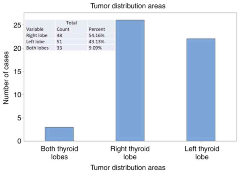
In 54.1% of cases, the malignant nodules were
distributed in the right thyroid lobe 43.1% in the left thyroid
lobe. In some cases (9%), the tumor areas were multiple, considered
multifocal, disseminated in both thyroid lobes (Fig. 1).
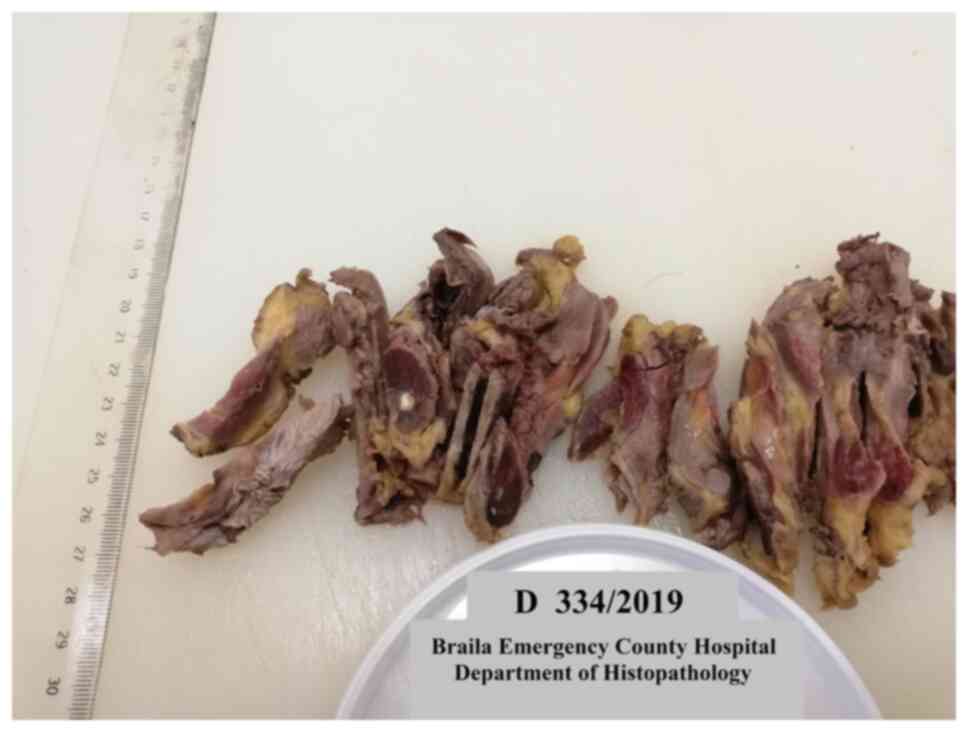
Approximately 92% of thyroid carcinomas were of
papillary type. Grossly, most papillary tumors were white-grayish
with infiltrative borders and firm surface (Fig. 2).
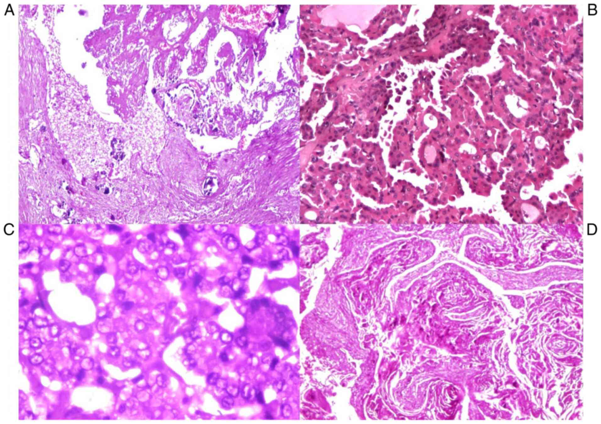
The microscopicarchitectural patterns were
micro-papillary, trabecular, solid and cystic types. The stroma
around the tumor was sometimes fibrotic and sclerotic.
Another typical, but not pathognomonic feature, was
the presence of psammoma bodies (5 cases). Distinct nuclear
features were crowded vesicular nuclei, optically clear (‘Orphan
Annie eyes’), irregular nuclear contours, nuclear grooves and
intranuclear cytoplasmic inclusions, marked crowding with
overlapping of adjacent nuclei, pale and clear chromatin (Fig. 3). The carcinomas were <1 cm,
so-called papillary microcarcinoma (in the present study the
dimensions were between 0.2 and 1 cm).
Squamous cell carcinoma involved both
lobes
The gross appearance was a firm consistency and
grayish-white color with areas of necrosis and extensive
infiltration of peri-thyroid soft tissue, vascular and peri-neural
invasion. Microscopically these tumors consisted of nests and
sheets of cells with squamous differentiation, with keratin ‘pearl’
formation.
Poorly differentiated carcinoma showed
evidence of follicular differentiation, fitting morphologically
between well-differentiated and undifferentiated thyroid carcinoma,
but WHO recognizes it as a separate entity (6). In the present study, we classified
them as undifferentiated thyroid carcinoma after Turin criteria and
applied IHC.
Grossly, these tumors presented as fleshy, large,
solid grey to white nodules with areas of hemorrhage and necrosis.
Microscopically, they were composed of a variable admixture of
spindle cells. The cells had eosinophilic cytoplasm, brisk mitotic
activity with abundant apoptosis.
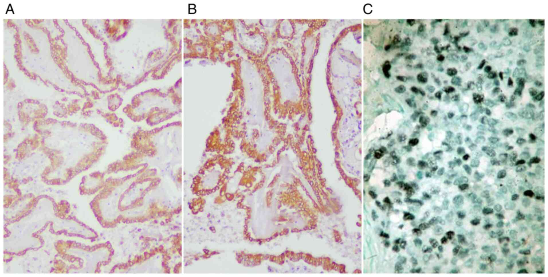
Overall, CK7 was diffusely expressed in papillary
carcinomas and focally expressed in squamous cell tumors and
undifferentiated tumors. EMA was diffusely positive in all thyroid
carcinomas, while TTF1 was negative in most of the cases (Fig. 4). Thyroglobulin was positive in all
cases, with variable expression in the cytoplasm of tumor
cells.
PCNA showed positive expression in ~7-8% of tumor
cells nuclei in undifferentiated carcinomas, in ~3-5% of tumor
cells nuclei in papillary carcinomas and ~20-25% of tumor cells
nuclei in squamous cell carcinoma. The PCNA proliferation index was
expressed in tumor cell nuclei, in 25% of well-differentiated
squamous tumor cells, both basal and parabasal; in the areas with
keratin and para-keratin formation, no expression was observed.
P53 was negative in all cases, including
undifferentiated and squamous cell carcinomas. Nuclear antibodies
were affected by autolysis and necrosis, therefore the
antigen-antibody reaction in the nucleus lost its expression.
Vimentin was negative in tumor cells, with expression only in
stroma and blood vessels.
Discussion
Thyroid carcinomas had an incidence in the present
study of 10%, which is consistent with the percentage reported by
studies in the literature (17,18).
Normal thyroid appeared in 40% of our cases, an incidence close to
that reported in the literature.
According to the WHO, the normal weight of the
thyroid has an upper limit of 40 g, anything exceeding this limit
is considered to be goiter. Our study showed a thyroid weight of
25-40 g, considered to be within normal limits.
Focal lymphocytic thyroiditis has been found in 39
cases. It is seen by some authors as a nonspecific inflammation
associated with etiological factors such as acute infections, local
trauma, chemicals, radiation, immune and with an uncertain
association with papillary thyroid cancer (19). In the current study, Hashimoto's
thyroiditis was associated with neoplasia in 1 case. Squamous
metaplasia had an incidence of 7.3% in our cases, with an
association of 20% of cases with thyroiditis. The incidence of
thyroid disease was not closely associated between sex, age and
pathological background.
Immunohistochemistry for CK7 and EMA was positive in
all three cases of undifferentiated carcinoma. These stains are
useful for diagnosis of tumors and to confirm that the neoplasm is
a carcinoma rather than a high-grade sarcoma (20). TTF-1 and thyreoglobulin are
typically negative in undifferentiated carcinoma. In the present
study, all 3 cases were also negative for TTF1.
Nuclear reactivity for PCNA has been reported in
~75-80% of anaplastic carcinoma (21). In our study, there were 2 cases of
anaplastic carcinoma, with 5-10% positivity in the tumor cell
nuclei and one case was negative.
Systematic detailed studies have demonstrated that a
thyroid, which is apparently normal on macroscopic examination, can
be the site of pathological manifestations, and sometimes
carcinomatous (22).
Thyroid nodules are quite common in the general
population, most often they are benign, about 10% of the detected
solitary nodules are malignant. Thyroid nodules detected in
polynodular goiter are generally colloidal, the risk of malignancy
being reduced (23).
In cases of thyroid carcinomas, the highest
frequency is occupied by papillary microcarcinomas, which have a
long evolution, these being detected incidentally during autopsies.
Papillary microcarcinoma is an extremely common incidental finding
and the vast majority of these tumors pursue a benign course
(24).
Regarding the association with the risk of thyroid
cancer in patients with pre-existing thyroid disease or
cardiovascular diseases (25), as
well as the association with overweight (26), our study did not show a direct
correlation.
The rise in incidence of papillary micro-carcinomas
creates management dilemmas. Therefore, the decision in the
management of these type of tumors begins at the time of surgery
and is associated with a more aggressive course. Younger age may be
predictive for radioactive iodine administration in the lowest-risk
patients (27,28).
In a cross-sectional study over an historical cohort
during a decade, conducted in Brazil, it was found that there was
an increase in the proportion of cases with malignant cytological
results among microcarcinomas, related to an enhancement in
preoperative diagnostic methods (29-31).
In another exhaustive retrospective study, performed
on more than 2,000 patients in an interval of 8 years, conducted in
Japan, it was also found that the oncological outcomes of the
immediate surgery and active surveillance groups were similarly
excellent, but the incidences of unfavorable events were higher in
the immediate surgery group (32).
In summary, papillary thyroid carcinomas are a
compact group with a heterogenous morpho-phenotypical and
immune-biological expression and behavior that are sometimes
overlooked (particularly the small size tumors, such as
micro-carcinomas). The majority of these carcinomas are
incidentally identified during a routine check or at autopsy.
Acknowledgements
Not applicable.
Funding
Funding: No funding was received.
Availability of data and materials
The datasets used and/or analyzed during the current
study are available from the corresponding author on reasonable
request.
Authors' contributions
ISM and MC performed the histological examinations
and IHC, and substantially contributed to the writing of the
manuscript. ZC and VM analyzed and interpreted the patient data.
BS, DS and AT searched the literature for similar work and articles
and contributed to writing the manuscript. All authors read and
approved the final manuscript.
Ethics approval and consent to
participate
The study was conducted according to the World
Medical Association Declaration of Helsinki, using a protocol
approved by the local Bioethics Committee from Brăila Emergency
County Hospital (Braila). All patients previously signed an
informed written consent about hospitalization, treatment and a
possible future publication of data.
Patient consent for publication
Not applicable.
Competing interests
The authors declare no conflict or competing
interests.
References
|
1
|
Pezzolla A, Lattarulo S, Milella M, Barile
G, Pascazio B, Ciampolillo A, Fabiano G and Palasciano N:
Incidental carcinoma in thyroid pathology: Our experience and
review of the literature. Ann Ital Chir. 81:165–169.
2010.PubMed/NCBI
|
|
2
|
Hurtado-López LM, Basurto-Kuba E, Montes
de Oca-Durán ER, Pulido-Cejudo A, Vázquez-Ortega R and
Athié-Gutiérrez C: Prevalence of thyroid nodules in the valley of
Mexico. Cir Cir. 79:114–117. 2011.PubMed/NCBI
|
|
3
|
Ramirez-Gonzalez LR, Sevilla-Vizcaino R,
Monge-Reyes P, Aldaz-Dorantes JE, Márquez-Valdez AR,
García-Martínez D, González-Ojeda A and Fuentes-Orozco C: Findings
of thyroid nodules in autopsies in western Mexico. Rev Med Inst Mex
Seguro Soc. 55:594–598. 2017.PubMed/NCBI(In Spanish).
|
|
4
|
Yu GP, Li JCL, Branovan D, McCormick S and
Schantz SP: Thyroid cancer incidence and survival in the national
cancer institute surveillance, epidemiology, and end results
race/ethnicity groups. Thyroid. 20:465–473. 2010.PubMed/NCBI View Article : Google Scholar
|
|
5
|
Park SY, Jung YS, Ryu CH, Lee CY, Lee YJ,
Lee EK, Kim SK, Kim TS, Kim TH, Jang J, et al: Identification of
occult tumors by whole-specimen mapping in solitary papillary
thyroid carcinoma. Endocr Relat Cancer. 22:679–686. 2015.PubMed/NCBI View Article : Google Scholar
|
|
6
|
Pinchera A: Thyroid incidentalomas. Horm
Res. 68 (Suppl 5):S199–S201. 2007.PubMed/NCBI View Article : Google Scholar
|
|
7
|
Vigneri R, Malandrino P and Vigneri P: The
changing epidemiology of thyroid cancer: Why is incidence
increasing? Curr Opin Oncol. 27:1–7. 2015.PubMed/NCBI View Article : Google Scholar
|
|
8
|
Vaideeswar P, Singaravel S and Gupte P:
The thyroid in ischemic heart disease: An autopsy study. Indian
Heart J. 70 (Suppl 3):S489–S491. 2018.PubMed/NCBI View Article : Google Scholar
|
|
9
|
Davies L, Morris LGT, Haymart M, Chen AY,
Goldenberg D, Morris J, Ogilvie JB, Terris DJ, Netterville J, Wong
RJ and Randolph G: AACE Endocrine Surgery Scientific Committee.
American association of clinical endocrinologists and American
college of endocrinology disease state clinical review: The
increasing incidence of thyroid cancer. Endocr Pract. 21:686–696.
2015.PubMed/NCBI View Article : Google Scholar
|
|
10
|
American Thyroid Association (ATA)
Guidelines Taskforce on Thyroid Nodules and Differentiated Thyroid
Cancer. Cooper DS, Doherty GM, Haugen BR, Hauger BR, Kloos RT, et
al: Revised American Thyroid Association management guidelines for
patients with thyroid nodules and differentiated thyroid cancer.
Thyroid Off J Am Thyroid Association. 19:1167–1214. 2009.PubMed/NCBI View Article : Google Scholar
|
|
11
|
Lee YS, Lim H, Chang HS and Park CS:
Papillary thyroid microcarcinomas are different from latent
papillary thyroid carcinomas at autopsy. J Korean Med Sci.
29:676–679. 2014.PubMed/NCBI View Article : Google Scholar
|
|
12
|
Janovsky CCPS, Bittencourt MS, de Novais
MAP, Maciel RMB, Biscolla RPM and Zucchi P: Thyroid cancer burden
and economic impact on the Brazilian public health system. Arch
Endocrinol Metab. 62:537–544. 2018.PubMed/NCBI View Article : Google Scholar
|
|
13
|
Kaliszewski K, Zubkiewicz-Kucharska A,
Kiełb P, Maksymowicz J, Krawczyk A and Krawiec O: Comparison of the
prevalence of incidental and non-incidental papillary thyroid
microcarcinoma during 2008-2016: A single-center experience. World
J Surg Oncol. 16(202)2018.PubMed/NCBI View Article : Google Scholar
|
|
14
|
Slijepcevic N, Zivaljevic V, Marinkovic J,
Sipetic S, Diklic A and Paunovic I: Retrospective evaluation of the
incidental finding of 403 papillary thyroid microcarcinomas in 2466
patients undergoing thyroid surgery for presumed benign thyroid
disease. BMC Cancer. 15(330)2015.PubMed/NCBI View Article : Google Scholar
|
|
15
|
Miyauchi A, Ito Y and Oda H: Insights into
the management of papillary microcarcinoma of the thyroid. Thyroid.
28:23–31. 2018.PubMed/NCBI View Article : Google Scholar
|
|
16
|
Volante M, Collini P, Nikiforov YE,
Sakamoto A, Kakudo K, Katoh R, Lloyd RV, LiVolsi VA, Papotti M,
Sobrinho-Simoes M, et al: Poorly differentiated thyroid carcinoma:
The turin proposal for the use of uniform diagnostic criteria and
an algorithmic diagnostic approach. Am J Surg Pathol. 31:1256–1264.
2007.PubMed/NCBI View Article : Google Scholar
|
|
17
|
Shah JP: Thyroid carcinoma: Epidemiology,
histology, and diagnosis. Clin Adv Hematol Oncol. 13 (Suppl
4):S3–S6. 2015.PubMed/NCBI
|
|
18
|
Olson E, Wintheiser G, Wolfe KM, Droessler
J and Silberstein PT: Epidemiology of thyroid cancer: A review of
the national cancer database, 2000-2013. Cureus.
11(e4127)2019.PubMed/NCBI View Article : Google Scholar
|
|
19
|
Lee I, Kim HK, Soh EY and Lee J: The
association between chronic lymphocytic thyroiditis and the
progress of papillary thyroid cancer. World J Surg. 44:1506–1513.
2020.PubMed/NCBI View Article : Google Scholar
|
|
20
|
Shvero J, Koren R, Shpitzer T, Feinmesser
R and Segal K: Immunohistochemical profile and treatment of
uncommon types of thyroid carcinomas. Oncol Rep. 10:2075–2078.
2003.PubMed/NCBI View Article : Google Scholar
|
|
21
|
Cvejic D, Savin S, Petrovic I, Selemetjev
S, Paunovic I, Tatic S and Havelka M: Galectin-3 and proliferating
cell nuclear antigen (PCNA) expression in papillary thyroid
carcinoma. Exp Oncol. 27:210–214. 2005.PubMed/NCBI
|
|
22
|
Brigante G, Monzani ML, Locaso M, Gnarini
VL, Graziadei L, Kaleci S, De Santis MC, Tagliavini S, Simoni M,
Rochira V and Madeo B: De novo lesions frequently develop in adult
normal thyroid over almost six years. Front Endocrinol (Lausanne).
11(18)2020.PubMed/NCBI View Article : Google Scholar
|
|
23
|
Dean DS and Gharib H: Epidemiology of
thyroid nodules. Best Pract Res Clin Endocrinol Metab. 22:901–911.
2008.PubMed/NCBI View Article : Google Scholar
|
|
24
|
Ito Y, Miyauchi A and Oda H: Low-risk
papillary microcarcinoma of the thyroid: A review of active
surveillance trials. Eur J Surg Oncol. 44:307–315. 2018.PubMed/NCBI View Article : Google Scholar
|
|
25
|
Gaman MA, Dobrica EC, Pascu EG, Cozma MA,
Epingeac ME, Gaman AM, Pantea Stoian A, Bratu OG and Diaconu CC:
Cardio metabolic risk factors for atrial fibrillation in type 2
diabetes mellitus: Focus on hypertension, metabolic syndrome and
obesity. J Mind Med Sci. 6:157–161. 2019.
|
|
26
|
Epingeac ME, Gaman MA, Diaconu C, Gad M
and Gaman AM: The evaluation of oxidative stress levels in obesity.
Rev Chim (Bucharest). 70:2241–2244. 2019.
|
|
27
|
Zimmermann MB and Galetti V: Iodine intake
as a risk factor for thyroid cancer: A comprehensive review of
animal and human studies. Thyroid Res. 8(8)2015.PubMed/NCBI View Article : Google Scholar
|
|
28
|
Haymart MR, Cayo M and Chen H: Papillary
thyroid microcarcinomas: Big decisions for a small tumor. Ann Surg
Oncol. 16:3132–3139. 2009.PubMed/NCBI View Article : Google Scholar
|
|
29
|
Girardi FM, Barra MB and Zettler CG:
Analysis of pattern of occurrence of thyroid carcinoma between 2001
and 2010. Braz J Otorhinolaryngol. 81:541–548. 2015.PubMed/NCBI View Article : Google Scholar
|
|
30
|
Pai SA: The Washington manual of surgical
pathology. J Clin Pathol. 62:766–767. 2009.
|
|
31
|
Dumitru N, Cocolos A, Caragheorgheopol A,
Dumitrache C, Bratu OG, Neagu TP, Diaconu CC and Ghemigian A:
Collagen-the ultrastructural element of the bone matrix. Rev Chim
(Bucharest). 69:1706–1709. 2018.
|
|
32
|
Oda H, Miyauchi A, Ito Y, Yoshioka K,
Nakayama A, Sasai H, Masuoka H, Yabuta T, Fukushima M, Higashiyama
T, et al: Incidences of unfavorable events in the management of
low-risk papillary microcarcinoma of the thyroid by active
surveillance versus immediate surgery. Thyroid. 26:150–155.
2016.PubMed/NCBI View Article : Google Scholar
|