1. Introduction
Tendons are the uniaxial connective tissue component
of the musculoskeletal system; they are responsible for
transmitting the contractile force between musculoskeletal tissues
and they are viscoelastic and sensitive to different degrees of
strain (1). When a tendon is
acutely injured or subjected to repeated excessive use
(collectively referred to as tendon disorders), its structure and
function may be altered, affecting the ability of millions of
individuals to perform physical exercise or work. Regrettably,
tendon disorders are common and the most frequent injuries include
the flexor and extensor tendons of the hand (incidence rates of
4.83 and 17.87/100,000 individuals per year, respectively), the
Achilles tendon (11.33/100,000 per year) and the rotator cuff
tendon (3.73/100,000 per year) (2).
The treatment options for tendon disorder mainly
include surgical repair, mechanical stimulation (sports
rehabilitation) and topical anti-inflammatory drugs, supplemented
by interventions such as physical therapy and tissue engineering.
Recent studies have indicated that platelet-rich plasma (PRP) is a
promising therapeutic approach for musculoskeletal injuries, as it
is simple, safe, cost-effective and minimally invasive (3-5).
PRP has been indicated to be an optimal autologous biological
blood-derived product that releases high concentrations of growth
factors and cytokines on injection, such as platelet-derived growth
factor (PDGF), vascular endothelial growth factor (VEGF), basic
fibroblast growth factor (bFGF), transforming growth factor-β
(TGF-β) and insulin-like growth factor-1 (IGF-1), which contribute
to tendon healing. The functions of growth factors are summarized
in Table I (6-9).
These growth factors perform their respective functions, where they
could promote tendon healing by promoting angiogenesis, cellular
proliferation, cellular differentiation and collagen production.
Previous research has indicated that PRP, including leukocyte- and
PRP (L-PRP) and pure PRP (P-PRP) (10), has a key function in promoting
tendon regeneration, repair and healing, as it may enhance the
formation and healing of the tendon tissue structure.
 | Table ISummary of the functions of growth
factors (6-9). |
Table I
Summary of the functions of growth
factors (6-9).
| Growth factor | Function |
|---|
| VEGF | • Angiogenesis |
| | • Migration and
mitosis of endothelial cells |
| | • Chemotactic for
macrophages and granulocytes |
| TGF-β | • Promotes cellular
proliferation and differentiation Angiogenesis |
| | • Stimulates matrix
and collagen synthesis |
| IGF-1 | • Promotes cellular
growth, proliferation and differentiation |
| | • Stimulates matrix
production |
| PDGF | • Angiogenesis |
| | • Triggers the
activities of neutrophils, fibroblasts and macrophages |
| | •
Chemoattractant/cell proliferator |
| | • Regulates the
expression of other growth factors |
| bFGF | • Angiogenesis |
| | • Promotes cellular
proliferation and migration |
| | • Promotes collagen
production and tissue repair |
Although several studies have reported favorable
clinical outcomes after the application of PRP, other studies have
indicated no significant improvement in pain or tendon functions
after L-PRP treatment (11-13).
These conflicting data may be mainly attributed to variations in
PRP preparation procedures, disease stage and patient-related
factors (12). A previous study
investigated the cellular components of PRP and indicated that the
leukocyte concentration varies significantly among different PRP
preparations as compared with platelets and fibrinogen (14). Furthermore, the leukocyte
concentration and composition strongly affect the quality of the
PRP. Most previous studies did not specify the type of PRP in terms
of whether it did or did not contain leukocytes (15,16).
However, due to the controversy regarding the efficacy of PRP, an
increasing number of studies have clarified the specific components
of PRP (17).
The present review provided a summary of the effects
of L-PRP on tendon disorders and the underlying mechanisms through
a comprehensive examination of the relevant published literature,
including clinical, animal and in vitro studies, with the
aim of providing evidence supporting the application of L-PRP in
tendon disorders in the future and resolving the controversy
surrounding the use of PRP to a certain extent.
2. Effects of L-PRP on tendon healing based
on basic studies
General
Tendon healing is a complex process, which largely
occurs through three overlapping phases: Inflammation,
proliferation and remodeling (1,3,18).
L-PRP accelerates the process of tendon healing, with the platelets
initiating wound repair by releasing locally acting growth factors
via degranulation of α-granules (6). The growth factors released from L-PRP
and their effects on the molecular structure of the tendon are
presented in Fig. 1. L-PRP can
promote angiogenesis, proliferation and differentiation of TSCs
into tenocytes, in addition to type I collagen production.
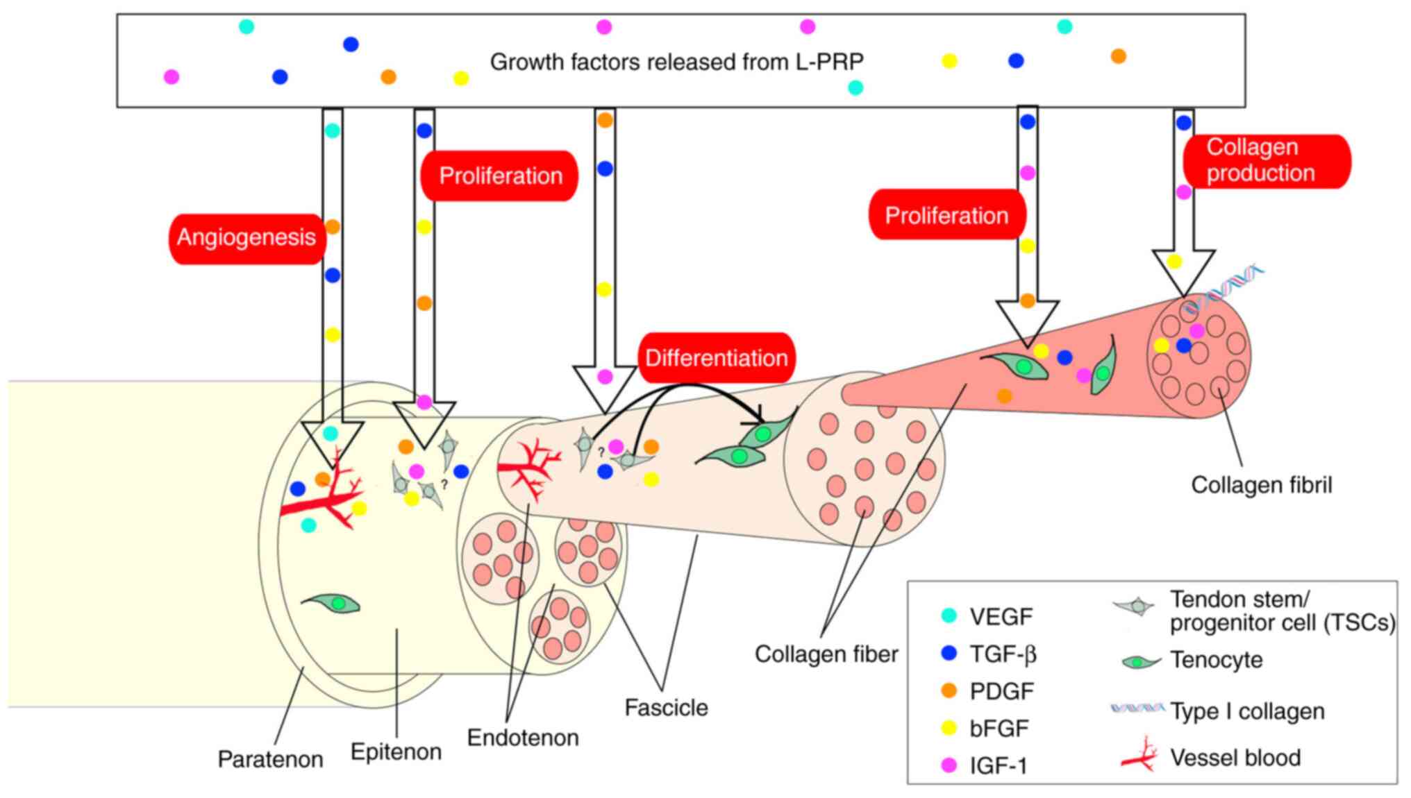 | Figure 1Key molecular, cellular and collagen
changes in tendon caused by growth factors released from L-PRP at
the early stage. L-PRP promotes angiogenesis, tendon cell
proliferation (both TSCs and tenocytes) and differentiation of TSCs
into tenocytes, as well as type I collagen production. The exact
location of TSCs is still debated (therefore indicated with ‘?’).
L-PRP, leukocyte- and platelet-rich plasma; VEGF, vascular
endothelial growth factor; PDGF, platelet-derived growth factor;
TSCs, tendon stem/progenitor cells; TGF-β, transforming growth
factor beta; IGF-1, insulin-like growth factor-1; bFGF, basic
fibroblast growth factor. |
Angiogenesis
A number of in vitro and animal studies have
reported that administration of L-PRP significantly increased
angiogenesis during the early phase of the tendon repair process
(19-24).
It has been suggested that poor vascularity is a major factor
limiting tendon healing capacity (25).
L-PRP contains higher concentrations of growth
factors involved in neovascularization compared with whole blood or
platelet-poor plasma, such as VEGF (6,20,26-29)
and PDGF (6,20,28,29),
which together promote vessel wall permeability, as well as the
growth and proliferation of vascular endothelial cells. Increased
growth factor concentration and expression may be an intrinsic
mechanism involved in inducing angiogenesis as part of the tissue
repair process. Following angiogenesis, the resumption of blood
flow promotes the recruitment of reparative cells from the
peripheral blood and bone marrow, so that the intrinsic tenocytes
and fibroblasts of surrounding tissues migrate to the injured site
and begin to proliferate and synthesize collagen (30).
Therefore, one of the mechanisms through which L-PRP
accelerates tendon healing is the enhancement of blood supply to
the injured soft tissue. The neovascularization proceeds along the
surface of the epitenon, passing through normal vascular areas and
providing the injured area with extrinsic cells, nutrients and
growth factors (7). This effect is
mediated by angiogenic growth factors, such as VEGF and PDGF, which
are contained in L-PRP. Furthermore, Kobayashi et al
(31) reported that the leukocyte
concentration in L-PRP was positively correlated with the
concentrations of PDGF and VEGF, while it was negatively correlated
with the concentration of bFGF (32). In conclusion, L-PRP accelerates the
angiogenic process and, subsequently, leads to the acceleration of
the tendon healing process.
Cell proliferation and
differentiation
There are two cell types in tendons: Tenocytes,
which are the predominant cell type, and the tendon stem/progenitor
cells (TSCs), which represent a small proportion (<5%) of the
total tendon cells. Although certain cell culture studies have
indicated that PRP treatment increases tenocyte proliferation
(26,27), the rate of tenocyte proliferation is
limited after tendon injury. On the contrary, TSCs have a high
proliferation rate, which may be further enhanced by PRP (33,34).
Also, similar to other adult stem cells, TSCs are able to
self-renew and differentiate into tenocytes that are responsible
for the maintenance and repair of the tendons (34,35).
Regarding the effect of L-PRP on tendon cell
proliferation and differentiation, studies have indicated that it
may be attributed to the high amount of growth factors in L-PRP,
including PDGF (6,20,28,29),
TGF-β (6,20,31,36)
and IGF-1(32). Those previous
in vitro or animal studies demonstrated that these growth
factors are present at high concentrations in L-PRP and have an
important role in cell proliferation and tenocyte differentiation
of TSCs (Table I). L-PRP may
modulate STAT3 and p27 expression to upregulate the expression of
cyclins and cyclin-dependent kinases to increase the proliferation
of tendon cells (37). Furthermore,
according to Zhou et al (35), L-PRP treatment induced significantly
higher proliferation and differentiation of tenocytes compared with
P-PRP in a cell culture experiment. However, Zhang et al
(28) reported opposite results,
namely that L-PRP exerts a harmful effect on TSCs, inhibiting their
proliferation, accelerating non-tenocyte differentiation and
inducing their apoptosis. Of note, TSCs in that study were isolated
from young healthy rabbits, which are different from the cells of
older human adults with chronic tendinopathy. A stricter
experimental design may be required to obtain evidence on this in
the future and research should particularly focus on whether the
TSCs were derived from healthy or injured tendons, and whether
tendon injury was acute or chronic.
Effect on metabolism
L-PRP has been indicated to affect the metabolism of
tendon cells involved in the wound healing process in in
vitro and animal studies (20,23,30,35,38).
In the proliferation phase, tendon fibroblasts promote the
synthesis of abundant type III collagen, granulation tissue and
other extracellular matrix (ECM) components (such as proteoglycans)
and deposit them to the wound site. In fact, normal tendon consists
of type I and type III collagen, with type I collagen being the
major component that is responsible for tensile strength (1). When tendons are injured, type I
collagen is downregulated and type III collagen synthesis is
intensified, and its synthesis occurs earlier in the progress of
tendon healing compared with that of type I collagen (39). PRP is known to enhance type I
collagen synthesis instead of type III collagen in order to
accelerate tendon healing (40).
However, leukocyte inclusion in L-PRP has been controversial, as
certain studies indicated a beneficial effect and others reported a
deleterious effect on tissue regeneration, as leukocytes may cause
more inflammation (28).
Certain studies have suggested that L-PRP exerts
both catabolic and anabolic effects (19,41),
whereas P-PRP exerts mainly anabolic effects on injured tissues.
Regarding the anabolic effects of L-PRP, the collagen fiber
arrangement time is shortened, which is caused by the direct
stimulation of local stem cells and ECM genes to accelerate
collagen synthesis via the profibrotic growth factors, such as
TGF-β and bFGF contained in the L-PRP (20,22,26).
Among these, TGF-β has been indicated to act in almost all phases
of tendon healing and serves as a stimulator of extrinsic cell
migration, regulation of proteinases and collagen production
(7).
With regard to the catabolic effects, the increased
numbers of leukocytes in L-PRP may stimulate fibroblasts to release
catabolic cytokines, such as interleukin-1β (IL-1β), tumor necrosis
factor-α (TNF-α) and matrix metalloproteinases (MMPs), which are
capable of degrading the ECM of various tissue types (26,30,35).
Regarding catabolic factors, the levels of MMP-1, MMP-3 and MMP-9
were indicated to be strongly correlated with the leukocyte
concentration (26,31,35)
and they may accelerate the proteolysis of the ECM through invading
blood vessel endothelial cells, resulting in impaired mechanical
stability. Based on the catabolic effect of leukocytes in PRP, it
may be inferred that an increased platelet concentration in L-PRP
would have a positive effect by suppressing the expression of
catabolic cytokines; however, it was reported that increasing the
platelet/leukocyte concentration ratio in L-PRP was not beneficial
(42). In addition, based on the
current knowledge, it is difficult to determine whether increased
gene expression levels of MMP1 and MMP3 may be beneficial for the
healing of degenerative or ruptured tendons (26).
Catabolism and anabolism are balanced in normal
tendons; however, this balance is disrupted in injured tendons.
Whether catabolism or anabolism prevails depends on the different
types of tendon injury and healing stage (23,30).
In a chronic rabbit Achilles tendinopathy model at 4 weeks after
collagenase induction, Yan et al (30) observed that both P-PRP and L-PRP
promoted the formation of larger collagen fibrils, but P-PRP
exerted a stronger effect than L-PRP. However, in the early model
at 1 week after collagenase induction, Li et al (23) detected higher collagen I and lower
collagen III content following L-PRP injection compared with that
following P-PRP injection.
In fact, the current opinion is that the effect of
L-PRP on metabolism is closely associated with the timing and the
phase of tendon healing. Application of L-PRP at the early stage of
tendon disorders or acute injury causes higher expression of
collagen I, while its delivery at a later stage results in higher
expression of collagen III (20,23,30,38).
In other words, early delivery of L-PRP promotes matrix maturation,
while late delivery impairs the matrix modeling process in tendon
repair.
Inflammatory response
Tissue healing is a process of inflammation
requiring leukocytes, which are contained in L-PRP. In the
inflammatory phase, inflammatory cells, including neutrophils,
monocytes and lymphocytes, migrate from surrounding tissues to the
wound site. Tendon healing begins with local hemostasis, followed
by the migration of neutrophils and phagocytes to the lesion site
to clear foreign bodies and necrotic tissues (43). Tissue macrophages are known to
become activated and polarized into M1 and M2 phenotypes under the
influence of the surrounding matrix and environmental factors; in
this paradigm, M1 macrophages predominate early and have a
proinflammatory function via the release of IL-1β, TNF-α and IL-6,
while M2 macrophages accumulate later and serve an
anti-inflammatory role via the release of IL-10 and TGF-β1(44). Macrophages have key roles in
promoting inflammation at the early stage and resolving
inflammation at the late stage of tendon healing.
The inclusion of leukocytes in L-PRP remain
controversial (14). Regarding the
positive effects, inflammation causes release of growth factors and
cytokines, which induce neovascularization and chemotaxis of
fibroblasts and stimulate collagen synthesis. Furthermore,
leukocytes in PRP recruit more macrophages through angiogenesis
during the early phase of tendon repair (38). Although leukocytes have key roles in
tissue repair and provide desirable protection against infectious
agents, their proinflammatory and immunological effects may also
induce undesirable local cell and tissue damage that compromises
the intended healing effect. With regard to the negative effects,
leukocytes are considered to be the major source of
pro-inflammatory cytokines (such as IL-1β, TNF-α and IL-6) and
catabolic enzymes, which may cause injury of therapeutic tissue
(45).
However, it is well known that controlled
inflammation has certain advantages in tissue repair, while
excessive or persistent inflammation may be harmful (46). Therefore, whether inflammation is
beneficial for tendon healing depends on the different stages of
tendon disorders. A previous study demonstrated that delivery of
L-PRP at the early rather than the late stage promoted the repair
of Achilles tendinopathy in rabbits and suggested that L-PRP may
alleviate inflammation at the early stage, whereas L-PRP exerted a
less prominent beneficial effect at the late stage of tendinopathy
(23). This is consistent with the
findings of Zhang et al (28), Zhou et al (35) and Jiang et al (38).
For the treatment of acute tendon injuries,
leukocytes in L-PRP may be helpful when inflammation is at a
critical stage by inducing a catabolic response, which may clear
foreign bodies and necrotic tissues by leukocyte recruitment to the
lesion site and by fighting off infectious agents. However, the
prolonged duration of infiltration by excessive numbers of
neutrophils may impair the healing process as chronic disease
replaces acute inflammation. Administration of L-PRP at the later
stage would lead to local inflammatory edema after tendon injury,
with scar tissue formation and angiofibroblastic dysfunction in
chronic tendinopathy homeostasis (30).
3. Effects of L-PRP on tendon healing based
on clinical studies
General
The clinical manifestations of tendon disease are
characterized by pain and dysfunction. The symptoms are well
defined and frequently long-lasting, limiting the patients'
functioning regarding physical exercise and daily activities.
Pain
L-PRP has been indicated to decrease the pain of
patients in the clinical setting (25,36,47-51).
According to the classic model, inflammation is responsible for the
pain associated with tendon disorders. However, chronically painful
tendons may exhibit no evidence of inflammation and there is no
associated pain in several intratendinous lesions detected on
magnetic resonance imaging (MRI) or ultrasound. It has been
proposed that pain may be caused by a combination of mechanical and
biochemical factors (52). Tendon
degeneration with mechanical breakdown of collagen may
theoretically cause pain, whereas chemical irritants and
neurotransmitters may also generate pain in tendon disorders.
In one of the largest studies, Mishra et al
(49) evaluated 230 patients with
lateral epicondylitis who failed to respond to conservative
treatment for at least 3 months. Patients treated with L-PRP
exhibited a significant improvement in pain compared with the
bupivacaine control at 24 weeks. More importantly, in a recent
study (36), Pearson's correlation
analysis was performed between the clinical score and the
biological components and the Pearson correlation coefficient was
calculated between the Visual Analogue Scale score and the
leukocyte concentration in L-PRP, demonstrating that L-PRP was
highly associated with pain relief in patients suffering from
tendon disorders. Of note, in the aforementioned studies, it was
observed that pain relief was first observed after 8 weeks after
L-PRP was applied to patients with chronic tendon injury for >3
months, while pain relief was first observed earlier (seven days
later) in patients with acute tendon injury treated with L-PRP
(25,36,47-51).
However, more research is required to explain this phenomenon.
Function
Certain studies have confirmed that L-PRP may
improve tendon function in terms of the range of joint movement
(ROM) and the ability to perform physical activities (36,48,53-55).
Lim et al (36) indicated
that L-PRP enhanced the Modified Mayo Clinic performance scores and
MRI grade in patients suffering from lateral epicondylitis for
>3 months. The study also used Pearson's correlation coefficient
to demonstrate the association between the scores and the growth
factor levels in PRP, indicating that the TGF-β level in L-PRP was
highly correlated with the Modified Mayo Clinic performance scores
and MRI grade improvement, whereas the VEGF level in L-PRP was
highly correlated with MRI grade improvement. Fitzpatrick et
al (53) reported that L-PRP
improved the modified Harris Hip Scores in patients suffering from
gluteus medius and minimus tendinopathy for >4 months. The
constant score and Disabilities of the Arm, Shoulder and Hand score
were tested in patients with rotator cuff tears/lateral
epicondylitis who were treated with L-PRP, with increasing scores
reflecting less pain, as well as improved ROM and ability to
perform physical activities (48,54).
Charousset et al (55) also
reported that, although there was no significant difference between
the L-PRP group and the blank group, the L-PRP group had smaller
iterative tears. Collectively, these results indicated that L-PRP
may promote the recovery of normal function following tendon
injury, as well as the restoration of mechanical properties.
4. Conclusion and perspectives
Tendon disorders are a frequently encountered
clinical problem that commonly affects athletes and middle-aged
patients who do not exercise much, accounting for 30-50% of all
sports-related injuries (56). With
the current widespread research indicating that PRP contains a
large number of cytokines and growth factors required for tendon
healing, PRP therapy has gradually gained popularity in the
clinical field (57).
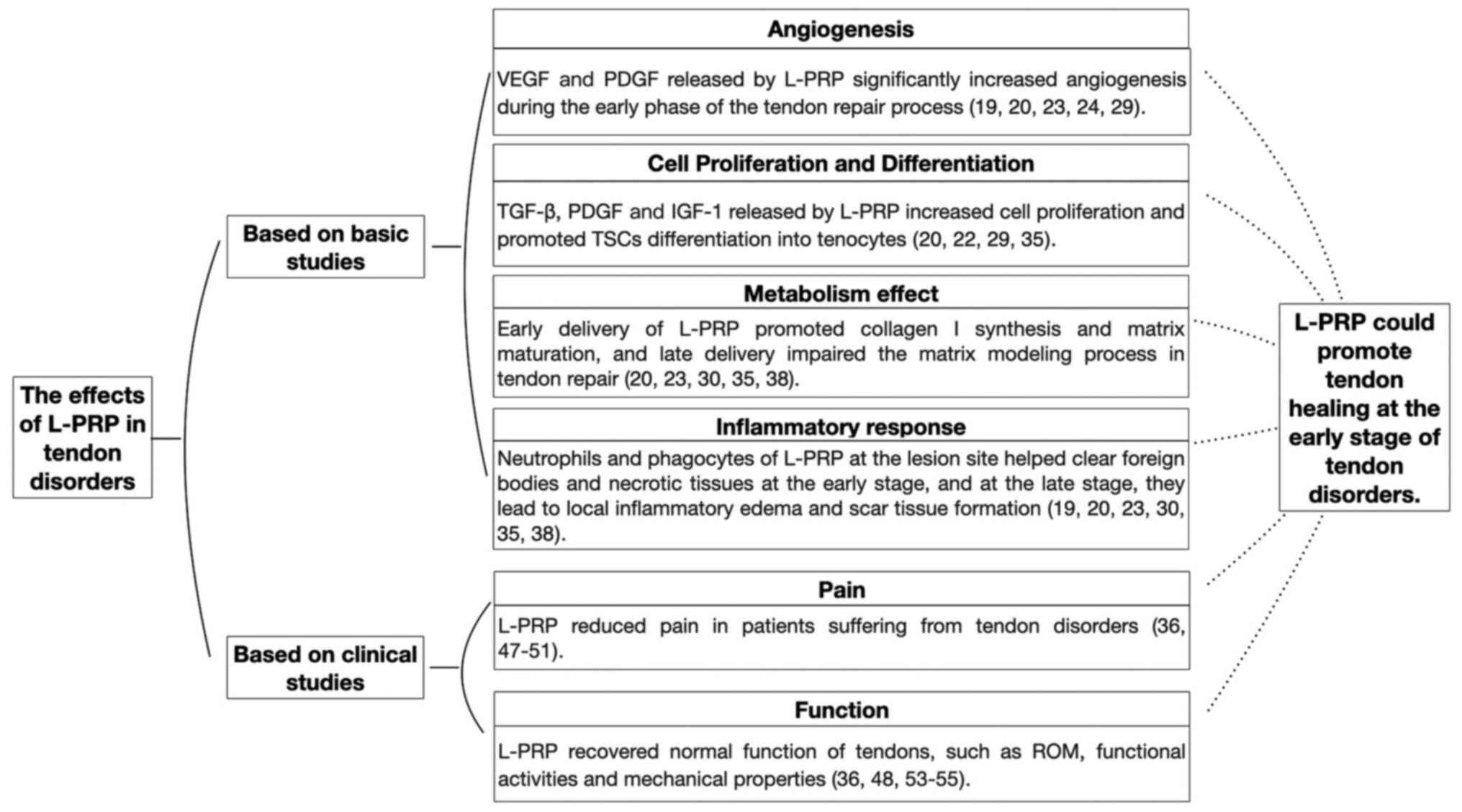
The main effects of L-PRP in tendon disorders
according to basic and clinical studies are summarized in Fig. 2. These clinical studies have
demonstrated that L-PRP is not only highly associated with pain
relief in patients suffering from tendon disorders, but may also
help the injured tendon to recover its normal functions, including
the ROM, functional activities and mechanical properties (36,47-51,53-55).
Based on basic studies, the underlying mechanisms
for the effects of L-PRP likely involve promoting angiogenesis,
cell proliferation and differentiation during the process of tendon
healing. Growth factors, such as VEGF and PDGF, released from
L-PRP, significantly increase angiogenesis during the early phase
of the tendon repair process (19,20,23,24,29).
Tenocyte proliferation and differentiation of TSCs into tenocytes
are crucial for tendon healing and a number of studies have
indicated that TGF-β, PDGF and IGF released by L-PRP accelerate the
process (20,22,29,35).
Furthermore, regarding metabolism and the inflammatory response,
early delivery of L-PRP induced additional inflammation and further
release of growth factors, which help clear foreign bodies and
necrotic tissues by neutrophils and phagocytes at the lesion site
at the early stage and accelerate the synthesis of the ECM by
transitioning from type III collagen fibers to type I collagen
fibers (19,20,23,30,35,38).
In addition, L-PRP may enhance antimicrobial functions and increase
the concentration of growth factors in platelet concentrate
(58).
From the current available literatures, leukocytes
in L-PRP are likely beneficial, but their effects depend on the
biological state of the injured tissue and its surrounding
microenvironment (38). The timing
of the L-PRP injection also appears to be important. The conclusion
of the present review is that L-PRP exerts beneficial effects by
promoting tendon healing at the early stage, whereas it is likely
to be detrimental to the tendon at a later stage due to the risk of
inducing excessive catabolic and inflammatory responses. Of note,
in the studies included, the effect of L-PRP depended on the
preparation method, the kit used to prepare L-PRP and the content
of leukocytes, as determined by cytology.
In the future, more basic and clinical research
should be performed to verify the effect of L-PRP and to clearly
determine the conditions under which L-PRP is beneficial for tendon
injury, with factors to be considered being the tendon disorder
type, stage of tendon healing and inflammation status. In addition,
the composition and contents of PRP preparations should be
accurately measured in future studies, as the majority of previous
clinical studies prepared PRP using different commercial kits and
the final product varies in terms of platelet and leukocyte
content. Furthermore, the immunomodulatory and metabolic effects of
all subpopulations of leukocytes included in the preparations
should be explored. Such efforts may help determine the optimal
composition of PRP preparations and improve the efficacy of PRP in
the treatment of tendon disorders.
Acknowledgements
Not applicable.
Funding
Funding: No funding was received.
Availability of data and materials
Not applicable.
Authors' contributions
XLL, RZ and XYL designed the present article. XLL
and RZ drawn the manuscript. BZ, YJL, SG and CLW a literature
search and selected the studies to be performed XLL, DXW and SL
revised including the manuscript. Data authentication is not
applicable. All authors approved the final version of the
article.
Ethics approval and consent to
participate
Not applicable.
Patient consent for publication
Not applicable.
Competing interests
The authors declare that they have no competing
interests.
References
|
1
|
Wang JH: Mechanobiology of tendon. J
Biomech. 39:1563–1582. 2006.PubMed/NCBI View Article : Google Scholar
|
|
2
|
Clayton RA and Court-Brown CM: The
epidemiology of musculoskeletal tendinous and ligamentous injuries.
Injury. 39:1338–1344. 2008.PubMed/NCBI View Article : Google Scholar
|
|
3
|
Nourissat G, Berenbaum F and Duprez D:
Tendon injury: From biology to tendon repair. Nat Rev Rheumatol.
11:223–233. 2015.PubMed/NCBI View Article : Google Scholar
|
|
4
|
Costa-Almeida R, Babo PS, Reis RL and
Gomes ME: Platelet-rich blood derivatives for tendon regeneration.
J Am Acad Orthop Surg. 28:e202–e205. 2020.PubMed/NCBI View Article : Google Scholar
|
|
5
|
Zhou Y and Wang JH: PRP treatment efficacy
for tendinopathy: A review of basic science studies. Biomed Res
Int. 2016(9103792)2016.PubMed/NCBI View Article : Google Scholar
|
|
6
|
Eppley BL, Woodell JE and Higgins J:
Platelet quantification and growth factor analysis from
platelet-rich plasma: Implications for wound healing. Plast
Reconstr Surg. 114:1502–1508. 2004.PubMed/NCBI View Article : Google Scholar
|
|
7
|
Molloy T, Wang Y and Murrell G: The roles
of growth factors in tendon and ligament healing. Sports Med.
33:381–394. 2003.PubMed/NCBI View Article : Google Scholar
|
|
8
|
Wu PI, Diaz R and Borg-Stein J:
Platelet-rich plasma. Phys Med Rehabil Clin N Am. 27:825–853.
2016.PubMed/NCBI View Article : Google Scholar
|
|
9
|
Jeong DU, Lee CR, Lee JH, Pak J, Kang LW,
Jeong BC and Lee SH: Clinical applications of platelet-rich plasma
in patellar tendinopathy. Biomed Res Int.
2014(249498)2014.PubMed/NCBI View Article : Google Scholar
|
|
10
|
Dohan Ehrenfest DM, Rasmusson L and
Albrektsson T: Classification of platelet concentrates: From pure
platelet-rich plasma (P-PRP) to leucocyte- and platelet-rich fibrin
(L-PRF). Trends Biotechnol. 27:158–167. 2009.PubMed/NCBI View Article : Google Scholar
|
|
11
|
Krogh TP, Fredberg U, Stengaard-Pedersen
K, Christensen R, Jensen P and Ellingsen T: Treatment of lateral
epicondylitis with platelet-rich plasma, glucocorticoid, or saline:
A randomized, double-blind, placebo-controlled trial. Am J Sports
Med. 41:625–635. 2013.PubMed/NCBI View Article : Google Scholar
|
|
12
|
de Vos RJ, Weir A, van Schie HT,
Bierma-Zeinstra SM, Verhaar JA, Weinans H and Tol JL: Platelet-rich
plasma injection for chronic Achilles tendinopathy: A randomized
controlled trial. JAMA. 303:144–149. 2010.PubMed/NCBI View Article : Google Scholar
|
|
13
|
de Jonge S, de Vos RJ, Weir A, van Schie
HT, Bierma-Zeinstra SM, Verhaar JA, Weinans H and Tol JL: One-year
follow-up of platelet-rich plasma treatment in chronic Achilles
tendinopathy: A double-blind randomized placebo-controlled trial.
Am J Sports Med. 39:1623–1629. 2011.PubMed/NCBI View Article : Google Scholar
|
|
14
|
Castillo TN, Pouliot MA, Kim HJ and Dragoo
JL: Comparison of growth factor and platelet concentration from
commercial platelet-rich plasma separation systems. Am J Sports
Med. 39:266–271. 2011.PubMed/NCBI View Article : Google Scholar
|
|
15
|
Baksh N, Hannon CP, Murawski CD, Smyth NA
and Kennedy JG: Platelet-rich plasma in tendon models: A systematic
review of basic science literature. Arthroscopy. 29:596–607.
2013.PubMed/NCBI View Article : Google Scholar
|
|
16
|
Chen X, Jones IA, Park C and Vangsness CT
Jr: The efficacy of platelet-rich plasma on tendon and ligament
healing: A systematic review and meta-analysis with bias
assessment. Am J Sports Med. 46:2020–2032. 2018.PubMed/NCBI View Article : Google Scholar
|
|
17
|
Fitzpatrick J, Bulsara M and Zheng MH: The
Effectiveness of Platelet-rich plasma in the treatment of
tendinopathy: A Meta-analysis of randomized controlled clinical
trials. Am J Sports Med. 45:226–233. 2017.PubMed/NCBI View Article : Google Scholar
|
|
18
|
Hope M and Saxby TS: Tendon healing. Foot
Ankle Clin. 12:553–567. 2007.PubMed/NCBI View Article : Google Scholar
|
|
19
|
Dragoo JL, Braun HJ, Durham JL, Ridley BA,
Odegaard JI, Luong R and Arnoczky SP: Comparison of the acute
inflammatory response of two commercial platelet-rich plasma
systems in healthy rabbit tendons. Am J Sports Med. 40:1274–1281.
2012.PubMed/NCBI View Article : Google Scholar
|
|
20
|
Kobayashi Y, Saita Y, Takaku T, Yokomizo
T, Nishio H, Ikeda H, Takazawa Y, Nagao M, Kaneko K and Komatsu N:
Platelet-rich plasma (PRP) accelerates murine patellar tendon
healing through enhancement of angiogenesis and collagen synthesis.
J Exp Orthop. 7(49)2020.PubMed/NCBI View Article : Google Scholar
|
|
21
|
Lyras DN, Kazakos K, Agrogiannis G,
Verettas D, Kokka A, Kiziridis G, Chronopoulos E and Tryfonidis M:
Experimental study of tendon healing early phase: Is IGF-1
expression influenced by platelet rich plasma gel? Orthop Traumatol
Surg Res. 96:381–387. 2010.PubMed/NCBI View Article : Google Scholar
|
|
22
|
Lyras DN, Kazakos K, Tryfonidis M,
Agrogiannis G, Botaitis S, Kokka A, Drosos G, Tilkeridis K and
Verettas D: Temporal and spatial expression of TGF-beta1 in an
Achilles tendon section model after application of platelet-rich
plasma. Foot Ankle Surg. 16:137–141. 2010.PubMed/NCBI View Article : Google Scholar
|
|
23
|
Li S, Wu Y, Jiang G, Tian X, Hong J, Chen
S, Yan R, Feng G and Cheng Z: Intratendon delivery of
leukocyte-rich platelet-rich plasma at early stage promotes tendon
repair in a rabbit Achilles tendinopathy model. J Tissue Eng Regen
Med. 14:452–463. 2020.PubMed/NCBI View Article : Google Scholar
|
|
24
|
Nishio H, Saita Y, Kobayashi Y, Takaku T,
Fukusato S, Uchino S, Wakayama T, Ikeda H and Kaneko K:
Platelet-rich plasma promotes recruitment of macrophages in the
process of tendon healing. Regen Ther. 14:262–270. 2020.PubMed/NCBI View Article : Google Scholar
|
|
25
|
Sharma P and Maffulli N: Tendon injury and
tendinopathy: Healing and repair. J Bone Joint Surg Am. 87:187–202.
2005.PubMed/NCBI View Article : Google Scholar
|
|
26
|
de Mos M, van der Windt AE, Jahr H, van
Schie HT, Weinans H, Verhaar JA and van Osch GJ: Can platelet-rich
plasma enhance tendon repair? A cell culture study. Am J Sports
Med. 36:1171–1178. 2008.PubMed/NCBI View Article : Google Scholar
|
|
27
|
Mazzocca AD, McCarthy MB, Chowaniec DM,
Dugdale EM, Hansen D, Cote MP, Bradley JP, Romeo AA, Arciero RA and
Beitzel K: The positive effects of different platelet-rich plasma
methods on human muscle, bone, and tendon cells. Am J Sports Med.
40:1742–1749. 2012.PubMed/NCBI View Article : Google Scholar
|
|
28
|
Zhang L, Chen S, Chang P, Bao N, Yang C,
Ti Y, Zhou L and Zhao J: Harmful effects of Leukocyte-rich
platelet-rich plasma on rabbit tendon stem cells in vitro. Am J
Sports Med. 44:1941–1951. 2016.PubMed/NCBI View Article : Google Scholar
|
|
29
|
Lyras DN, Kazakos K, Verettas D, Botaitis
S, Agrogiannis G, Kokka A, Pitiakoudis M and Kotzakaris A: The
effect of platelet-rich plasma gel in the early phase of patellar
tendon healing. Arch Orthop Trauma Surg. 129:1577–1582.
2009.PubMed/NCBI View Article : Google Scholar
|
|
30
|
Yan R, Gu Y, Ran J, Hu Y, Zheng Z, Zeng M,
Heng BC, Chen X, Yin Z, Chen W, et al: Intratendon delivery of
leukocyte-poor platelet-rich plasma improves healing compared with
leukocyte-rich platelet-rich plasma in a rabbit Achilles
tendinopathy model. Am J Sports Med. 45:1909–1920. 2017.PubMed/NCBI View Article : Google Scholar
|
|
31
|
Kobayashi Y, Saita Y, Nishio H, Ikeda H,
Takazawa Y, Nagao M, Takaku T, Komatsu N and Kaneko K: Leukocyte
concentration and composition in platelet-rich plasma (PRP)
influences the growth factor and protease concentrations. J Orthop
Sci. 21:683–689. 2016.PubMed/NCBI View Article : Google Scholar
|
|
32
|
Denapoli PM, Stilhano RS, Ingham SJ, Han
SW and Abdalla RJ: Platelet-rich plasma in a murine model:
Leukocytes, growth factors, Flt-1, and muscle healing. Am J Sports
Med. 44:1962–1971. 2016.PubMed/NCBI View Article : Google Scholar
|
|
33
|
Zhang J and Wang JH: Characterization of
differential properties of rabbit tendon stem cells and tenocytes.
BMC Musculoskelet Disord. 11(10)2010.PubMed/NCBI View Article : Google Scholar
|
|
34
|
Zhang J and Wang JH: Platelet-rich plasma
releasate promotes differentiation of tendon stem cells into active
tenocytes. Am J Sports Med. 38:2477–2486. 2010.PubMed/NCBI View Article : Google Scholar
|
|
35
|
Zhou Y, Zhang J, Wu H, Hogan MV and Wang
JH: The differential effects of leukocyte-containing and pure
platelet-rich plasma (PRP) on tendon stem/progenitor
cells-implications of PRP application for the clinical treatment of
tendon injuries. Stem Cell Res Ther. 6(173)2015.PubMed/NCBI View Article : Google Scholar
|
|
36
|
Lim W, Park SH, Kim B, Kang SW, Lee JW and
Moon YL: Relationship of cytokine levels and clinical effect on
platelet-rich plasma-treated lateral epicondylitis. J Orthop Res.
36:913–920. 2018.PubMed/NCBI View Article : Google Scholar
|
|
37
|
Yu TY, Pang JH, Wu KP, Lin LP, Tseng WC
and Tsai WC: Platelet-rich plasma increases proliferation of tendon
cells by modulating Stat3 and p27 to up-regulate expression of
cyclins and cyclin-dependent kinases. Cell Prolif. 48:413–420.
2015.PubMed/NCBI View Article : Google Scholar
|
|
38
|
Jiang G, Wu Y, Meng J, Wu F, Li S, Lin M,
Gao X, Hong J, Chen W, Yan S, et al: Comparison of leukocyte-rich
platelet-rich plasma and leukocyte-poor platelet-rich plasma on
achilles tendinopathy at an early stage in a rabbit model. Am J
Sports Med. 48:1189–1199. 2020.PubMed/NCBI View Article : Google Scholar
|
|
39
|
Gaut L and Duprez D: Tendon development
and diseases. Wiley Interdiscip Rev Dev Biol. 5:5–23.
2016.PubMed/NCBI View Article : Google Scholar
|
|
40
|
Alsousou J, Thompson M, Harrison P,
Willett K and Franklin S: Effect of platelet-rich plasma on healing
tissues in acute ruptured Achilles tendon: A human
immunohistochemistry study. Lancet. 385 (Suppl
1)(S19)2015.PubMed/NCBI View Article : Google Scholar
|
|
41
|
Filardo G, Kon E, Pereira Ruiz MT, Vaccaro
F, Guitaldi R, Di Martino A, Cenacchi A, Fornasari PM and Marcacci
M: Platelet-rich plasma intra-articular injections for cartilage
degeneration and osteoarthritis: Single-versus double-spinning
approach. Knee Surg Sports Traumatol Arthrosc. 20:2082–2091.
2012.PubMed/NCBI View Article : Google Scholar
|
|
42
|
McCarrel TM, Minas T and Fortier LA:
Optimization of leukocyte concentration in platelet-rich plasma for
the treatment of tendinopathy. J Bone Joint Surg Am.
94(e143)2012.PubMed/NCBI View Article : Google Scholar
|
|
43
|
Butler DL, Juncosa N and Dressler MR:
Functional efficacy of tendon repair processes. Annu Rev Biomed
Eng. 6:303–329. 2004.PubMed/NCBI View Article : Google Scholar
|
|
44
|
Stolk M, Klatte-Schulz F, Schmock A,
Minkwitz S, Wildemann B and Seifert M: New insights into
tenocyte-immune cell interplay in an in vitro model of
inflammation. Sci Rep. 7(9801)2017.PubMed/NCBI View Article : Google Scholar
|
|
45
|
Sunwoo JY, Eliasberg CD, Carballo CB and
Rodeo SA: The role of the macrophage in tendinopathy and tendon
healing. J Orthop Res. 38:1666–1675. 2020.PubMed/NCBI View Article : Google Scholar
|
|
46
|
Dakin SG, Dudhia J and Smith RK: Resolving
an inflammatory concept: The importance of inflammation and
resolution in tendinopathy. Vet Immunol Immunopathol. 158:121–127.
2014.PubMed/NCBI View Article : Google Scholar
|
|
47
|
D'Ambrosi R, Palumbo F, Paronzini A,
Ragone V and Facchini RM: Platelet-rich plasma supplementation in
arthroscopic repair of full-thickness rotator cuff tears: A
randomized clinical trial. Musculoskelet Surg. 100:25–32.
2016.PubMed/NCBI View Article : Google Scholar
|
|
48
|
Zhang Z, Wang Y and Sun J: The effect of
platelet-rich plasma on arthroscopic double-row rotator cuff
repair: A clinical study with 12-month follow-up. Acta Orthop
Traumatol Turc. 50:191–197. 2016.PubMed/NCBI View Article : Google Scholar
|
|
49
|
Mishra AK, Skrepnik NV, Edwards SG, Jones
GL, Sampson S, Vermillion DA, Ramsey ML, Karli DC and Rettig AC:
Efficacy of platelet-rich plasma for chronic tennis elbow: A
double-blind, prospective, multicenter, randomized controlled trial
of 230 patients. Am J Sports Med. 42:463–471. 2014.PubMed/NCBI View Article : Google Scholar
|
|
50
|
Mishra A and Pavelko T: Treatment of
chronic elbow tendinosis with buffered platelet-rich plasma. Am J
Sports Med. 34:1774–1778. 2006.PubMed/NCBI View Article : Google Scholar
|
|
51
|
Thanasas C, Papadimitriou G, Charalambidis
C, Paraskevopoulos I and Papanikolaou A: Platelet-rich plasma
versus autologous whole blood for the treatment of chronic lateral
elbow epicondylitis: A randomized controlled clinical trial. Am J
Sports Med. 39:2130–2134. 2011.PubMed/NCBI View Article : Google Scholar
|
|
52
|
Khan KM, Cook JL, Bonar F, Harcourt P and
Astrom M: Histopathology of common tendinopathies: Update and
implications for clinical management. Sports Med. 27:393–408.
1999.PubMed/NCBI View Article : Google Scholar
|
|
53
|
Fitzpatrick J, Bulsara MK, O'Donnell J and
Zheng MH: Leucocyte-rich platelet-rich plasma treatment of gluteus
medius and minimus tendinopathy: A double-blind randomized
controlled trial with 2-year follow-up. Am J Sports Med.
47:1130–1137. 2019.PubMed/NCBI View Article : Google Scholar
|
|
54
|
Gosens T, Peerbooms JC, van Laar W and den
Oudsten BL: Ongoing positive effect of platelet-rich plasma versus
corticosteroid injection in lateral epicondylitis: A double-blind
randomized controlled trial with 2-year follow-up. Am J Sports Med.
39:1200–1208. 2011.PubMed/NCBI View Article : Google Scholar
|
|
55
|
Charousset C, Zaoui A, Bellaiche L and
Piterman M: Does autologous leukocyte-platelet-rich plasma improve
tendon healing in arthroscopic repair of large or massive rotator
cuff tears? Arthroscopy. 30:428–435. 2014.PubMed/NCBI View Article : Google Scholar
|
|
56
|
Jarvinen TA, Kannus P, Maffulli N and Khan
KM: Achilles tendon disorders: Etiology and epidemiology. Foot
Ankle Clin. 10:255–266. 2005.PubMed/NCBI View Article : Google Scholar
|
|
57
|
Wasterlain AS, Braun HJ, Harris AH, Kim HJ
and Dragoo JL: The systemic effects of platelet-rich plasma
injection. Am J Sports Med. 41:186–193. 2013.PubMed/NCBI View Article : Google Scholar
|
|
58
|
Moojen DJ, Everts PA, Schure RM,
Overdevest EP, van Zundert A, Knape JT, Castelein RM, Creemers LB
and Dhert WJ: Antimicrobial activity of platelet-leukocyte gel
against Staphylococcus aureus. J Orthop Res. 26:404–410.
2008.PubMed/NCBI View Article : Google Scholar
|