Introduction
Heat shock proteins (HSPs) are encoded by a large
family of genes (1-3).
The essential function of these proteins is to serve as molecular
chaperones that maintain numerous cellular proteins in their normal
three-dimensional structures, in order to continue performing their
normal functions when the ambient temperature is increased
(1-3).
However, the majority of heat shock proteins also have numerous
other functions and are often overexpressed in various situations
of cellular stress (1,2,4-7).
For example, when normal cells evolve into cancer cells, the
expression of HSPs is often increased, which is usually a mechanism
by which cancer cells become resistant to various therapies
(5,7).
It is well known that the majority of genes in the
human, mouse or rat genome can be expressed as multiple protein
isoforms to suit a variety of developmental, physiological or
pathological situations (8-11).
As we have previously described (10-14),
multiple mechanisms for the protein multiplicity exist, including
alternative use of transcription start sites to yield different RNA
transcripts, alternative use of splicing sites of the same RNA
transcript to produce different mRNA variants, and alternative use
of different open reading frames (ORFs) of the same mRNA for
protein translation. In our previous studies (12-14),
proteins were fractionated from several human cell lines using
SDS-PAGE, proteins were isolated at certain positions on the gels,
and the isolated proteins were identified using a routine liquid
chromatography with tandem mass spectrometry (LC-MS/MS) approach.
Notably, it was found that proteins of numerous genes were not
supposed to be detected at these positions due to their theoretical
molecular masses being either too large or too small for them to
appear at the SDS-PAGE positions. Furthermore, proteins produced
from the same genes could be simultaneously detected at two or more
SDS-PAGE positions. These data suggested that numerous genes may
have additional protein isoform(s) in addition to the wild-type
(Wt) or the canonical proteins (12-14).
To determine how HSP genes respond to heat stress
in vitro at the RNA level, the RNA expression of eight
arbitrarily selected HSP genes was detected in three human cell
lines cultured at 39˚C for three days. This is a less febrile
temperature, but severe enough for the majority of patients to
decide to see a doctor. As numerous HSP genes are transcriptionally
activated by the heat shock transcription factor 1 (HSF1) protein
(4,15), the RNA expression of the HSF1 gene
was also studied. As some of these genes have multiple RNA variants
listed in the National Center for Biotechnology Information (NCBI)
database, certain variants other than the Wt or the canonical form
were also studied. The results demonstrated that the HSF1 and
several HSP genes yielded multiple RNA variants, some of which
demonstrate changes in the expression levels when compared with
their counterparts at 37˚C. A total of four new mRNA variants of
the HSP27-encoding HSPB1 gene that encode three short HSP27
isoforms were identified. Furthermore, reanalysis of certain
previous proteomics data also revealed that proteins of certain HSP
genes may be detected simultaneously at multiple positions on
SDS-PAGE, suggesting that these genes may express multiple protein
isoforms.
Materials and methods
Cell lines and cell culture for RNA
analyses
The human embryonic kidney (HEK) 293 cell line, the
human cervical cancer HeLa cell line and the non-small cell lung
cancer H1650 cell line were obtained from the American Type Culture
Collection (ATCC). The cells were cultured in water-jacket
incubators at 5% CO2 that were recalibrated regularly to
ensure accuracy. All three cell lines were cultured at 37˚C or 39˚C
in a Dulbecco's modified Eagle's medium supplemented with 10% fetal
bovine serum. A thermometer was placed in the incubator to monitor
and guarantee the accuracy and stability of the temperature. After
72 h of culture, the cells reached 70-80% confluence and were
harvested with TRIzol® (Invitrogen; Thermo Fisher
Scientific, Inc.; cat. no. 15596-026) for RNA isolation.
Reverse transcription-polymerase chain
reaction (RT-PCR) assay
Immediately after cells were lysed in TRIzol
reagent, total RNA samples were extracted from the lysates
according to the instructions for the TRIzol reagent. An aliquot of
the RNA was primed with random hexamers or a poly-dT primer during
reverse transcription to complementary DNA (cDNA) using MMLV
Reverse Transcriptase (Promega Corporation; cat. no. M1705;
www.promega.com) according to the reagent manual.
The cDNA was amplified using a PCR, in which the cDNA template was
melted at 95˚C for 4 min, followed by 34-38 cycles of 95, 58 and
72˚C with each step for 20 sec. Each PCR was ended by a final
extension at 72˚C for 5 min. The primers were all designed to be
20-mers with AT to GC ratios around 1:1, which allowed the PCR
conditions to be unified for all target amplicons. However, the
number of PCR cycles needed to be optimized based on the general
expression level of each individual gene, so that the reaction was
terminated within the linear portion of the amplification. Each
primer pair used for PCR was determined in such a way that the two
primers were separated by one or more large introns to avoid
amplification of extant genomic DNA. All the PCR primers used are
listed in Table I. In most cases,
forward (F) primers were named by calculating the distance (number)
from their first nucleotide (nt) to the first nt of the RNA,
whereas reverse (R) primers were named by calculating the number of
nts from their last nt to the first nt of the RNA. This method of
primer naming usually allows for an easy calculation of the PCR
amplicon size by subtracting the F primer number from the R primer
number, but this calculation will be inaccurate in cases where
alternative splicing results in addition or deletion of exon(s).
For semi-quantification of the expression level, cDNA loading was
normalized using the HRPT1 gene.
 | Table IHSF1 and HSP gene primers used. |
Table I
HSF1 and HSP gene primers used.
| Gene | Primer name | Sequence |
Primers/amplicon |
Detecteda |
|---|
| HSF1 | HSF1F574 |
5'-ACAGCGTCACCAAGCTGCTG-3' | F574/R1312=739
bps | NM_005526.2;
XM_005272315.1; XM_005272316.1; |
| | HSF1R1312 |
5'-TTGTCCAGGCAGGCTACGCT-3' | | XR_246618.2;
XM_005272317.1 |
| | HSF1XM17R |
5'-TGGCTGGACTTGGCCATGCG-3' | F574/RXM17R=642
bps | XM_005272317.1 |
| | HSF1R1576 |
5'-GTGTAGTGCACCAGCTGCTT-3' | F574/R1576=824 bps;
1003 bp | XR_246618.2;
NM_005526.2 |
| | HSF1XR26 |
5'-TAGACATCTGTGGAGTGCGA-3' | F574/XR26=586
bps | XM_005272317.1 |
| HSP90AA1 | HSP90F459 |
5'-AGGAAGCCCCTCTGAAGCCT-3' | F459/R1232=774
bps | NM_001017963.2;
XM011536718.2 |
| | HSP90R1232 |
5'-GTCCTCACTGTGAATGATCC-3' | | |
| | HSP90VF97 |
5'-GTCGCTATATAAGGCAGGCG-3' | VF97/R1232=614
bps | NM_005348.3 |
| HSPA1A | A1AF171 |
5'-CTTCTCGCGGATCCAGTGTT-3' | F171/R1104=934
bps | NM_005345.5 |
| | A1AR1104 |
5'-CAGGGAGTCGATCTCCAGGC-3' | | |
| HSPA1B | A1BF146 |
5'-CTTGTCGCGGATCCCGTCCG-3' | F146/R1077=932
bps | NM_005346.4 |
| | A1BR1077 |
5'-CAGGGAGTCGATCTCCAGGC-3' | | |
| HSPA6 | HSP6AF280 |
5'-GTGCGGAAAGGTTCGCGAAA-3' | F280/R1047=768
bps | NM_002155.3 |
| | HSP6AR1047 |
5'-GAACCGACACATCGAAGGTG-3' | | |
| HSPA1L | A1L-F120 |
5'-GCTGCGTAATCTGGACGTTT-3' | F120/R899=780
bps | NM_005527.3;
XM_005249070.2 |
| | A1L-R899 |
5'-AGCCTGTTGTCAAAGTCCTC-3' | | |
| | A1LV-F79 |
5'-GTGCAGTTTGATATTGAGGG-3' | VF79/R899=821
bps | NM_005527.3;
XM_005249070.2 |
| HSPB1 | HSPB1F19 |
5'-CTCAAACGGGTCATTGCCAT-3' | F19/R845=827
bps | NM_001540.3 |
| | HSPB1R845 |
5'-CAAAAGAACACACAGGTGGC-3' | | |
| | HSPB1R313 |
5'-AGTGTGCCGGATCTCCGAGA-3' | used in
5'-RACE | |
| | HSPB1F43 |
5'-AGAGACCTCAAACACCGCCT-3' | used in nested
PCR | new variants |
| | HSPB1F70 |
5'-ATACCCGACTGGAGGAGCAT-3' | | |
| | HSPB1R788 |
5'-ATCCGGGCTAAGGCTTTACT-3' | | |
Nested or semi-nested PCR for
preliminary identification of PCR amplicons
PCR products were routinely fractionated in a 1%
agarose gel that contained 1 µg/ml of ethidium bromide to visualize
DNA in the gel during electrophoresis. For those RNAs that
demonstrated a questionable or additional band in the gel, a
semi-nested or nested PCR was performed to preliminarily determine
whether the band was derived from the target gene. For such a PCR,
the template DNA was obtained from the DNA band using one of the
two methods we described previously (16). In one method, a small blade used for
eye surgery was used to excise a small piece (1 mm3 or
smaller) of the agarose with the DNA, which was put directly into a
20-µl volume of PCR reagents as the template. The DNA would be
released from the gel during the first step of the PCR, which was
an incubation at 95˚C for 4 min to melt the DNA, as the agarose gel
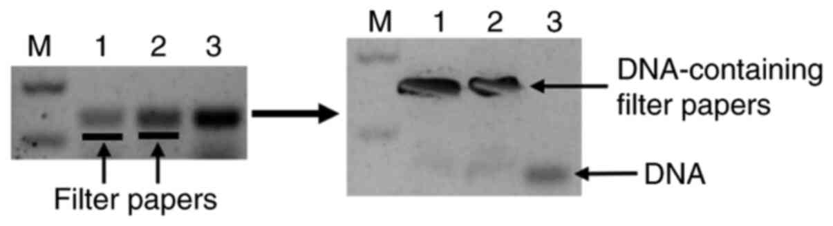
melts at a temperature of approximately 87˚C. In the second method,
a blade was used to make an incision in the gel immediately below
the band in question, followed by insertion of a small piece of
Whatman filter paper into the incision (Fig. 1). Electrophoresis was then continued
for a few more minutes until the DNA had entered into the filter
paper. The paper was then removed and placed into an Eppendorf tube
containing 25-30 µl of a 10-mT TE [Tris(hydroxymethyl)aminomethane
and Ethylene Diamine Tetraacetic Acid] buffer (pH 7.4), followed by
elution of the DNA by vortexing the tube for a short time (16). A semi-nested or nested PCR was
performed using one or two inner primers and 1-2 µl of the eluted
DNA as the template. After the template DNA was melted at 95˚C for
4 min, PCR was allowed for 35 cycles of 95, 58 and 72˚C with each
step for 20 sec, followed by a 5-min final extension at 72˚C, as
its purpose was to preliminarily determine the identity, but not
the expression level, of the template DNA.
T-A cloning, DNA sequencing and
sequence analyses
For direct sequencing of a PCR amplicon, the desired
DNA band in the agarose gel was excised and purified using a simple
method previously described by us, which in principle isolates DNA
from an agarose gel by centrifuging the DNA-containing gel slice at
~15,743.9 x g for 10 min at room temperature (17). Following being isolated from the
gel, the DNA was precipitated with ethanol and then dissolved with
a small volume of water or a 1-mM or 10-mM TE buffer (pH 7.4) to
ensure a high concentration of the DNA, which was sent to Genewiz,
Inc. (www.genewiz.com) for direct sequencing
from both ends using the gene-specific primers used for the PCR. If
the data showed a mixture of different sequences, the purified DNA
would be cloned into a T-A vector and then transfected into
bacteria for amplification, as described in our previous studies
(18-21).
The desired bacterial clones resulting from the T-A cloning were
selected using the ‘dirty plasmid’ method described previously
(22); plasmid DNA was purified
from each selected bacterial clone and was sequenced. DNA sequences
were analyzed using a ClustalW software (https://www.genome.jp/tools-bin/clustalw), NCBI BLAST
(http://blast.ncbi.nlm.nih.gov/Blast.cgi) and UCSC's
BLAT (http://genome.ucsc.edu/cgi-bin/hgBlat). DNAStar 7.1
software (https://www.dnastar.com) was used to
identify the start and stop codons, as well as the associated ORFs.
Multiple sequence alignments were performed using the Mega-X
(https://megasoftware.net/) or ClustalW
software and then edited using the BioEditor 7.2 software
(https://bioedit.software.informer.com/7.2), followed
by manual editing in a Word document.
5'-RACE assay
A total RNA sample isolated from H1650 cells
cultured at 37˚C was used for 5'-RACE assays using a FirstChoice
RLM-RACE kit (cat. no. LSAM1700M; Ambion Inc.) according to the
manufacturer's protocols and as described previously (18,23).
HSP90VF97 and HSPB1R313 were used as the reverse primers for the
HSP90AA1 gene and the HSPB1 gene, respectively (Table I). Following fractionation and
visualization of the 5'-RACE products in a 1% agarose gel, the band
of interest was purified using the method described earlier for the
purpose of T-A cloning (17). The
purified DNA was directly sequenced; if the sequencing quality was
unsatisfactory, the DNA would be cloned into a T-A vector and the
selected bacterial clones would then be sequenced as described
earlier.
SDS-PAGE and LC-MS/MS analyses
The proteomics data presented in the present study
were derived from two previously reported studies, including
detailed materials and methods (13,14).
The present study reanalyzed the raw LC-MS/MS data from these
studies. One study involved HEK293 cells and the metastatic breast
cancer MDA-MB231 cell line (obtained directly from the MD Anderson
Cancer Center) (14). The other
study involved MB231 cells and another human breast cancer cell
line, MCF7 (obtained directly from the Michigan Cancer Foundation)
(13).
In the two studies, cells were cultured at 37˚C
under the same conditions as described earlier. Cells at ~80%
confluence were lysed using a buffer (24) containing 1x protease inhibitor
cocktail (Sigma-Aldrich; Merck KGaA) (13,14),
followed by centrifugation at 18,892.7 x g for 20 min at 4˚C. The
supernatant was collected as the protein sample and its
concentration was determined using bicinchoninic acid reagents
(Pierce; Thermo Fisher Scientific, Inc.). The proteins were diluted
with a 5X Western blotting loading buffer (250 mM Tris-HCl, 500 mM
DTT, 10% SDS, 2% 2-mercaptoethanol, 50% glycerol, and 0.5%
bromophenol blue). After boiling for 4 min, the proteins were
loaded at 50 µg (14) or 70 µg
(13) per lane into a 10% SDS-PAGE
gel that was 2-cm longer than other gels made using ordinary
mini-gel casting systems (13,14).
Electrophoresis was then performed to fractionate the proteins. In
one study, gel strips approximately 2-mm wide were excised at the
26-kDa and 40-kDa positions, respectively, guided by pre-staining
protein markers loaded also into the gel (14,19).
Similarly, in another study gel strips were excised at the 72-kDa,
55-kDa and 48-kDa positions (13).
Inside the gel strips, the proteins were dehydrated using
acetonitrile (ACN), reduced, alkylated with 10 mM dithiothreitol
and 55 mM iodoacetamide, and then digested to peptides using
trypsin. The peptides were extracted with ACN containing 0.1%
formic acid (FA), and were then vacuum-dried. The peptides were
dissolved in 0.1% FA, loaded into a nano RP column (5-µm Hypersil
C18, 75 mm x 100 mm; Thermo Fisher Scientific, Inc.), and eluted
using ACN. Different eluted fractions were delivered into a
Q-Exactive mass spectrometer (Thermo Fisher Scientific, Inc.) as
described previously (13,14). With the higher-energy collisional
dissociation (HCD) as the MS/MS acquisition method, raw MS/MS data
were converted into an MGF format using Proteome Discoverer 1.2
(Thermo Fisher Scientific, Inc.). With Mascot v2.3.01 (www.matrixscience.com), the MGF files were matched to
the human SwissProt database for protein identification.
Results
Complicated changes in the ratios of
expression levels between RNA variants of HSP genes and HSF1 at
39˚C
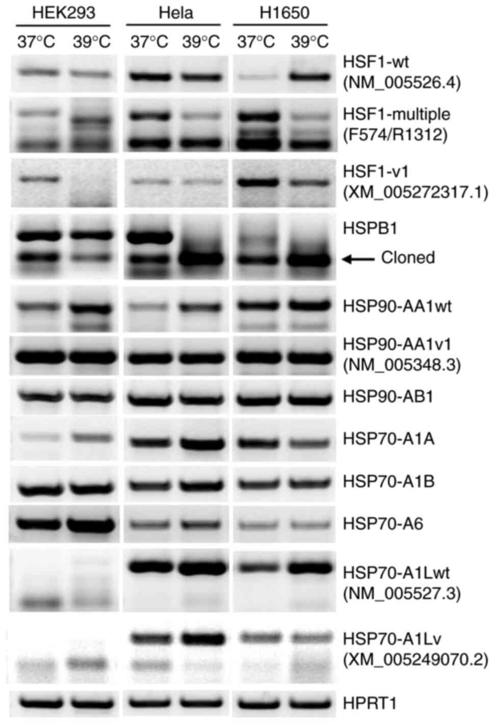
As shown in Fig. 2,
RT-PCR detected the RNAs of the HSP90AB1, HSP70A1A, HSP70A1B and
HSP70A6 genes as a single band at the anticipated molecular weights
indicated in Table I. However, one
or more additional bands of the HSF1, HSPB1, HSP90AA1 and HSP70A1L
genes were also detected (Fig.
2).
The NCBI database currently lists 11 HSF1 RNAs. The
NM_005526.4 RNA is the only NM (normalized) sequence that is
designated herein as the Wt. Whether the remaining 10 RNAs really
exist remains unknown, as they are either XM (predicted mRNA) or XR
(predicted non-coding RNA) sequences. The primers used in the
present study were designed based on a previous version of the NM
sequence (NM_005526.2). RT-PCR using the primer pair F574/R1576
detected the Wt RNA, which was further confirmed by direct
sequencing of the PCR amplicon. Notably, this RNA exhibited a
markedly increased expression at 39˚C only in H1650 cells, while
exhibiting a slight decrease in expression in HEK293 and Hela
cells. With the primer pair F574/R1312 designed to amplify several
RNA variants, RT-PCR produced multiple bands that likely included,
besides possible artifacts, some of the predicted RNAs or their
alternative splicing variants that are currently unknown. Notably,
the responses of these PCR amplicons to the increased temperature
were quite different from each another and from one cell line to
another, and the results were too complex to distinguish one
amplicon from another (Fig. 2).
RT-PCR using primer pair F574/XR26 resulted in a single band, which
was confirmed by direct sequencing as XM_005272317.1 RNA
(designated herein as variant 1). This RNA showed different extents
of decrease in the three cell lines (Fig. 2). In all three cell lines, the
HSP90AA1 gene demonstrated increased expression in its Wt form,
while the expression of its variant remained unchanged (Fig. 2).
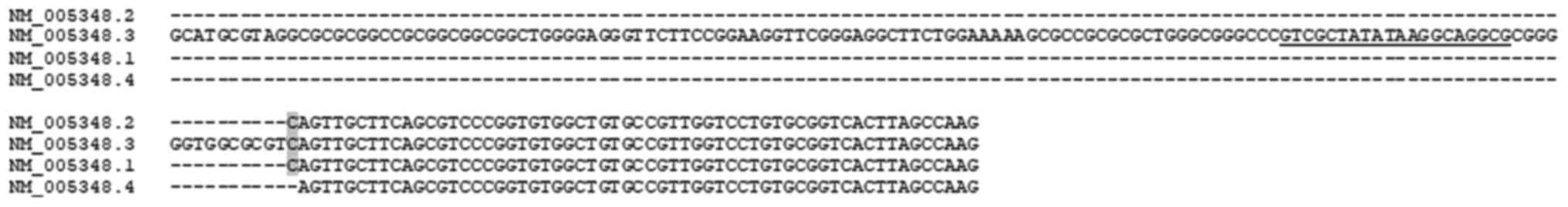
The first exon of the current version (NM_005348.4)
of HSP90AA1 mRNA is much shorter than its counterpart in a previous
version (NM_005348.3), and lacks the sequence of the primer, FV97
(Table I), which was designed
several years ago. However, RT-PCR using the primer pair VF97/R1232
yielded an amplicon whose size was anticipated based on the
NM_005348.3. Direct sequencing of this amplicon confirmed that it
contained the VF97 sequence, and the 5'-RACE results further
confirmed that its 5'-end matched with that of the NM_005348.3
(Fig. 3). These data together
indicated that although the current version is also authentic and
should be designated as Wt RNA, the previous version remains as a
variant despite having been abandoned by NCBI.
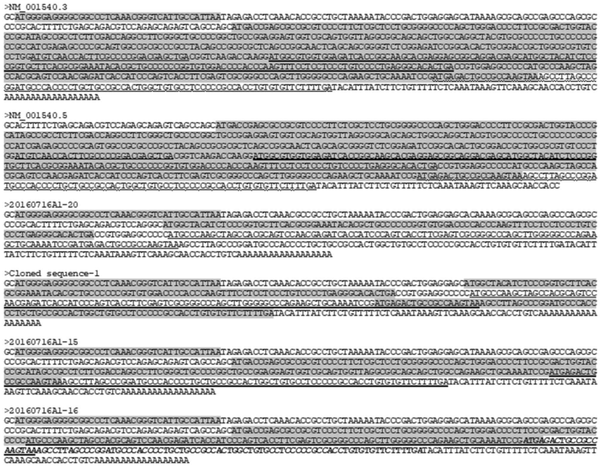
Identification of four HSPB1 RNA
variants that encode HSP27 protein isoforms
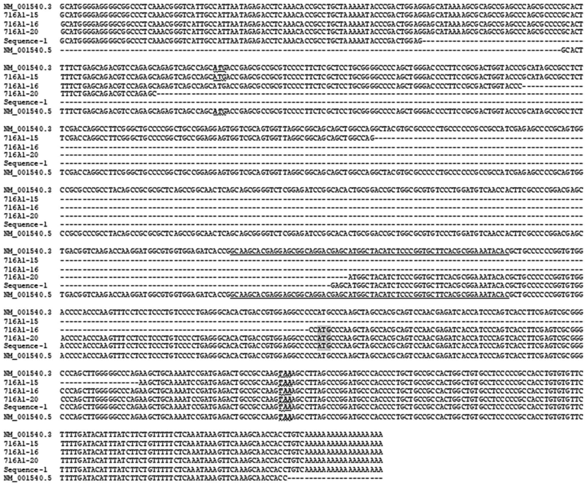
At present, only one RNA of the HSPB1 gene is listed
in the NCBI database. Its current version is NM_001540.5 (Fig. 4), which lacks the first 115 nts of a
previous version (NM_001540.3) that contains three of our forward
primers (F19, F43 and F70) designed a few years ago (Table I). However, in the majority of the
samples studied, RT-PCR using the primer pair F19/R845 yielded not
only the anticipated band, but also an additional band below it,
which was ~400 base pairs smaller (Fig.
2). Compared with its counterpart at 37˚C, this smaller
amplicon showed decreased expression in HEK293 cells but increased
expression in HeLa and H1650 cells at 39˚C. Below this smaller
amplicon is another weak and even smaller band in the HEK293 and
HeLa cells cultured at 37˚C. Nested RT-PCRs with inner primer pairs
F43-R778 and F70-R778 (Table I) may
continue to result in these two smaller bands (data not shown),
suggesting that they may be unreported HSPB1 RNA variants.
The second band was excised from H1650 cells
cultured at 39˚C from an agarose gel, the DNA was purified and it
was cloned into a T-A vector. Sequencing over 20 resultant plasmid
clones yielded five different HSPB1 RNA sequences, each of which
appeared in at least two clones, including the Wt RNA and four
previously unreported variants coined as 716A1-15 (MW881014),
716A1-16 (MW881015), 716A1-20 (MW881016) and cloned sequence-1
(MW881017) (Fig. 5). These RT-PCR
and sequencing results involving F19, F43 and F70 primers suggested
that NM_01540.3 RNA is actually a variant of HSPB1 RNA relative to
NM_001540.5, and that the cloned band is a mixture of different
heterodimers formed between these five HSPB1 RNAs. A 5'-RACE assay
using R313 as the reverse primer also confirmed that the 5'-end of
the HSPB1 RNAs is identical to that of the NM_001540.3 sequence.
Furthermore, NM_01540.5 lacks the last four nts and the poly-A tail
encompassed by NM_01540.3, but the PCR using cDNA samples primed by
a poly-dT primer detected the Wt form and the cloned band, which
also supported the conclusion that NM_01540.3 is an authentic RNA
variant.
The four new HSPB1 RNA variants were lacking a part
of the middle region of the NM_01540.3 sequence (Fig. 4). The 716A1-15 RNA has a deletion of
426 nts, including 211 nts from exon 1, the whole 64-nt exon 2 and
151 nts from exon 3. The 716A1-16 RNA has a deletion of 431 nts,
including 293 nts from exon 1, the whole 64-nt exon 2 and 74 nts
from exon 3. The 716A1-20 RNA has 404 nts deleted, including 377
nts from exon 1 and 27 nts from exon 2, whereas the cloned
sequence-1 has 459 nts deleted, including 436 nts from exon 1 and
23 nts from exon 2.
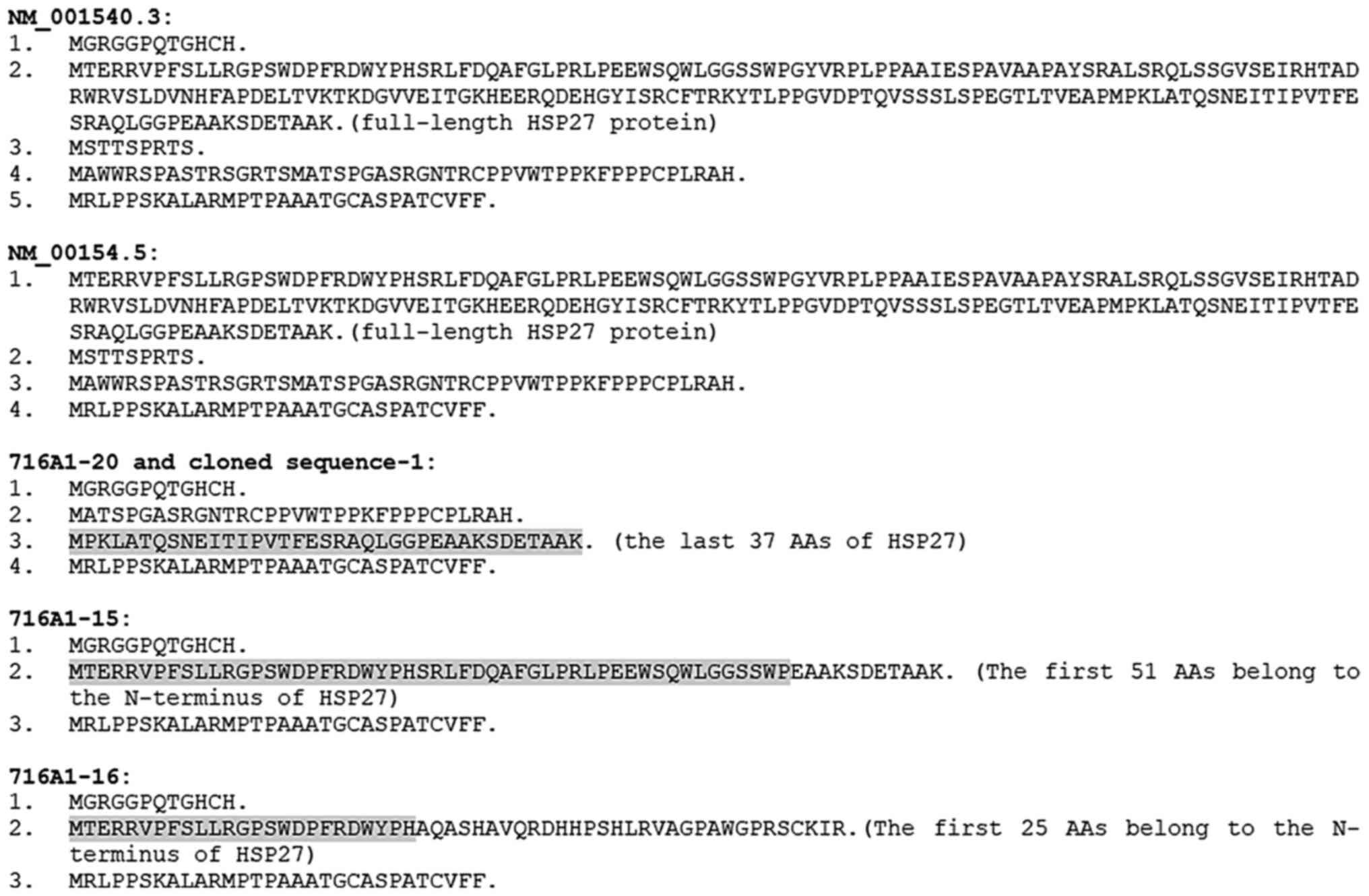
The ORF encoding the Wt HSP27 protein in NM_001540.3
has two additional short ORFs encoding non-HSP27 peptides (Figs. 5 and 6). Furthermore, there is an additional ORF
that begins with an ATG start codon at the 3'-end, but extends
beyond the stop codon of the ORF for the HSP27 (Fig. 5). The peptide sequences encoded by
these short ORFs are shown in Fig.
6. NM_001540.5 lacks the first 115 nts and thus lack the first
short ORF (Fig. 5). All four novel
RNAs lack the canonical ATG for translation of the Wt HSP27
protein. However, deletion of the middle region leads to the
formation of one or two new ORFs that encode new HSP27 isoforms
containing either the N- or C-terminal region of the HSP27 protein.
The 716A1-15 and the cloned sequence-1 encode the same HSP27
isoform, whereas the 7161A-16 encodes two isoforms (Figs. 5 and 6). Furthermore, each of these four novel
RNAs encodes several other short ORFs, some of which also appear in
the NM_001540.3 RNA.
Detection of proteins from the same
HSP genes at multiple positions of SDS-PAGE
If HSP genes respond to increased ambient
temperatures primarily by changes in the ratios of expression
levels between different RNA variants (including some new ones),
they may express multiple protein isoforms. Therefore, the
proteomics data from two previous studies was reanalyzed to
determine whether certain HSP or associated genes express multiple
protein isoforms. In one study, proteins from 12 HSP genes were
detected in MB231 and MCF7 cells at the 72-kDa position of SDS-PAGE
(Table II), of which the HSP90AA1,
STIP1, SHPA8, HSP90AB1, TRAP1 and HSPH1 genes have multiple protein
isoforms listed in the NCBI database, with their numbers of amino
acids (AAs) presented in Table II.
The largest and smallest isoforms of these proteins range between
approximately 98 kDa (HSP90AA1) and 53.5 kDa (HSPA8). However, it
remains unknown whether the largest or the smallest are truly
expressed and detected because, following excision from the
SDS-PAGE gel at the indicated position (72-kDa), our approach uses
short peptide(s) to predict the existence of the proteins. Notably,
several detected genes have only one protein form listed in the
NCBI database, which is too small to be detected at 72-kDa. For
instance, the HSPD1 gene was detected, but NCBI demonstrates that
its protein has only 573 AAs (less than 61.1 kDa), indicating that
either HSPD1 has an unidentified larger isoform or the canonical
protein has undergone substantial post-translational modifications
that greatly hinder its migration in the SDS-PAGE, as previously
inferred (12-14).
| Table IIHeat-shock and associated proteins
detected at different positions of SDS-PAGE. |
Table II
Heat-shock and associated proteins
detected at different positions of SDS-PAGE.
|
Positiona | Cell line | Proteins detected
(isoform variation)b |
|---|
| 72-kD (65-80
kD) | MB231 | HSPD1(573);
HSP90AA1 (854,732,853); STIP1 (590,543,519); HSPA9(679);
HSPA8 (493,646); HSP90B1(803); HSPA5(654); HSP90AB1
(724,714,676); HSPA1A (641); TRAP1 (651,704,564); HSPA4(840);
HSPA1L (641) |
| | MCF7 | HSPD1; TRAP1;
HSPA2(639); HSP90AA1; HSP90AB1; STIP1; HSPA9; HSPA5; HSPA8;
HSPA1A; HSP90B1; HSPA6(643); HSPH1
(851,858,782,860,814,809,853,816,736,780,807) |
| 55-kD (50-60
kD) | MB231 | HSPD1;
HSP90AA1; HSP90AB1; HSPE1 (102); HSP90B1; HSPA8;
HSPA14(509); HSPA5; CDC37 (429,378); HSPA9; HSPA1A; AHSA1
(203,338) |
| | MCF7 | HSPD1; STIP1;
HSPA5; HSPA8; HSP90AB1; HSPH1; TRAP1; HSPA1A; HSPA1L;
HSP90AA1; HSPA9 |
| 48-kD (43-53
kD) | MB231 | HSPD1;
HSP90AA1; HSP90AB1; HSPE1; HSPA5; HSPA8; CDC37;
HSP90B1; HSPA1A or HSPA1L; HSPA9 |
| | MCF7 | HSPD1; HSP90AA1;
HSP90AB1; HSPA1A; HSPA8; HSPA9; HSPA14(509); HSPH1;
HSPA1L |
| 40-kD | MB231 | HSPA8;
HSPD1; HSPA9; HSPA5; HSPBP1 (359,405,431,327) |
| (36-44 kD) | HEK293 | HSPD1; AHSA1;
HSP90B1; TRAP1; HSP90AB1; HSPA9; HSPA5; HSP90AA1; HSPA8;
HSPBP1; STIP1; HSPA1A; HSPA4; HSPH1; CDC37 |
| 26-kD (22-30
kD) | HEK293 | HSPD1; HSP90AB1;
HSPB1(205); HSP90B1; HSP90AA1; TRAP1; HSPA8; HSPA5; HSPA1A;
HSPA1L; HSPA9; HSPE1; AHSA1 |
The protein products of 9 to 13 HSP or associated
genes that were detected at 72-kDa were also detected at the 55-kDa
and/or 48-kDa positions in MB231 or MCF7 cells. Notably, the
majority of these genes have isoforms much larger than 55 or 48 kDa
and were already detected at the 72-kDa position (Table II), implying that they are detected
simultaneously at two or three positions on SDS-PAGE. On the other
hand, proteins from the HSPE1 gene were also detected at the 55-kDa
and 48-kDa positions, despite the fact that the gene has only one
protein of 102 AAs (10.9 kDa) listed in the NCBI database (Table II).
In a separate experiment, proteins from 8-12 HSP or
associated genes were detected in MB231 and HEK293 cells at the
40-kDa and/or 26-kDa positions. However, according to the NCBI
database, these proteins varied between 98-53.5 kDa (Table II). The majority of these genes
were also detected in MB231 and MCF7 cells at the 72-kDa, 55-kDa
and/or 48-kDa positions in the aforementioned study (Table II). By contrast, HSPE1, whose
protein should be 10.9 kDa, was also detected at the 26-kDa in
HEK293 cells.
Discussion
Although cells may behave differently in
vitro compared with in vivo, it was worth noting that at
39˚C, the Wt forms of the majority of the genes studied did not
show a pronounced increase at the RNA level. Furthermore, even the
Wt mRNA of the HSF1 gene, whose protein is the master
transactivator of the majority of HSP genes, did not demonstrate a
universal rise. Associated with these findings were complex changes
in the ratios of expression levels between different RNA variants
of HSF1 and several HSP genes. These results collectively suggested
that regulation of alternative transcriptional initiation to
generate different RNA transcripts and regulation of alternative
splicing to convert the same transcripts into different mature RNA
variants are two important mechanisms by which cells adapt to
increased ambient temperatures. These mechanisms collectively lead
to complex changes in the ratios of expression levels between
different RNA variants of the same gene, including certain new ones
that may not be expressed under physiological conditions. Given
that HSPs have versatile functions, these mechanisms may confer
onto cells tactics to promptly adapt to a sudden increase in
ambient temperature and other microenvironmental changes under
hostile conditions. These RNA changes, as well as other molecular
mechanisms that are not discussed herein, render cells more
flexible in dealing with stressful situations.
Over recent decades, NCBI has updated its database
by deleting the part of an RNA sequence that houses our RT-PCR
primers, despite the existence of the missing part being confirmed
by nested RT-PCR and/or sequencing of the RT-PCR amplicons,
including in the cases of HSP90AA1 and HSPB1 shown in the present
study. A suggestion is made to our RNA research peers that numerous
older versions of RNA sequences may be expressed in certain
situations and thus should be regarded as RNA variants. Since these
earlier versions may still be retrieved from NCBI in the majority
of cases, it is strongly recommended that these earlier versions
should be retrieved and consulted when studying RNA sequences.
In the realm of RNA research, certain studies
consider arbitrarily those RNAs with their largest ORF shorter than
100 codons or 300 nts as long non-coding RNAs (25-29),
while other studies define long non-coding RNAs more stringently
with 200 nts as the arbitrary cut-off (30-32).
The present study (8,10,33),
along with many others (34,35),
argue against these existing definitions as numerous studies have
demonstrated that very short peptides [as short as 11 AAs encoded
by only 33 nts (36-39)]
have important functions. Therefore, numerous RNAs, including
virtually all mRNAs, may function through translation into proteins
or short peptides and through one of the several known non-coding
mechanisms (including miRNA or siRNA mechanisms), with the steroid
receptor RNA activator being the first known example of such dual
mechanisms (40). The number of
pure long non-coding RNAs, defined as those that function only via
a non-coding mechanism, is much smaller than has been claimed in
the literature. In fact, few reports to date have carefully studied
long non-coding RNAs for short peptide expression. The dichotomy of
RNAs into long non-coding ones and coding ones is too arbitrary
(41) and may be greatly
misleading. These general conceptions lead us to consider that the
four HSPB1 RNA variants identified herein should be classified as
mRNAs if the deduced peptides are expressed, although the longest
ORF they encompass is shorter than 200 nts.
The possible translation of an HSP27-containing
peptide from the 716A1-16 RNA raises concerns about the definition
of ‘protein isoforms’ used in the present. This is because only 25
of the 56 AAs encoded by the 716A1-16 belong to HSP27, and this
short peptide (if it is expressed) may have quite a different
function compared with Wt HSP27, which encompasses 205 AAs. By
contrast, it is not uncommon for two protein isoforms encoded by
two differently spliced mRNAs of the same gene to have opposite
functions. For example, the Bcl-xL (the long form of Bcl-X)
promotes cell survival, while the Bcl-xS (the short form lacking
one exon) promotes cell death; however, the long and short forms of
the FANCL gene exhibit the opposite phenomenon (42). Therefore, this deduced
HSP27-containing peptide may be considered by others as a non-HSP27
protein, despite it being considered an HSP27 isoform in the
present study. This problem requires further discussion in the RNA
research fraternity to reach a common definition of ‘protein
isoforms’, since the human genome may encode an astounding number
of such similar/dissimilar ORFs.
Reanalysis of previous proteomics data has revealed
that numerous HSP or associated genes have their protein products
detected simultaneously at multiple positions on SDS-PAGE,
including some positions with much larger or smaller molecular
masses than their theoretical counterparts (Table II). Explanations for those
appearing at a relatively low position on the SDS-PAGE include that
either they are isoform(s) smaller than the Wt or they are degraded
debris. Explanations for those appearing at a much larger position
include that either they are unknown isoforms or they are known
isoforms, but have undergone multiple post-translational
modifications, including the recently reported lysine lactation
(43). At present, there is still
lacking a reliable high-throughput technique to rectify the
authenticity of those proteins that appear at the ‘wrong’ positions
on SDS-PAGE and to rectify the three putative HSP27 isoforms
described in the present study. These technical hurdles include the
lack of isoform-specific antibodies to determine protein identity
using Western blotting, and the fact that different isoforms
sharing parts of the AA sequence may differ in conformation and
thus have different antibody epitopes.
In conclusion, the RT-PCR results demonstrated that
HSF1 and several HSP genes respond to increased ambient
temperatures in cell culture through complex changes in the ratios
of expression levels between different RNA variants, including
certain previously unreported variants and older versions of the
RNAs that have been replaced by newer versions. Among these
variants are four novel RNA variants of the HSPB1 gene that may
encode three short HSP27 protein isoforms. In line with these
observations, reanalysis of proteomics data from two previous
studies also reveals that proteins of certain HSP or associated
genes may be detected simultaneously at multiple positions on
SDS-PAGE, suggesting that these genes are likely to be expressed in
multiple isoforms. Among the limitations of the present study is
the lack of determination of the time course of the alterations in
the expression of individual HSP genes at the full range of febrile
temperatures, i.e. from 38˚C or 42˚C. Using alternative
transcription-initiation sites and alternative RNA splicing to
produce different HSF1 RNAs and HSP genes may be important
molecular mechanisms that allow cells to respond to an elevated
ambient temperature in vitro.
Acknowledgements
Not applicable.
Funding
Funding: The present study was supported by a grant (grant no.
81660501) to Dezhong Joshua Liao and two grants (grant nos.
81460364 and 81760429) to Hai Huang from the National Natural
Science Foundation of China, as well as a grant (grant no.
2019-5610) to Hai Huang from Guizhou Provincial Innovative Talents
Team of China.
Availability of data and materials
The sequences cloned in the present study have been
documented in public databases: MW881014 for 7161A-15, MW881015 for
7161A-16, MW881017 for 7161A-20, and MW881017 for cloned sequence
1. The sequences can be retrieved by entering the corresponding
access number into the NCBI website for nucleotides (www.ncbi.nlm.nih.gov/nuccore/?term=)
Authors' contributions
XG performed part of the study and drafted the
manuscript. KYZ and HYZ performed part of sequence analysis and
bioinformatics analysis while preparing the figures and tables. LZ
reanalyzed and corrected all open reading frames of the RNA
sequences as well as helped draw conclusions while performing
English editing of the manuscript. CFY performed some bench
experiments and helped conceptualize the manuscript. HH
participated in conceptualization and discussion of the study and
helped draw conclusions. DZL conceptualized and finalized the
manuscript.
Ethics approval and consent to
participate
Not applicable.
Patient consent for publication
Not applicable.
Competing interests
The authors declare that they have no competing
interests.
References
|
1
|
Chaudhury S, Keegan BM and Blagg BSJ: The
role and therapeutic potential of Hsp90, Hsp70, and smaller heat
shock proteins in peripheral and central neuropathies. Med Res Rev.
41:202–222. 2021.PubMed/NCBI View Article : Google Scholar
|
|
2
|
Dabbaghizadeh A and Tanguay RM: Structural
and functional properties of proteins interacting with small heat
shock proteins. Cell Stress Chaperones. 25:629–637. 2020.PubMed/NCBI View Article : Google Scholar
|
|
3
|
Bascos NAD and Landry SJ: A history of
molecular chaperone structures in the protein data bank. Int J Mol
Sci. 20(6195)2019.PubMed/NCBI View Article : Google Scholar
|
|
4
|
Masser AE, Ciccarelli M and Andreasson C:
Hsf1 on a leash-controlling the heat shock response by chaperone
titration. Exp Cell Res. 396(112246)2020.PubMed/NCBI View Article : Google Scholar
|
|
5
|
Ahmed K, Zaidi SF, Mati-Ur-Rehman Rehman R
and Kondo T: Hyperthermia and protein homeostasis: Cytoprotection
and cell death. J Therm Biol. 91(102615)2020.PubMed/NCBI View Article : Google Scholar
|
|
6
|
Inia JA and O'Brien ER: Role of heat shock
protein 27 in modulating atherosclerotic inflammation. J Cardiovasc
Transl Res. 14:3–12. 2021.PubMed/NCBI View Article : Google Scholar
|
|
7
|
Prince TL, Lang BJ, Guerrero-Gimenez ME,
Fernandez-Munoz JM, Ackerman A and Calderwood SK: HSF1: Primary
factor in molecular chaperone expression and a major contributor to
cancer morbidity. Cells. 9(1046)2020.PubMed/NCBI View Article : Google Scholar
|
|
8
|
He Y, Yuan C, Chen L, Lei M, Zellmer L,
Huang H and Liao DJ: Transcriptional-Readthrough RNAs reflect the
phenomenon of ‘A Gene Contains Gene(s)’ or ‘Gene(s) within a Gene’
in the human genome, and thus are not Chimeric RNAs. Genes (Basel).
9(40)2018.PubMed/NCBI View Article : Google Scholar
|
|
9
|
He Y, Yuan C, Chen L, Liu Y, Zhou H, Xu N
and Liao DJ: While it is not deliberate, much of today's biomedical
research contains logical and technical flaws, showing a need for
corrective action. Int J Med Sci. 15:309–322. 2018.PubMed/NCBI View Article : Google Scholar
|
|
10
|
Jia Y, Chen L, Ma Y, Zhang J, Xu N and
Liao DJ: To know how a gene works, we need to redefine it first but
then, more importantly, to let the cell itself decide how to
transcribe and process its RNAs. Int J Biol Sci. 11:1413–1423.
2015.PubMed/NCBI View Article : Google Scholar
|
|
11
|
Liu X, Wang Y, Yang W, Guan Z, Yu W and
Liao DJ: Protein multiplicity can lead to misconduct in western
blotting and misinterpretation of immunohistochemical staining
results, creating much conflicting data. Prog Histochem Cytochem.
51:51–58. 2016.PubMed/NCBI View Article : Google Scholar
|
|
12
|
Qu J, Zhang J, Zellmer L, He Y, Liu S,
Wang C, Yuan C, Xu N, Huang H and Liao DJ: About three-fourths of
mouse proteins unexpectedly appear at a low position of SDS-PAGE,
often as additional isoforms, questioning whether all protein
isoforms have been eliminated in gene-knockout cells or organisms.
Protein Sci. 29:978–990. 2020.PubMed/NCBI View
Article : Google Scholar
|
|
13
|
Yan R, Zhang J, Zellmer L, Chen L, Wu D,
Liu S, Xu N and Liao JD: Probably less than one-tenth of the genes
produce only the wild type protein without at least one additional
protein isoform in some human cancer cell lines. Oncotarget.
8:82714–82727. 2017.PubMed/NCBI View Article : Google Scholar
|
|
14
|
Zhang J, Lou X, Shen H, Zellmer L, Sun Y,
Liu S, Xu N and Liao DJ: Isoforms of wild type proteins often
appear as low molecular weight bands on SDS-PAGE. Biotechnol J.
9:1044–1054. 2014.PubMed/NCBI View Article : Google Scholar
|
|
15
|
Taga A and Cornblath DR: A novel HSPB1
mutation associated with a late onset CMT2 phenotype: Case
presentation and systematic review of the literature. J Peripher
Nerv Syst. 25:223–229. 2020.PubMed/NCBI View Article : Google Scholar
|
|
16
|
Gao X, Zhang K, Lu T, Zhao Y, Zhou H, Yu
Y, Zellmer L, He Y, Huang H and Joshua Liao D: A reassessment of
several erstwhile methods for isolating DNA fragments from agarose
gels. 3 Biotech. 11(138)2021.PubMed/NCBI View Article : Google Scholar
|
|
17
|
Sun Y, Sriramajayam K, Luo D and Liao DJ:
A quick, cost-free method of purification of DNA fragments from
agarose gel. J Cancer. 3:93–95. 2012.PubMed/NCBI View
Article : Google Scholar
|
|
18
|
Sun Y, Cao S, Yang M, Wu S, Wang Z, Lin X,
Song X and Liao DJ: Basic anatomy and tumor biology of the RPS6KA6
gene that encodes the p90 ribosomal S6 kinase-4. Oncogene.
32:1794–1810. 2013.PubMed/NCBI View Article : Google Scholar
|
|
19
|
Sun Y, Lou X, Yang M, Yuan C, Ma L, Xie
BK, Wu JM, Yang W, Shen SX, Xu N and Liao DJ: Cyclin-dependent
kinase 4 may be expressed as multiple proteins and have functions
that are independent of binding to CCND and RB and occur at the S
and G 2/M phases of the cell cycle. Cell Cycle. 12:3512–3525.
2013.PubMed/NCBI View
Article : Google Scholar
|
|
20
|
Yang M, Sun Y, Ma L, Wang C, Wu JM, Bi A
and Liao DJ: Complex alternative splicing of the smarca2 gene
suggests the importance of smarca2-B variants. J Cancer. 2:386–400.
2011.PubMed/NCBI View Article : Google Scholar
|
|
21
|
Yang M, Wu J, Wu SH, Bi AD and Liao DJ:
Splicing of mouse p53 pre-mRNA does not always follow the ‘first
come, first served’ principle and may be influenced by cisplatin
treatment and serum starvation. Mol Biol Rep. 39:9247–9256.
2012.PubMed/NCBI View Article : Google Scholar
|
|
22
|
Yang W, Chen L, Yan F, Xie BK, Liao Y and
Liao DJ: Dirty plasmid used as a quick and low-cost method to
identify bacterial colonies with a recombinant plasmid. Indian J
Applied Res. 6(649)2016.
|
|
23
|
Wu J, Wu SH, Bollig A, Thakur A and Liao
DJ: Identification of the cyclin D1b mRNA variant in mouse. Mol
Biol Rep. 36:953–957. 2009.PubMed/NCBI View Article : Google Scholar
|
|
24
|
Bollig-Fischer A, Thakur A, Sun Y, Wu J
and Liao DJ: The predominant proteins that react to the MC-20
estrogen receptor alpha antibody differ in molecular weight between
the mammary gland and uterus in the mouse and rat. Int J Biomed
Sci. 8:51–63. 2012.PubMed/NCBI
|
|
25
|
Bazzini AA, Johnstone TG, Christiano R,
Mackowiak SD, Obermayer B, Fleming ES, Vejnar CE, Lee MT, Rajewsky
N, Walther TC and Giraldez AJ: Identification of small ORFs in
vertebrates using ribosome footprinting and evolutionary
conservation. EMBO J. 33:981–993. 2014.PubMed/NCBI View Article : Google Scholar
|
|
26
|
Cheng H, Chan WS, Li Z, Wang D, Liu S and
Zhou Y: Small open reading frames: Current prediction techniques
and future prospect. Curr Protein Pept Sci. 12:503–507.
2011.PubMed/NCBI View Article : Google Scholar
|
|
27
|
Kageyama Y, Kondo T and Hashimoto Y:
Coding vs non-coding: Translatability of short ORFs found in
putative non-coding transcripts. Biochimie. 93:1981–1986.
2011.PubMed/NCBI View Article : Google Scholar
|
|
28
|
Landry CR, Zhong X, Nielly-Thibault L and
Roucou X: Found in translation: Functions and evolution of a
recently discovered alternative proteome. Curr Opin Struct Biol.
32:74–80. 2015.PubMed/NCBI View Article : Google Scholar
|
|
29
|
Pauli A, Valen E and Schier AF:
Identifying (non-)coding RNAs and small peptides: Challenges and
opportunities. Bioessays. 37:103–112. 2015.PubMed/NCBI View Article : Google Scholar
|
|
30
|
Signal B, Gloss BS and Dinger ME:
Computational approaches for functional prediction and
characterisation of long noncoding RNAs. Trends Genet. 32:620–637.
2016.PubMed/NCBI View Article : Google Scholar
|
|
31
|
Jia H, Osak M, Bogu GK, Stanton LW,
Johnson R and Lipovich L: Genome-wide computational identification
and manual annotation of human long noncoding RNA genes. RNA.
16:1478–1487. 2010.PubMed/NCBI View Article : Google Scholar
|
|
32
|
Samudyata , Castelo-Branco G and
Bonetti A: Birth, coming of age and death: The intriguing life of
long noncoding RNAs. Semin Cell Dev Biol. 79:143–152.
2018.PubMed/NCBI View Article : Google Scholar
|
|
33
|
Yuan C, Han Y, Zellmer L, Yang W, Guan Z,
Yu W, Huang H and Liao DJ: It is imperative to establish a pellucid
definition of chimeric RNA and to clear up a lot of confusion in
the relevant research. Int J Mol Sci. 18(714)2017.PubMed/NCBI View Article : Google Scholar
|
|
34
|
Reisacher C and Arbibe L: Not lost in host
translation: The new roles of long noncoding RNAs in infectious
diseases. Cell Microbiol. 21(e13119)2019.PubMed/NCBI View Article : Google Scholar
|
|
35
|
Ruiz-Orera J, Messeguer X, Subirana JA and
Alba MM: Long non-coding RNAs as a source of new peptides. Elife.
3(e03523)2014.PubMed/NCBI View Article : Google Scholar
|
|
36
|
Chu Q, Ma J and Saghatelian A:
Identification and characterization of sORF-encoded polypeptides.
Crit Rev Biochem Mol Biol. 50:134–141. 2015.PubMed/NCBI View Article : Google Scholar
|
|
37
|
Kondo T, Hashimoto Y, Kato K, Inagaki S,
Hayashi S and Kageyama Y: Small peptide regulators of actin-based
cell morphogenesis encoded by a polycistronic mRNA. Nat Cell Biol.
9:660–665. 2007.PubMed/NCBI View Article : Google Scholar
|
|
38
|
Kondo T, Plaza S, Zanet J, Benrabah E,
Valenti P, Hashimoto Y, Kobayashi S, Payre F and Kageyama Y: Small
peptides switch the transcriptional activity of Shavenbaby during
Drosophila embryogenesis. Science. 329:336–339. 2010.PubMed/NCBI View Article : Google Scholar
|
|
39
|
Ladoukakis E, Pereira V, Magny EG,
Eyre-Walker A and Couso JP: Hundreds of putatively functional small
open reading frames in Drosophila. Genome Biol.
12(R118)2011.PubMed/NCBI View Article : Google Scholar
|
|
40
|
Chooniedass-Kothari S, Emberley E,
Hamedani MK, Troup S, Wang X, Czosnek A, Hube F, Mutawe M, Watson
PH and Leygue E: The steroid receptor RNA activator is the first
functional RNA encoding a protein. FEBS Lett. 566:43–47.
2004.PubMed/NCBI View Article : Google Scholar
|
|
41
|
Bogard B, Francastel C and Hube F:
Multiple information carried by RNAs: Total eclipse or a light at
the end of the tunnel? RNA Biol. 17:1707–1720. 2020.PubMed/NCBI View Article : Google Scholar
|
|
42
|
Yuan C, Xu N and Liao J: Switch of FANCL,
a key FA-BRCA component, between tumor suppressor and promoter by
alternative splicing. Cell Cycle. 11(3356)2012.PubMed/NCBI View Article : Google Scholar
|
|
43
|
Zhang D, Tang Z, Huang H, Zhou G, Cui C,
Weng Y, Liu W, Kim S, Lee S, Perez-Neut M, et al: Metabolic
regulation of gene expression by histone lactylation. Nature.
574:575–580. 2019.PubMed/NCBI View Article : Google Scholar
|