Introduction
Lung cancer is one of the most frequent causes of
cancer-related death, with an incidence rate of 1.3 million cases
per year and a 5-year survival rate of <15% (1-3).
Among the various subtypes of lung cancer, non-small cell lung
cancer (NSCLC) is the most common type, which is mostly detected at
an advanced stage (4). The low
survival rate of NSCLC results from the limited efficacy of
treatment and difficulties in the early detection of advanced
disease (5). Targeted therapy and
early detection are the most effective approaches to significantly
reduce the mortality of patients with NSCLC. Therefore, it is
urgently required to identify novel biomarkers that participate in
the progression of NSCLC.
MicroRNAs (miRNAs/miRs) are short, noncoding RNAs
with vital roles in the post-transcriptional regulation of gene
expression and various biological processes (6). Dysregulated miRNAs have attracted
special attention and have roles in tumorigenesis and cancer
progression. For instance, miR-654-3p suppresses hepatocellular
carcinoma progression (7).
miR-188-5p promotes the proliferation and migration of gastric
cancer cells (8). Accumulating
evidence has indicated that certain miRNAs, such as miR-650,
miR-195-5p and miR-30, regulate cell proliferation, migration,
invasion and apoptosis in NSCLC (9-11).
The deregulation of specific miRNAs may serve as a biomarker for
the progression and prognosis of NSCLC.
miR-1181 was reported to be abnormally expressed in
several cancer types, including ovarian cancer, pancreatic cancer,
hepatocellular carcinoma and NSCLC, and was reported to be related
to the development of these cancers (12-15).
It was hypothesized that miR-1181 may have a vital role in the
development and prognosis of NSCLC, but data on the function of
miR-1181 in NSCLC have remained limited. In the present study, the
expression of miR-1181 in NSCLC and adjacent tissues, as well as
the association between miR-1181 expression and the clinical
features and survival rate of patients were analyzed to evaluate
the prognostic value of miR-1181. The function of miR-1181 in NSCLC
cell proliferation, migration and invasion was also investigated
through Cell Counting Kit-8 (CCK8) and Transwell assays.
Materials and methods
Specimens
Paired NSCLC and adjacent non-tumor tissues
confirmed by at least three pathological experts were collected
from 118 patients with NSCLC treated at Nanjing Chest Hospital
(Nanjing, China) between January 2012 and December 2014. All
patients provided written informed consent for participating in the
present study and did not received any chemotherapy or radiotherapy
prior to the surgery. The survival information of the patients with
NSCLC was obtained in a 5-year follow-up survey. All tissues were
frozen in liquid nitrogen immediately after collection and stored
at -80˚C. The present study was approved by the Ethics Committee of
Nanjing Chest Hospital (Nanjing, China).
Cell culture and transfection
The human NSCLC cell lines A549, H1299, H2009 and
NE18, as well as the normal human lung epithelial cell line
BEAS-2B, were purchased from the American Type Culture Collection.
Cells were cultured in RPMI-1640 medium (Invitrogen; Thermo Fisher
Scientific, Inc.) containing 10% FBS (Invitrogen; Thermo Fisher
Scientific, Inc.), with maintenance at 37˚C in a humidified
atmosphere with 5% CO2.
miR-1181 mimics (50 nM;
5'-CCGUCGCCGCCACCCGAGCCG-3'), miR-1181 inhibitor (50 nM;
5'-CGGCUCGGGUGGCGGCGACGG-3') and corresponding negative control
[mimics NC (5'-GGACCAAATCTCGAGATTTGG-3') and inhibitor NC
(5'-UCUACUCUUUCUAGGAGGUUGUGA-3'); Guangzhou RiboBio Co., Ltd.] were
transfected into cells using the Lipofectamine® 2000
reagent (Invitrogen; Thermo Fisher Scientific, Inc.) at room
temperature for 24 h according to the manufacturer's protocol to
regulate the expression of miR-1181. After 24 h of transfection,
the cells were ready for use in the subsequent experiments.
RNA isolation and reverse
transcription-quantitative (RT-q) PCR
Total RNA was isolated from collected tissues and
cell lines using TRIzol reagent (Invitrogen; Thermo Fisher
Scientific, Inc.) according to the manufacturer's protocol and
reverse-transcribed into complementary DNA at 42˚C for 15 min and
at 85˚C for 2 min using a TaqMan MicroRNA Reverse Transcription Kit
(Applied Biosystems; Thermo Fisher Scientific, Inc.). qPCR was
performed with a SYBR-Green I Master Mix kit (Invitrogen; Thermo
Fisher Scientific, Inc.) in the 7300 Real-Time PCR System (Applied
Biosystems; Thermo Fisher Scientific, Inc.). The primer sequences
were as follows: 5'-
GTGCAGGGTCCGAGGTCAGAGCCACCTGGGCAATTTTTTTTTTTCGGCTC-3' for
reverse-transcription, the forward primer was
5'-CCGGGCCGTCGCCGCCACCC-3', and the reverse primer was
5'-GTGCAGGGTCCGAGGTCA-3' for miR-1181 qPCR. U6 (forward,
5'-CTCGCTTCGGCAGCACA-3' and reverse, 5'-AACGCTTCACGAATTTGCGT-3')
was used as an internal control and the 2-ΔΔCq method
was used to quantify the expression of miR-1181(16). The thermocycling conditions were set
as 94˚C for 5 min and then 35 cycles of denaturation for 30 sec at
94˚C, annealing for 30 sec at 50˚C and extension for 45 sec at
72˚C.
CCK8 assay
According to the protocol for the CCK-8 Assay kit
(Dojindo Molecular Technologies Inc.), the CCK8 assay was performed
to analyze the proliferation of NSCLC cells. In brief, NSCLC cells
at a density of 5x10³ cells per well were seeded into
96-well plates in triplicates. After incubating for 0, 24, 48 and
72 h at 37˚C, CCK8 reagent was added to each well, followed by
further incubation for 4 h at 37˚C. The absorbance of each well at
450 nm was detected using a microplate reader (Thermo Fisher
Scientific, Inc.) to evaluate the proliferation of NSCLC cells.
Transwell assay
NSCLC cells in serum-free medium were seeded into
the upper wells of a 24-well Transwell chamber with 8-µm pore size
(Corning, Inc.) at 2x10⁵ cells per well. Fresh culture
medium with 10% FBS was added to the bottom chamber as the
chemoattractant. After 48 h of incubation at 37˚C, cells on the
upper surface of the membrane were removed and those that had
transgressed to the bottom chamber were fixed with 4%
paraformaldehyde for 20 min at room temperature and stained with
0.1% crystal violet for 10 min at room temperature. To assess cell
invasion, the upper chamber was precoated with 0.5% Matrigel (BD
Biosciences) at 37˚C overnight. Migrated and invaded cells were
counted by a light microscope (magnification, x400). The images
were captured randomly from at least 5 fields and analyzed by
ImageJ software (version 1.49; National Institutes of Health).
Luciferase reporter assays
Potential targets of miR-1181 were first predicted
in silico using targetscan (http://www.targetscan.org/vert_72/) and then confirmed
using a luciferase reporter assay. A fragment of the 3'
untranslated region (3'UTR) of axin 1 (AXIN1) containing a
prospective binding site for miR-1181 was amplified with the
genomic DNA extracted from A549 cells using the same reverse
transcription protocol as aforementioned used as a template and the
AmpFlSTR Identifiler PCR Amplification Kit (Applied Biosystems;
Thermo Fisher Scientific, Inc.) according to the protocol. The
amplification conditions were: Initial denaturation for 10 min at
95˚C, followed by 45 cycles of denaturation at 95˚C for 10 sec,
annealing for 20 sec at 60˚C and extension at 72˚C for 20 sec. The
amplified fragment was then inserted into the reporter vector
pmirGLO (Promega Corporation) to construct the luciferase reporter
plasmid AXIN1 wild-type (WT) according to previous studies
(17,18). Furthermore, its mutant (MT) sequence
was also synthesized with mutations in the binding sites introduced
using the QuikChange Site-Directed Mutagenesis kit (Stratagene;
Agilent Technologies, Inc.) to construct the luciferase reporter
plasmid AXIN1 MT. A549 cells were co-transfected with pGL3-AXIN1-WT
(10 nM) or pGL-AXIN1-MT (10 nM) and miR-1181 mimics (50 nM),
miR-1181 inhibitor (50 nM) or negative controls (50 nM) using
Lipofectamine® 2000 reagent (Invitrogen; Thermo Fisher
Scientific, Inc.). The luciferase activity levels were analyzed by
the Dual-Luciferase Reporter Assay Kit (Promega Corporation) with
Renilla as the normalization control according to the
manufacturer's protocol.
Statistical analysis
Statistical analyses were performed with GraphPad
Prism 5.0 software (GraphPad Software, Inc.) and SPSS 20.0 software
(IBM, Corp.). All values are expressed as the mean ± standard
deviation obtained from ≥ 3 experiments. Significant differences
between groups were determined by a paired Student's t-test or
one-way ANOVA followed by Tukey's post hoc test. The χ2
test was used to evaluate the association between miR-1181
expression and the clinical features of patients. The prognostic
value of miR-1181 expression was assessed by Kaplan-Meier followed
by long-rank test and Cox regression analysis. P<0.05 was
considered to indicate statistical significance.
Results
miR-1181 is significantly upregulated
in NSCLC and associated with lymph node metastasis and TNM
stage
The recruited patients were consisted of 69 males
and 49 females with an average age of 56.3±9.33 years. The
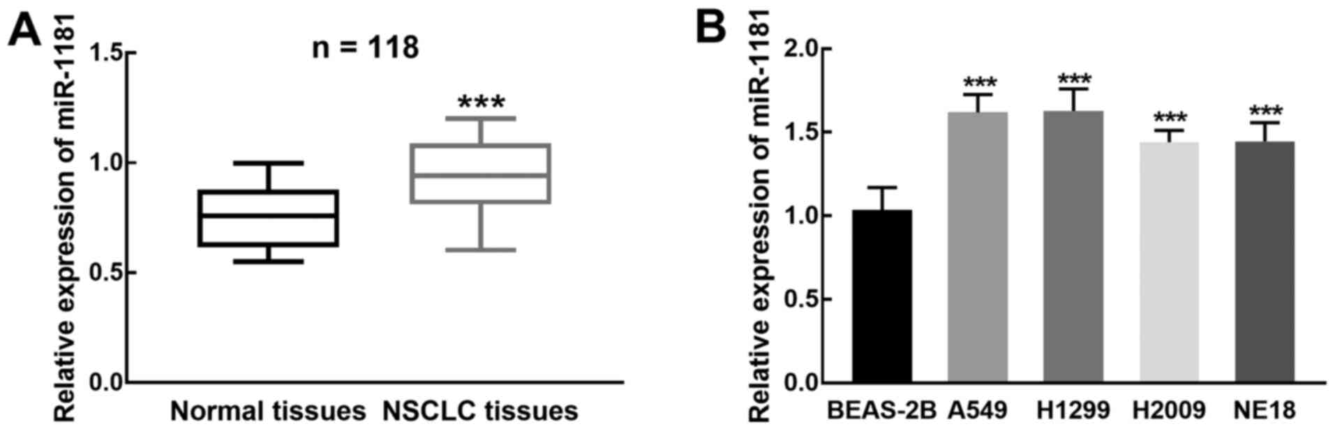
expression of miR-1181 was significantly increased in NSCLC tissues
compared with that in adjacent normal tissues (P<0.001, Fig. 1A). This upregulation of miR-1181 was
also observed in NSCLC cells compared with that in the normal
BEAS-2B cells (P<0.001, Fig.
1B).
Using the mean relative miR-1181 expression levels
in NSCLC tissues (0.939) as the cut-off, patients with NSCLC were
divided into the low and high miR-1181 expression groups. The
expression level of miR-1181 was significantly associated with
lymph node metastasis (P=0.025) and TNM stage of patients
(P=0.013), but no significant relationship with any of the other
clinical features was identified (P>0.05, Table I).
 | Table IAssociation between miR-1181
expression and clinical features of patients with non-small cell
lung cancer. |
Table I
Association between miR-1181
expression and clinical features of patients with non-small cell
lung cancer.
| | Expression of
miR-1181 | |
|---|
| Parameter | Total patients
(n=118) | High (n=67) | Low (n=51) | P-value |
|---|
| Age (years) | | | | 0.509 |
|
<60 | 63 | 35 | 28 | |
|
≥60 | 55 | 32 | 23 | |
| Sex | | | | 0.492 |
|
Male | 69 | 39 | 30 | |
|
Female | 49 | 28 | 21 | |
| Tumor size
(cm) | | | | 0.172 |
|
<4 | 64 | 33 | 31 | |
|
≥4 | 54 | 34 | 20 | |
|
Differentiation | | | | 0.102 |
|
Well+moderate | 71 | 30 | 41 | |
|
Poor | 47 | 37 | 10 | |
| Lymph node
metastasis | | | | 0.025 |
|
Negative | 67 | 38 | 29 | |
|
Positive | 51 | 29 | 22 | |
| TNM stage | | | | 0.013 |
|
I/II | 68 | 42 | 26 | |
|
III/IV | 50 | 25 | 25 | |
High expression of miR-1181 is
associated with poor prognosis
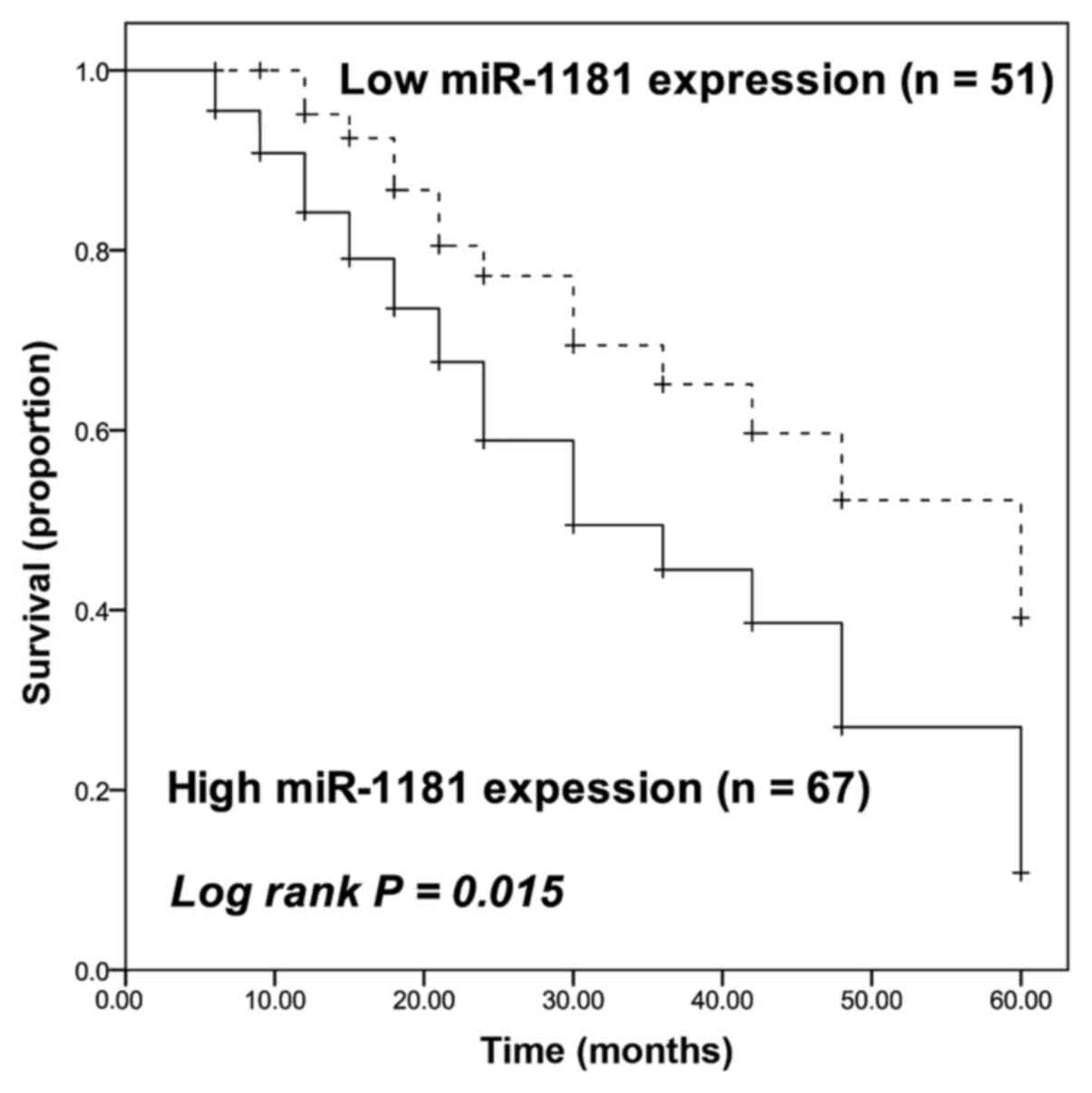
The survival information of patients with NSCLC was
obtained and data were plotted as Kaplan-Meier curves (Fig. 2). The patients with high miR-1181
expression had a poorer prognosis than those with low miR-1181
expression (log-rank P=0.015). To further assess the prognostic
value of miR-1181, Cox regression analysis was used to determine
the influence of miR-1181 expression on the survival probability of
the patients. miR-1181 was significantly associated with the
survival of patients with NSCLC, with a hazard ratio (HR) of 2.169
(95% CI=1.101-4.272, P=0.025). In addition to the TNM stage
(HR=2.014, 95% CI=1.060-3.828, P=0.033), miR-1181 expression in
tumor tissues may be considered as an independent prognostic
indicator for NSCLC (Table
II).
| Table IIMultivariate Cox regression analysis
between miR-1181 expression and survival rate of patients with
non-small cell lung cancer after adjusting for the cofounding
factors. |
Table II
Multivariate Cox regression analysis
between miR-1181 expression and survival rate of patients with
non-small cell lung cancer after adjusting for the cofounding
factors.
| Factor | Hazards ratio | 95% confidence
interval | P-value |
|---|
| miR-1181 (high vs.
low) | 2.169 | 1.101-4.272 | 0.025 |
| Age (≥60 vs.
<60) | 1.134 | 0.640-2.010 | 0.665 |
| Sex (male vs.
female) | 1.185 | 0.657-2.136 | 0.572 |
| Tumor size (≥4 cm
vs. <4 cm) | 1.572 | 0.827-2.989 | 0.167 |
| Differentiation
(poor vs. well+moderate) | 1.660 | 0.875-3.151 | 0.121 |
| Lymph node
metastasis (positive vs. negative) | 1.670 | 0.884-3.156 | 0.114 |
| TNM stage (III-IV
vs. I-II) | 2.014 | 1.060-3.828 | 0.033 |
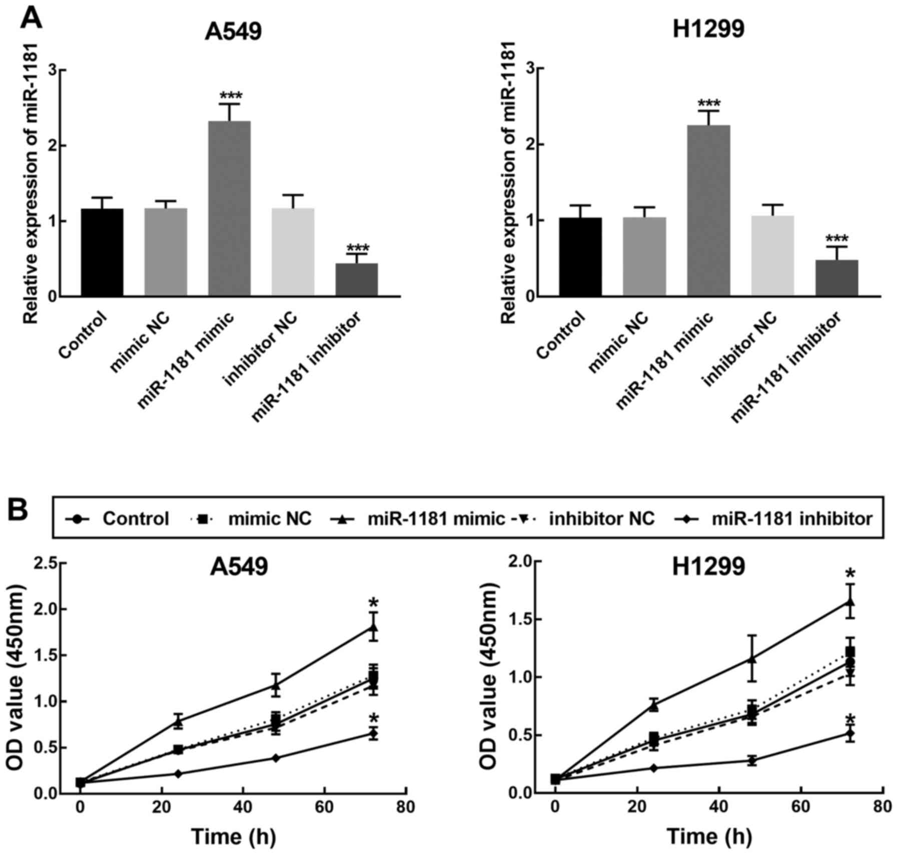
Overexpression of miR-1181 promotes
NSCLC cell proliferation
miR-1181 mimics, miR-1181 inhibitor or the
corresponding NC were transfected into A549 and H1299 cells to
regulate the expression level of miR-1181. The expression of
miR-1181 in A549 and H1299 cells was confirmed to be significantly
increased after transfection with miR-1181 mimics and to be reduced
in the miR-1181 inhibitor-transfected cells (P<0.001, Fig. 3A). To assess the proliferation of
the transfected cells, the CCK8 assay was applied, revealing that
the proliferation of NSCLC cells with knockdown of miR-1181 was
significantly reduced compared with that of the cells with
overexpression of miR-1181 (P<0.05, Fig. 3B).
Overexpression of miR-1181 promotes
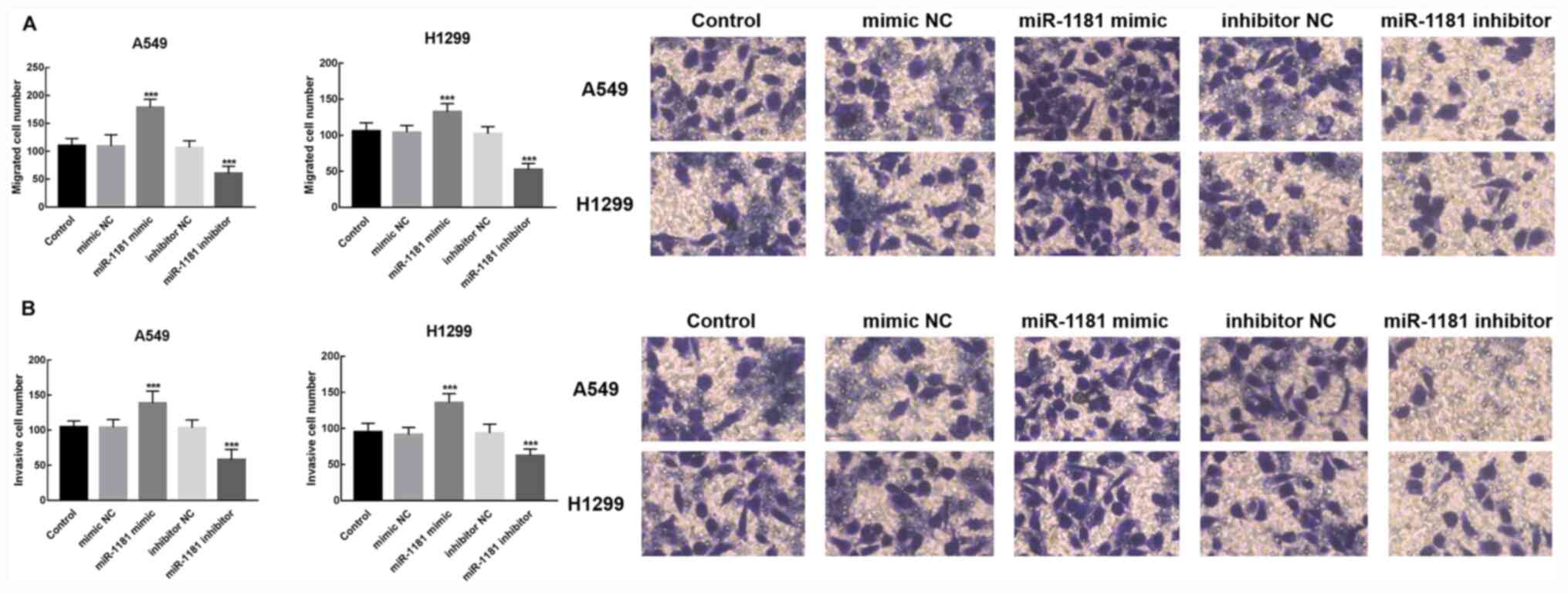
migration and invasion of NSCLC cells
To evaluate the role of miR-1181 in NSCLC cell
migration and invasion, Transwell assays were performed on A549 and
H1299 cells transfected with miR-1181 mimics or inhibitor. The
results suggested that the number of migrated cells in the group of
NSCLC cells with miR-1181 overexpression was significantly
elevated, while that of the NSCLC cells with knockdown of miR-1181
was significantly decreased in comparison with the respective
controls (P<0.001, Fig. 4A).
Similarly, the invasion of NSCLC cells was inhibited by knockdown
of miR-1181 and promoted by overexpression of miR-1181 (P<0.001,
Fig. 4B). Hence, these results
indicated the regulatory function of miR-1181 in the migration and
invasion of NSCLC cells.
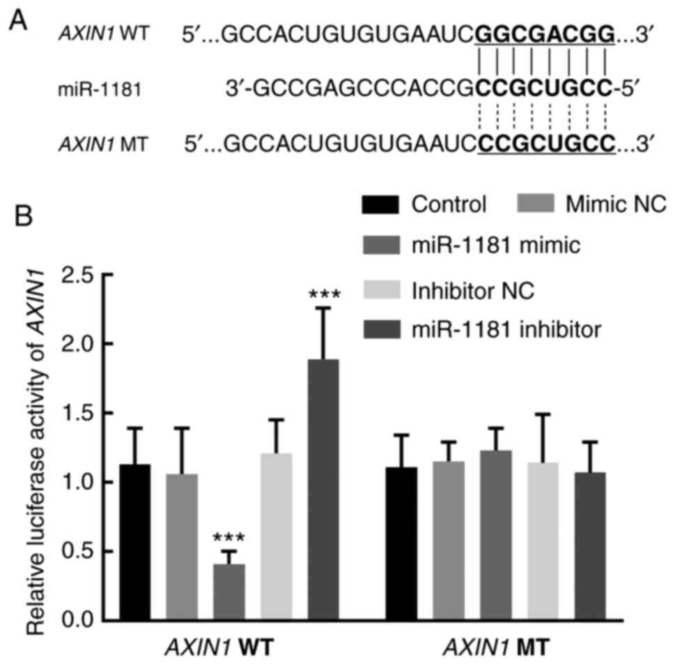
AXIN1 is a direct target of
miR-1181
The binding sites between miR-1181 and the 3'UTR of
AXIN1 were predicted in silico and are presented in Fig. 5A. This predicted binding interaction
was then confirmed with a luciferase reporter assay. The relative
luciferase activity of the AXIN1 WT reporter plasmid was inhibited
by overexpression of miR-1181 in the miR-1181 mimics group and
enhanced by the silencing of miR-1181 in the miR-1181 inhibitor
group (P<0.001, Fig. 5B).
However, the relative luciferase activity of the AXIN1 MT was not
significantly affected by the changes in miR-1181 expression
(P>0.05, Fig. 5B).
Discussion
NSCLC remains the leading cause of cancer-related
death with a low 5-year survival rate due to the lack of effective
diagnostic methods and therapeutic targets (19). miRNAs have been indicated to be
critical regulators in the pathogenesis of cancer, as certain
miRNAs have critical roles in the tumorigenesis and development of
various cancer types (20,21). For instance, the expression of
miR-21 was significantly associated with the progression of
hepatocellular carcinoma (22). In
addition, miR-182 promotes the progression of prostate cancer by
activating the Wnt/β-catenin signaling pathway (23). In NSCLC, miR-675 was reported to
enhance the disease progression by activating the NF-κB signaling
pathway (24) and miR-125a-5p
suppressed NSCLC progression by targeting suppressor of variegation
3-9 homolog 1, as the overexpression of miR-125a-5p inhibited cell
proliferation, migration and invasion and activated cell apoptosis
in NSCLC (25).
A previous study focusing on the miRNA expression
profiling of NSCLC indicated that miR-1181 was differentially
expressed in NSCLC (12), and thus,
it was speculated in the present study that miR-1181 participates
in the progression of NSCLC. In the present study, data derived
from tissues of patients with NSCLC and NSCLC cell lines suggested
significant upregulation of miR-1181, consistent with the previous
study. Furthermore, the upregulation of miR-1181 exhibited a
significant association with lymph node metastasis and TNM stage in
patients with NSCLC. miR-1181 was considered to be able to predict
the prognosis of NSCLC, as its upregulation was associated with a
poorer survival rate of patients with NSCLC. Accumulating evidence
has demonstrated the prognostic value of miRNAs in several cancer
types (26-28),
and a variety of miRNAs, such as miR-519a, miR-378 and miR-1246,
have been considered as prognostic biomarkers for NSCLC (29-31).
In the present study, the results of the Cox regression analysis
indicated that miR-1181 and the TNM stage were independent
prognostic indicators for NSCLC. These results indicated that
miR-1181 may be involved in the progression of NSCLC and predict
poor prognosis of affected patients.
miR-1181 has been reported to be differentially
expressed in various cancer types. In hepatocellular carcinoma,
miR-1181 was indicated to be upregulated and serve as an oncogene
due to its positive effect on the progression of hepatocellular
carcinoma by repressing AXIN1(15).
miR-1181 inhibited the migration, invasion and proliferation of
pancreatic cancer by targeting STAT3 and inhibited stem cell-like
phenotypes by downregulating SOX2 and STAT3 in pancreatic cancer
(13,32). A previous study also reported that
miR-1181 promoted mesenchymal-epithelial transition in ovarian
cancer cells, which is closely associated with the migration and
invasion of cancer cells (14). The
present results indicated that the miR-1181 mimics increased the
expression of miR-1181, which promoted cell proliferation,
migration and invasion of NSCLC cells, while miR-1181 inhibitor
reduced miR-1181 expression and decreased cell proliferation,
migration and invasion. These results demonstrated that miR-1181
may serve as a biomarker for the progression and prognosis of
NSCLC.
Previously, AXIN1 was reported to be a specific
target of miR-1181 during the regulation of miR-1181 in
hepatocellular carcinoma (15). It
was speculated that the tumor promoter role of miR-1181 was a
result of targeting AXIN1. The results of the luciferase reporter
assay of the present study suggested that overexpression of
miR-1181 significantly inhibited the relative luciferase activity
of the AXIN1 WT reporter plasmid, while it was enhanced by
knockdown of miR-1181. These results indicated that AXIN1 is a
direct target of miR-1181 with involvement in the progression of
NSCLC.
However, more work is still required in future
studies. In vivo experiments are important parts in the
investigation of tumor development (33,34).
The results of the present study revealed the tumor promoter role
of miR-1181 in NSCLC in vitro, while in vivo
experiments, such as in vivo migration and invasion assays
and tumor growth assays, are also needed to verify the function of
miR-1181 in the progression of NSCLC.
In conclusion, the present study indicated that the
upregulation of miR-1181 in NSCLC tissues was associated with lymph
node metastasis and TNM stage of patients and predicted poor
prognosis. miR-1181 may be involved in the progression of NSCLC due
to enhancing the proliferation, migration and invasion of NSCLC
cells. These results indicated that miR-1181 may serve as a
potential novel therapeutic target for the treatment of NSCLC.
Acknowledgements
Not applicable.
Funding
Funding: No funding was received.
Availability of data and materials
The datasets used and/or analyzed during the current
study are available from the corresponding author on reasonable
request.
Authors' contributions
FL, GM, BW, XW and CH conceived the study. FL GM,
and BW performed the experiments and wrote the manuscript. FL, XW
and CH analyzed the data. All authors read and approved the final
version of the manuscript. FL and GM checked and approved the
authenticity of the raw data.
Ethics approval and consent to
participate
This study was approved by the Ethics Committee of
Nanjing Chest Hospital (Nanjing, China). All patients provided
written informed consent.
Patient consent for publication
Not applicable.
Competing interests
The authors declare that they have no competing
interests.
References
|
1
|
Parkin DM, Bray FI and Devesa SS: Cancer
burden in the year 2000. The global picture. Eur J Cancer. 37
(Suppl 8):S4–S66. 2001.PubMed/NCBI View Article : Google Scholar
|
|
2
|
Bray F, Ferlay J, Soerjomataram I, Siegel
RL, Torre LA and Jemal A: Global cancer statistics 2018: GLOBOCAN
estimates of incidence and mortality worldwide for 36 cancers in
185 countries. CA Cancer J Clin. 68:394–424. 2018.PubMed/NCBI View Article : Google Scholar
|
|
3
|
Bilfinger T, Keresztes R, Albano D and
Nemesure B: Five-year survival among stage IIIA lung cancer
patients receiving two different treatment modalities. Med Sci
Monit. 22:2589–2594. 2016.PubMed/NCBI View Article : Google Scholar
|
|
4
|
Siegel RL, Miller KD and Jemal A: Cancer
statistics, 2018. CA Cancer J Clin. 68:7–30. 2018.PubMed/NCBI View Article : Google Scholar
|
|
5
|
Minna JD, Roth JA and Gazdar AF: Focus on
lung cancer. Cancer Cell. 1:49–52. 2002.PubMed/NCBI View Article : Google Scholar
|
|
6
|
Lin S and Gregory RI: MicroRNA biogenesis
pathways in cancer. Nat Rev Cancer. 15:321–333. 2015.PubMed/NCBI View
Article : Google Scholar
|
|
7
|
Yang J, Zhang Z, Chen S, Dou W, Xie R and
Gao J: miR-654-3p predicts the prognosis of hepatocellular
carcinoma and inhibits the proliferation, migration, and invasion
of cancer cells. Cancer Biomark. 28:73–79. 2020.PubMed/NCBI View Article : Google Scholar
|
|
8
|
Wang M, Qiu R, Gong Z, Zhao X, Wang T,
Zhou L, Lu W, Shen B, Zhu W and Xu W: miR-188-5p emerges as an
oncomiRNA to promote gastric cancer cell proliferation and
migration via upregulation of SALL4. J Cell Biochem.
120:15027–15037. 2019.PubMed/NCBI View Article : Google Scholar
|
|
9
|
Tang X, Ding Y, Wang X, Wang X, Zhao L and
Bi H: miR-650 promotes non-small cell lung cancer cell
proliferation and invasion by targeting ING4 through
Wnt-1/β-catenin pathway. Oncol Lett. 18:4621–4628. 2019.PubMed/NCBI View Article : Google Scholar
|
|
10
|
Luo J, Pan J, Jin Y, Li M and Chen M:
miR-195-5p Inhibits proliferation and induces apoptosis of
non-small cell lung cancer cells by targeting CEP55. Onco Targets
Ther. 12:11465–11474. 2019.PubMed/NCBI View Article : Google Scholar
|
|
11
|
Zhong K, Chen K, Han L and Li B:
MicroRNA-30b/c inhibits non-small cell lung cancer cell
proliferation by targeting Rab18. BMC Cancer.
14(703)2014.PubMed/NCBI View Article : Google Scholar
|
|
12
|
Hu Y, Wang L, Gu J, Qu K and Wang Y:
Identification of microRNA differentially expressed in three
subtypes of non-small cell lung cancer and in silico functional
analysis. Oncotarget. 8:74554–74566. 2017.PubMed/NCBI View Article : Google Scholar
|
|
13
|
Wang J, Guo XJ, Ding YM and Jiang JX:
miR-1181 inhibits invasion and proliferation via STAT3 in
pancreatic cancer. World J Gastroenterol. 23:1594–1601.
2017.PubMed/NCBI View Article : Google Scholar
|
|
14
|
Zhang HY, Li JH, Li G and Wang SR:
Activation of ARK5/miR-1181/HOXA10 axis promotes
epithelial-mesenchymal transition in ovarian cancer. Oncol Rep.
34:1193–1202. 2015.PubMed/NCBI View Article : Google Scholar
|
|
15
|
Song Z, Yu Z, Chen L, Zhou Z, Zou Q and
Liu Y: MicroRNA-1181 supports the growth of hepatocellular
carcinoma by repressing AXIN1. Biomed Pharmacother.
119(109397)2019.PubMed/NCBI View Article : Google Scholar
|
|
16
|
Livak KJ and Schmittgen TD: Analysis of
relative gene expression data using real-time quantitative PCR and
the 2(-Delta Delta C(T)) method. Methods. 25:402–408.
2001.PubMed/NCBI View Article : Google Scholar
|
|
17
|
Cai Z, Li J, Zhuang Q, Zhang X, Yuan A,
Shen L, Kang K, Qu B, Tang Y, Pu J, et al: MiR-125a-5p ameliorates
monocrotaline-induced pulmonary arterial hypertension by targeting
the TGF-β1 and IL-6/STAT3 signaling pathways. Exp Mol Med. 50:1–11.
2018.PubMed/NCBI View Article : Google Scholar
|
|
18
|
Benderska N, Dittrich AL, Knaup S, Rau TT,
Neufert C, Wach S, Fahlbusch FB, Rauh M, Wirtz RM, Agaimy A, et al:
miRNA-26b overexpression in ulcerative colitis-associated
carcinogenesis. Inflamm Bowel Dis. 21:2039–2051. 2015.PubMed/NCBI View Article : Google Scholar
|
|
19
|
Pao W: New approaches to targeted therapy
in lung cancer. Proc Am Thorac Soc. 9:72–73. 2012.PubMed/NCBI View Article : Google Scholar
|
|
20
|
Liu CG, Calin GA, Meloon B, Gamliel N,
Sevignani C, Ferracin M, Dumitru CD, Shimizu M, Zupo S, Dono M, et
al: An oligonucleotide microchip for genome-wide microRNA profiling
in human and mouse tissues. Proc Natl Acad Sci USA. 101:9740–9744.
2004.PubMed/NCBI View Article : Google Scholar
|
|
21
|
Volinia S, Calin GA, Liu CG, Ambs S,
Cimmino A, Petrocca F, Visone R, Iorio M, Roldo C, Ferracin M, et
al: A microRNA expression signature of human solid tumors defines
cancer gene targets. Proc Natl Acad Sci USA. 103:2257–2261.
2006.PubMed/NCBI View Article : Google Scholar
|
|
22
|
Yoon JS, Kim G, Lee YR, Park SY, Tak WY,
Kweon YO, Park JG, Lee HW, Han YS, Ha HT, et al: Clinical
significance of microRNA-21 expression in disease progression of
patients with hepatocellular carcinoma. Biomark Med. 12:1105–1114.
2018.PubMed/NCBI View Article : Google Scholar
|
|
23
|
Wang D, Lu G, Shao Y and Xu D: MiR-182
promotes prostate cancer progression through activating
Wnt/β-catenin signal pathway. Biomed Pharmacother. 99:334–339.
2018.PubMed/NCBI View Article : Google Scholar
|
|
24
|
Feng Y, Yang C, Hu D, Wang X and Liu X:
miR-675 promotes disease progression of non-small cell lung cancer
via activating NF-κB signaling pathway. Cell Mol Biol
(Noisy-le-grand). 63:7–10. 2017.PubMed/NCBI View Article : Google Scholar
|
|
25
|
Liu H, Ma Y, Liu C, Li P and Yu T: Reduced
miR-125a-5p level in non-small-cell lung cancer is associated with
tumour progression. Open Biol. 8(180118)2018.PubMed/NCBI View Article : Google Scholar
|
|
26
|
Shao C, Yang F, Qin Z, Jing X, Shu Y and
Shen H: The value of miR-155 as a biomarker for the diagnosis and
prognosis of lung cancer: A systematic review with meta-analysis.
BMC Cancer. 19(1103)2019.PubMed/NCBI View Article : Google Scholar
|
|
27
|
An JX, Ma ZS, Ma MH, Shao S, Cao FL and
Dai DQ: MiR-1236-3p serves as a new diagnostic and prognostic
biomarker for gastric cancer. Cancer Biomark. 25:127–132.
2019.PubMed/NCBI View Article : Google Scholar
|
|
28
|
Peng X, Pan X, Liu K, Zhang C, Zhao L, Li
H, Guan X, Xu W, Xu J, Zhang F and Lai Y: miR-142-3p as a novel
biomarker for predicting poor prognosis in renal cell carcinoma
patients after surgery. Int J Biol Markers. 34:302–308.
2019.PubMed/NCBI View Article : Google Scholar
|
|
29
|
Wang Y, Jiang F, Wang J, Fu Y, Li Y and Li
F: MiR-519a functions as a tumor suppressor and is negatively
associated with poor prognosis of non-small cell lung cancer.
Cancer Biomark. 28:121–128. 2020.PubMed/NCBI View Article : Google Scholar
|
|
30
|
Zhang Y and Xu H: Serum exosomal miR-378
upregulation is associated with poor prognosis in non-small-cell
lung cancer patients. J Clin Lab Anal. 34(e23237)2020.PubMed/NCBI View Article : Google Scholar
|
|
31
|
Fan L, Wang J, Cao Q, Ding X and Li B:
Aberrant miR-1246 expression promotes radioresistance in non-small
cell lung cancer: A potential prognostic biomarker and radiotherapy
sensitization target. Am J Cancer Res. 10:314–335. 2020.PubMed/NCBI
|
|
32
|
Jiang J, Li Z, Yu C, Chen M, Tian S and
Sun C: MiR-1181 inhibits stem cell-like phenotypes and suppresses
SOX2 and STAT3 in human pancreatic cancer. Cancer Lett.
356:962–970. 2015.PubMed/NCBI View Article : Google Scholar
|
|
33
|
Ning L, Zhang M, Zhu Q, Hao F, Shen W and
Chen D: miR-25-3p inhibition impairs tumorigenesis and invasion in
gastric cancer cells in vitro and in vivo. Bioengineered. 11:81–90.
2020.PubMed/NCBI View Article : Google Scholar
|
|
34
|
Soheilyfar S, Velashjerdi Z, Sayed
Hajizadeh Y, Fathi Maroufi N, Amini Z, Khorrami A, Haj Azimian S,
Isazadeh A, Taefehshokr S and Taefehshokr N: In vivo and in vitro
impact of miR-31 and miR-143 on the suppression of metastasis and
invasion in breast cancer. J BUON. 23:1290–1296. 2018.PubMed/NCBI
|