Introduction
Intervertebral disc degeneration is a debilitating
condition of the joints that affects millions of individuals and
remains an unresolved problem (1).
Intervertebral disc degeneration presents a global public health
and economic burden, causing pain in the back or neck and
frequently in the arms and legs, leading to increased morbidity and
disability (2). It is reported that
~5.9 million people are affected by intervertebral disc
degeneration (3) and 1-2% of the
adult population becomes disabled secondarily to back pain in the
USA per year (4). Intervertebral
disc degeneration is thought to be mainly characterized by
degenerative spinal changes and the gradual formation of
osteophytes, disc narrowing and spinal stenosis (5).
Conservative therapies for intervertebral disc
degeneration include non-invasive therapies, such as physical
therapy, weight reduction and anti-inflammatory medication, or
corticosteroid injection; the non-invasive treatments frequently
fail and further result in chronic lower back pain (6). Surgical interventions include lumbar
spinal fusion surgery and artificial disc replacement surgery
(7).
Due to their ease of harvesting, chondrogenic
differentiation capacity and high proliferation ability, as well as
their paracrine and immune-modulating effects through the
production of cytokines and growth factors, mesenchymal stem cells
(MSCs) represent a promising option as a regenerative medical
treatment that may potentially replenish defective tissue and
regenerate healthy tissue in the degenerated intervertebral disc
(8,9). Yang et al (10) indicated that bone marrow-derived
stem cells (BMSCs) were able to arrest the degeneration of the
murine notochordal nucleus pulposus (NP) and contribute to the
augmentation of the extracellular matrix in the NP by both
autonomous differentiation and stimulatory action on endogenous
cells. Centeno et al (11)
reported that patients with degenerative disc disease treated with
autologous cultured MSCs for lower back pain with radicular
symptoms demonstrated significant improvements in pain and
function, as well as overall subjective improvement with only minor
adverse events through 6 years of follow-up.
However, studies have reported a series of risks
that limit the direct application of MSCs in tissue repair and
tissue regeneration. The intrinsic risk factors include cellular
rejection and immunosuppression, infusion toxicity and the risk of
iatrogenic tumor formation, and the extrinsic risks caused by human
handling, such as culture conditions, cryopreservation and various
other cell manipulations, remain a big challenge (12). In addition, the ossification of
neo-cartilage differentiated from stem cells, as well as
abnormalities in the proliferation and differentiation ability and
cell phenotype (undesired hypertrophy), remain unresolved (13).
It was demonstrated that the repair and regeneration
potency of MSCs in injured tissues or organs may not be attributed
to their differentiation ability but rather to their paracrine
signaling effects (14). Evidence
has indicated that extracellular nanovesicles (40-100 nm in
diameter) released from cells carry microRNAs (miRNAs), mRNAs and
proteins that mediate cell-cell microcommunication and the
transport of paracrine factors, which may have a significant role
during tissue repair and tissue regeneration, as well as immune
regulation, leading to a therapeutic effect (15).
However, the effect of adipose-derived stem cell
(ADSC)-derived extracellular nanovesicles on intervertebral disc
degeneration has remained largely unexplored. The present study
aimed to evaluate the effectiveness of extracellular nanovesicles
isolated from ADSCs on NP cells from patients with intervertebral
disc degeneration. It was indicated that ADSC-derived extracellular
nanovesicles were able to stimulate chondrocyte migration and
proliferation, and downregulate intervertebral disc
degeneration-related inflammatory cytokines, indicating their
potential therapeutic utility in intervertebral disc
degeneration.
Materials and methods
Cell isolation and culture
The ADSCs were harvested from patients undergoing
liposuction surgery for abdominoplasty (patients in good condition
without hypertension, hyperlipidemia, diabetes and coronary heart
disease; 3 male and 3 female patients; age range, 40-60 years). In
total, 2,000-4,000 ml was taken from each patient, and the samples
from all patients were not pooled together. ADSCs were isolated by
digestion and centrifugation at 300 x g, and were re-suspended and
cultured in DMEM (Gibco; Thermo Fisher Scientific, Inc.) containing
10% (v/v) FBS (Hyclone; GE Healthcare Life Sciences) in
100-mm2 tissue culture flasks at 37˚C in a humidified
incubator with 5% CO2 (16). After 24 h, the non-adherent cells
were removed. After 10-14 days, adherent cells were trypsinized and
subcultured.
NP cells were harvested from intervertebral disc
degeneration patients undergoing lumbar spinal fusion surgery to
relieve lower back pain (patients in good condition without
hypertension, hyperlipidemia, diabetes and coronary heart disease;
3 male and 3 female patients; age range, 40-60 years) and the
tissues were treated with 0.1% collagenase (Sigma-Aldrich; Merck
KGaA) and 2 U/ml hyaluronidase (Sigma-Aldrich; Merck KGaA) for 6-8
h in the incubator with 5% CO2 at 37˚C. Next, the
digested tissues were passed through a 200-µm filter to remove any
undigested particles. The suspension was centrifuged at 1,000 x g
for 5 min at 4˚C, washed three times with sterile PBS and cultured
in DMEM with 10% FBS and antibiotics (1% penicillin/streptomycin)
in the incubator with 5% CO2 at 37˚C. When confluent,
the NP cells were harvested using 0.25% trypsin-EDTA (Gibco; Thermo
Fisher Scientific, Inc.) and re-seeded onto 10-cm2
culture plates at a density of 1x10⁶ cells per plate.
The complete medium was replenished every other day (17). ADSCs and NP cells at passage 2 were
used in the present study.
Extracellular nanovesicle isolation
and identification
ADSC-derived extracellular nanovesicles were
isolated as described previously (18). In brief, when ADSCs at passage 2
reached 75% confluence, they were cultured in α-MEM (Gibco; Thermo
Fisher Scientific, Inc.) with extracellular nanovesicle-depleted
FBS for 48 h, and extracellular vesicle-depleted FBS was also used
in the subsequent functional assays. Following the incubation, the
supernatant was obtained and centrifuged at 500 x g for 10 min
twice, at 2,000 x g for 15 min twice, and at 10,000 x g for 30 min
twice, all at 4˚C. The supernatant was then transferred to
Ultra-Clear tubes (Beckman Coulter, Inc.) and centrifuged at 70,000
x g for 1 h at 4˚C. The pellet containing extracellular
nanovesicles was washed with PBS and centrifuged at 70,000 x g for
1 h at 4˚C. The pellet was then carefully resuspended in 200 µl PBS
and used immediately or stored at -80˚C.
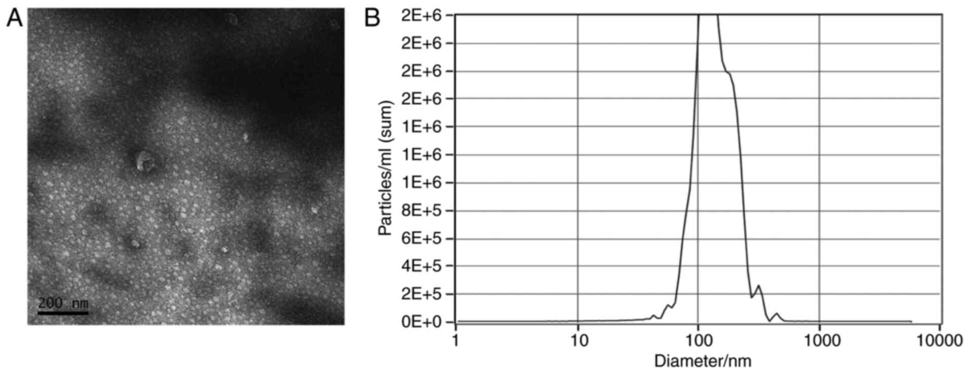
The ultrastructure of the ADSC-derived extracellular
nanovesicles was analyzed using transmission electron microscopy.
ADSC-derived extracellular nanovesicle pellets were transferred to
carbon-coated 200-mesh copper electron microscopy grids and
incubated for 10 min at room temperature. Subsequently, the
extracellular nanovesicles were stained with uranyl acetate and
washed with PBS three times. After drying at room temperature, they
were observed under a transmission electron microscope (19).
Particle size distributions were measured via
NanoSight analysis. The concentration and size distribution of
ADSC-derived extracellular nanovesicles were measured using tunable
resistive pulse sensing analysis with qNano (Izon Science, Ltd.).
ADSC-derived extracellular nanovesicles were placed in the Nanopore
(NP150; cat. no. A37355; Izon Science, Ltd.) at a 47.0-mm stretch
with a voltage of 0.6 V. Izon Control Suite software v.2.2 (Izon
Science, Ltd.) was used for data analysis.
Cell proliferation and migration
A Cell Counting Kit-8 (CCK-8; Dojindo Molecular
Technologies, Inc.) was used to evaluate NP cell proliferation. In
brief, 103 NP cells in 100 µl were seeded into each well
of a 96-well plate. After 12 h, 50 µg/ml extracellular nanovesicles
were added to the wells in the experimental group, while no
extracellular nanovesicles were added to the wells of the control
group. The medium was replaced every 2 days using fresh medium with
or without extracellular nanovesicles and medium with extracellular
vesicle-depleted FBS was used in the functional assays. At
different time-points (1, 3, 5 and 7 days), 10 µl of CCK-8 solution
was added to each well, along with 90 µl of high-glucose DMEM
without FBS (Gibco; Thermo Fisher Scientific, Inc.). After another
3 h of incubation, the absorbance was measured
spectrophotometrically at a wavelength of 450 nm with a microplate
reader.
Scratch wound assays were used to evaluate the
effects of extracellular nanovesicles on NP-cell migration, as
described previously (20). In
brief, 2x10⁵ cells/well were seeded into 6-well plates
and incubated for 12 h. A sterile 200-µl pipette tip was used to
make a scratch when the cells were 80% confluent and non-adherent
cells were removed by washing with PBS. The cells were cultured
with or without 50 µg/ml extracellular nanovesicles. Images were
acquired at 0, 12 and 24 h, and Image-Pro Plus 6.0 software (Media
Cybernetics) was used to measure the scratched areas.
Chondrocytic gene expression
The total RNA was extracted from each specimen using
TRIzol® reagent and complementary DNA was obtained by
reverse transcription (RT) using the SuperScript First-Strand
Synthesis system (Thermo Fisher Scientific, Inc.) for PCR.
RT-quantitative PCR was performed to evaluate the expression levels
of collagen-II (forward primer, 5'-GAGCCAAAGGATCTGCTGGT-3') and
reverse primer, 5'-TTGGGGCCTTGTTCACCTTT-3'), aggrecan (forward
primer, 5'-AAAAGGAGGCCACAGTGCTT-3' and reverse primer,
5'-GGCCGTACCAATCTCACACA-3') and Sox-9 (forward primer,
5'-AGGAGAACCCCAAGATGCAC-3' and reverse primer,
5'-GAGGCGTTTTGCTTCGTCAA-3'). The expression of GAPDH (forward
primer, 5'-GAGAAGGCTGGGGCTCATTT-3' and reverse primer,
5'-AGTGATGGCATGGACTGTGG-3) was quantified as an internal control.
The PCR was performed using SYBR Green PCR master mix (Thermo
Fisher Scientific, Inc.) and an ABI Prism 7000 Sequence Detection
system with ABI Prism 7000 software (Applied Biosystems; Thermo
Fisher Scientific, Inc.). The PCR thermocycling conditions were as
follows: 95˚C for 15 min; 35 cycles of 94˚C for 1 min, 59˚C for 1
min and 72˚C for 1 min; and 72˚C for 10 min. The specificity of the
amplification of the expected DNA fragments was confirmed using 2%
agarose gel electrophoresis and by analysis of the melting curves.
An amplification reaction control with no RT enzyme was performed
in order to assess the interference of potential genomic DNA in the
RNA solution. The relative gene expression was calculated using the
2-∆∆Cq method (21).
Telomerase activity detected by
PCR-ELISA
Telomerase PCR-ELISA (Thermo Fisher Scientific,
Inc.) was used to determine the telomerase activity following the
manufacturer's protocols and as described previously (22). After 7 days of incubation, the NP
cells cultured with or without extracellular nanovesicles were
digested, collected and homogenized in a lysis buffer, and
centrifuged at 100 x g for 10 min at 4˚C. The extracts were sent to
an internal department for telomeric repeat amplification.
Subsequently, the elongated fragments were amplified by PCR and the
PCR products were quantified by ELISA, as previously described
(22). The results were normalized
to those obtained for a standard.
Levels of inflammatory cytokines
ELISAs were performed to evaluate the expression
levels of inflammatory cytokines in the two groups. Media were
collected and centrifuged at 168 x g for 5 min at 4˚C to remove
cellular debris, and subsequently, IL-1α (cat. no. SLA50, R&D
Systems), IL-1β (cat. no. SLB50, R&D), IL-6 (cat. no. S6050,
R&D), IL-17 (cat. no. S1700, R&D), NF-κB-p65 (cat. no.
ab176648, Abcam) and TNF-α (cat. no. STA00D, R&D) in the
supernatant were measured using Quantikine ELISA kits according to
the manufacturer's protocols.
Statistical analysis
Cell proliferation, gene expression and protein
levels are presented as the mean ± standard deviation and were
analyzed by one-way ANOVA followed by Tukey's post-hoc test.
Statistical analysis was performed using GraphPad Prism version 5.0
(GraphPad Software, Inc.). P<0.05 was considered to indicate
statistical significance.
Results
Characteristics of exosomes
Observation by transmission electron microscopy
indicated that ADSC-derived extracellular nanovesicles were
characterized by diameters in the range of 40-200 nm and most of
them presented with a round shape (Fig.
1A). It was observed by NanoSight analysis that the peak of the
distribution of diameters of extracellular nanovesicles was 120 nm
(Fig. 1B).
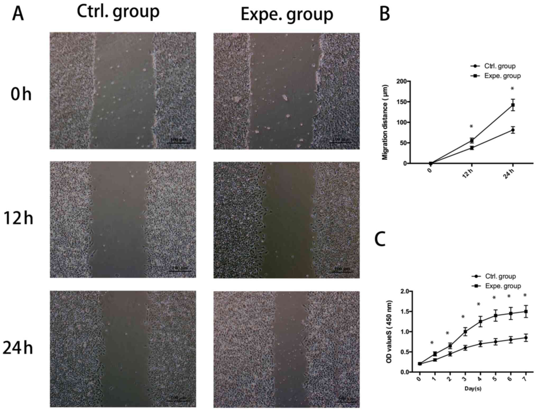
Effects of ADSC-derived extracellular
nanovesicles on the proliferation and migration of human NP
cells
As presented in Fig.
2A and B, the migration of NP
cells cultured in the presence of ADSC-extracellular nanovesicles
in the experimental group was increased compared with that of the
control group (P<0.05). It was also indicated that human NP-cell
proliferation was significantly increased in the presence of
extracellular nanovesicles from days 1-7 (Fig. 2C).
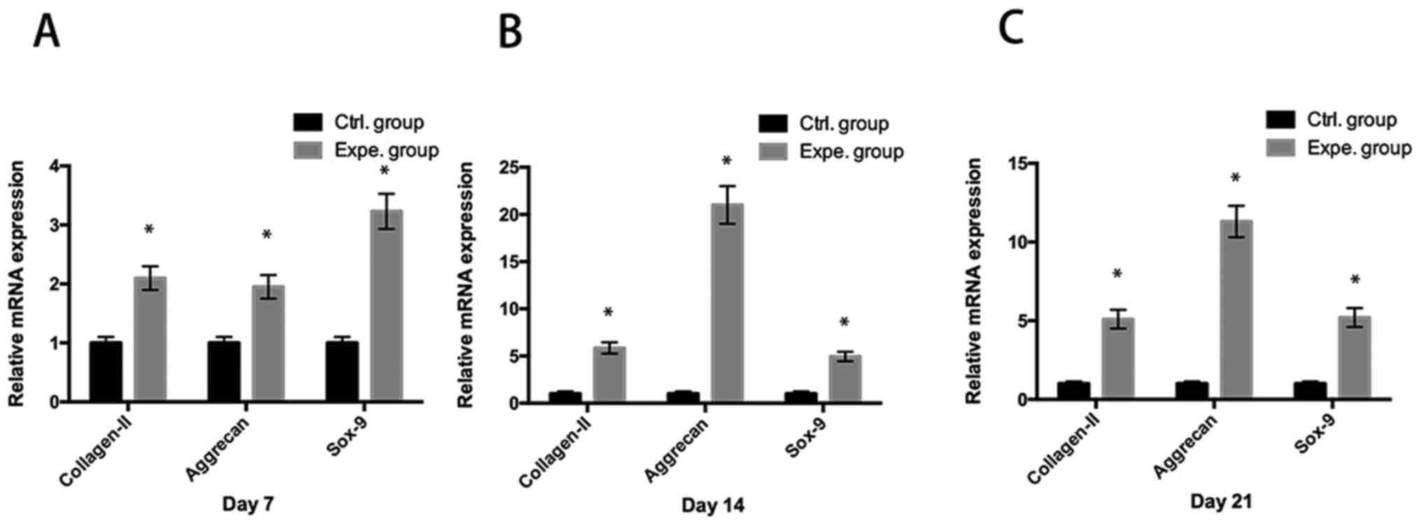
ADSC-derived extracellular
nanovesicles promote the chondrocytic gene expression in human NP
cells
It was observed that ADSC-derived extracellular
nanovesicles significantly promoted the mRNA expression of
chondrocytic genes (collagen-II, aggrecan and Sox-9) in human NP
cells in the experimental group compared with that in the control
group (P<0.05) at day 7 (Fig.
3A), day 14 (Fig. 3B) and day
21 (Fig. 3C).
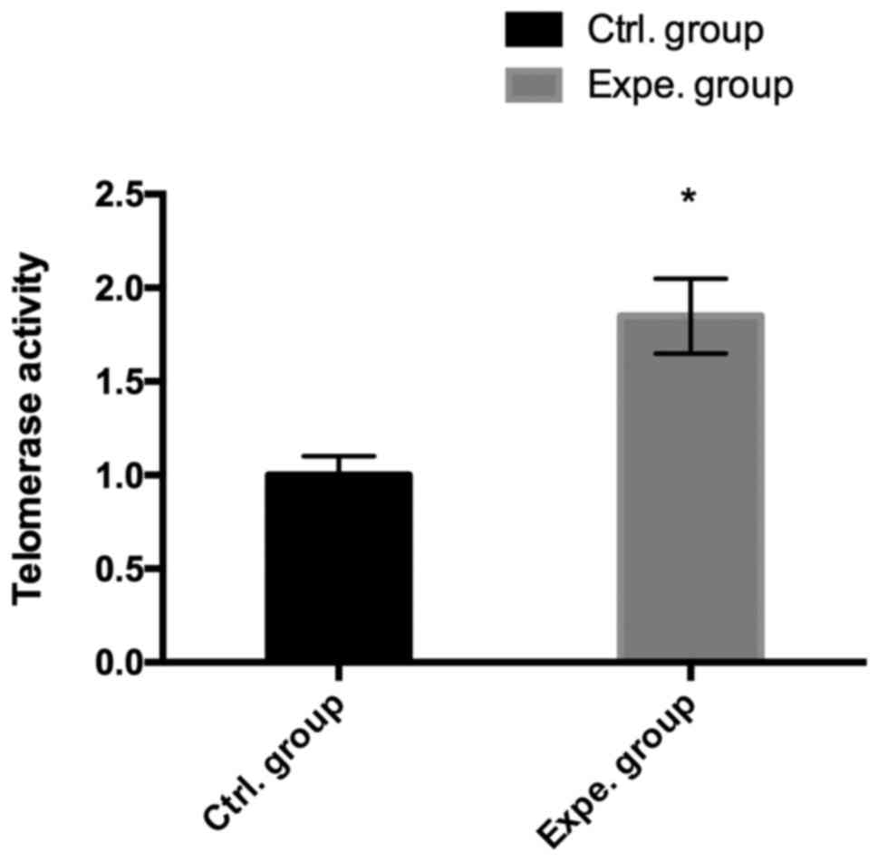
Telomerase activity in human NP
cells
The effects of ADSC-derived extracellular
nanovesicles on the telomerase activity in human NP cells were
examined. Compared with that in the control group, a significant
increase in telomerase activity was observed in human NP cells
treated with extracellular nanovesicles in the experimental group
(P<0.05; Fig. 4).
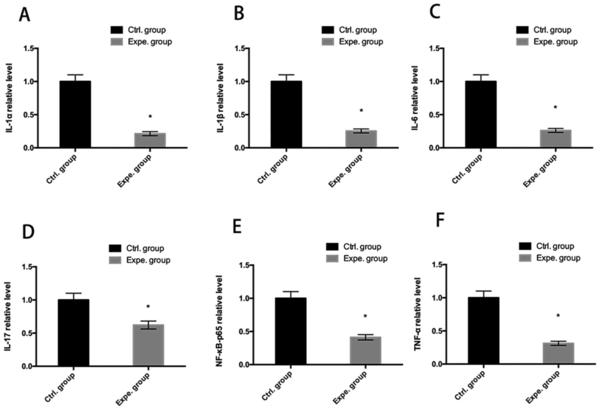
Levels of inflammatory cytokines
Human NP cells were cultured with or without
extracellular nanovesicles for 7 days. Inflammatory cytokine levels
were measured by ELISAs. As indicated in Fig. 5, IL-1α, IL-1β, IL-6, IL-17,
NF-κB-p65 and TNF-α were significantly reduced after 7 days culture
with exosomes (P<0.05), indicating that the extracellular
nanovesicles inhibited the secretion of inflammatory cytokines
(Fig. 5).
Discussion
Emerging evidence has indicated that MSCs are
promising options for the treatment of human intervertebral disc
degeneration (11,23). Cao et al (24) reported that co-culture of BMSCs may
be able to delay NP-cell matrix degeneration and co-culturing BMSCs
with NP cells appeared to result in the promotion of the gene
expression of aggrecan, collagen-II and Sox-9. Leung et al
(25) demonstrated that MSCs are
able to suppress abnormal deposition of collagen-I in the NP and
modulate the profibrotic mediators matrix metalloproteinase-12 and
heat shock protein 47, thus reducing collagen aggregation and
maintaining proper fibrillar properties and function. However, the
mechanisms underlying the therapeutic effects of MSCs have remained
elusive. A recent study indicated that the therapeutic action of
MSCs is not dependent on the engraftment of MSCs at the site of
injury or the differentiation capability of the transplanted MSCs,
but rather the secretion of paracrine factors (26).
It has been reported that the therapeutic efficacy
is attributed to the secretion of extracellular nanovesicles,
secreted bilipid membrane vesicles of 40-100 nm in diameter that
have the natural ability to transmit intercellular signals, such as
carrying small RNAs, DNA and proteins (27). In the present study, the isolated
ADSC-derived extracellular nanovesicles were characterized and
transmission electron microscopy and laser diffraction demonstrated
the cup-shaped morphology of the isolated extracellular
nanovesicles with diameters of 40-100 nm. NP cells have been
reported to have a low capacity for proliferation and migration
(28) and the effect of
extracellular nanovesicles on the proliferation and migration of NP
cells was evaluated in the present study. The results suggested
that ADSC-derived extracellular nanovesicles improved the
proliferation and migration activity of the degenerated NP cells.
Lu et al (29) reported that
in their experiment, BM-MSC-derived exosomes promoted NP-cell
proliferation.
Intervertebral disc degeneration is characterized by
decreased expression of chondrogenic genes, such as aggrecan,
collagen-II and Sox-9. Kim et al (30) demonstrated that co-culture with
adipose-derived stem cells restored the chondrogenic properties of
degenerative NP cells. In the present study, the expression levels
of chondrogenic genes were significantly higher in extracellular
nanovesicle-treated degenerative NP cells than those in the
degenerative NP cells cultured alone, indicating that extracellular
nanovesicles improved the viability of degenerative NP cells. This
was further supported by the results for telomerase activity, which
suggested that extracellular nanovesicles improved telomerase
activity.
Intervertebral disc degeneration is a chronic
degenerative disease characterized by back, neck and/or radicular
pain (31). It is well known that
increases in the levels of inflammatory cytokines secreted by
intervertebral disc cells have a role in the occurrence of disc
degeneration disease, such as IL-1α, IL-1β, IL-6, IL-17, NF-κB-p65
and TNF-α (32). The release of
these inflammatory cytokines stimulates chemokine production and
extracellular matrix degradation and then causes degeneration of
the intervertebral disc tissues, further resulting in lumbar disc
herniation and radicular pain (33). Recent evidence has demonstrated that
MSCs have great potential for application in the treatment of
intervertebral disc degeneration, as MSCs have chondrogenic
differentiation potential, but also have a vital role in
immunoregulation and tissue repair/regeneration through the
secretion of various soluble factors, such as extracellular
nanovesicles (34). The present
study indicated that ADSC-derived extracellular nanovesicle
treatment decreased the secretion of inflammatory cytokines (IL-1α,
IL-1β, IL-6, IL-17, NF-κB-p65 and TNF-α).
As a limitation to the present study, only one dose
of extracellular vesicles was used when performing functional
assays, as the quantity of extracellular vesicles that may be
isolated from MSCs is low. Multiple doses of extracellular vesicles
should be used when performing functional assays in future
studies.
In conclusion, ADSC-derived extracellular
nanovesicles improved the proliferation and migration of
degenerative NP cells and inhibited inflammatory activity, which
may provide a promising option for the treatment of intervertebral
disc degeneration disease.
Acknowledgements
Not applicable.
Funding
This study was supported by the Jiangsu Provincial Medical
Innovation Team (grant no. CXTDB2017004), the Natural Science
Foundation of Yangzhou (grant no. YZ2017116) and Key Supporting
Technical Projects by Norhern Jiangsu People's Hospital (grant no.
fcjs202004).
Availability of data and materials
The datasets used and/or analyzed during the current
study are available from the corresponding author on reasonable
request.
Authors' contributions
ZZ contributed to the conception and design of the
study, the acquisition, analysis and interpretation of the data of
the study. XMF contributed to the acquisition, analysis and
interpretation of data of the study. LZ contributed to the analysis
and interpretation of data of the study. JY contributed to the
analysis and interpretation of data of the study, and drafting of
the work and its critical revision for important intellectual
content. JH contributed to the conception and design of the study.
JC contributed to the conception and design of the study. SZ
contributed to the conception and design of the study, the
acquisition, analysis and interpretation of the data of the study.
QW contributed to the analysis and interpretation of data of the
study. All authors contributed to the drafting of the work and its
critical revision for important intellectual content, read and
approved the final version of the manuscript and agreed to be
accountable for all aspects of the study in ensuring that questions
related to the accuracy or integrity of any part of the work are
appropriately investigated and resolved. ZZ and QW confirm the
authenticity of all the raw data.
Ethics approval and consent to
participate
This study was approved by the Ethics Committee of
Northern Jiangsu People's Hospital (Yangzhou, China) and informed
consent was obtained from all patients.
Patient consent for publication
Not applicable.
Competing interests
The authors declare that they have no competing
interests.
References
|
1
|
Fernandez-Moure J, Moore CA, Kim K, Karim
A, Smith K, Barbosa Z, Van Eps J, Rameshwar P and Weiner B: Novel
therapeutic strategies for degenerative disc disease: Review of
cell biology and intervertebral disc cell therapy. SAGE Open Med.
6(2050312118761674)2018.PubMed/NCBI View Article : Google Scholar
|
|
2
|
Petit A and Roquelaure Y: Low back pain,
intervertebral disc and occupational diseases. Int J Occup Saf
Ergon. 21:15–19. 2015.PubMed/NCBI View Article : Google Scholar
|
|
3
|
Steelman T, Lewandowski L, Helgeson M,
Wilson K, Olsen C and Gwinn D: Population-based risk factors for
the development of degenerative disc disease. Clin Spine Surg.
31:E409–E412. 2018.PubMed/NCBI View Article : Google Scholar
|
|
4
|
Jakoi AM, Pannu G, D'Oro A, Buser Z, Pham
MH, Patel NN, Hsieh PC, Liu JC, Acosta FL, Hah R and Wang JC: The
clinical correlations between diabetes, cigarette smoking and
obesity on intervertebral degenerative disc disease of the lumbar
spine. Asian Spine J. 11:337–347. 2017.PubMed/NCBI View Article : Google Scholar
|
|
5
|
Teraguchi M, Yoshimura N, Hashizume H,
Muraki S, Yamada H, Minamide A, Oka H, Ishimoto Y, Nagata K,
Kagotani R, et al: Prevalence and distribution of intervertebral
disc degeneration over the entire spine in a population-based
cohort: The wakayama spine study. Osteoarthritis Cartilage.
22:104–110. 2014.PubMed/NCBI View Article : Google Scholar
|
|
6
|
Clouet J, Fusellier M, Camus A, Le Visage
C and Guicheux J: Intervertebral disc regeneration: From cell
therapy to the development of novel bioinspired endogenous repair
strategies. Adv Drug Deliv Rev. 146:306–324. 2019.PubMed/NCBI View Article : Google Scholar
|
|
7
|
Alvi MA, Kerezoudis P, Wahood W, Goyal A
and Bydon M: Operative approaches for lumbar disc herniation: A
systematic review and multiple treatment meta-analysis of
conventional and minimally invasive surgeries. World Neurosurg.
114:391–407 e2. 2018.PubMed/NCBI View Article : Google Scholar
|
|
8
|
Ayala-Cuellar AP, Kang JH, Jeung EB and
Choi KC: Roles of mesenchymal stem cells in tissue regeneration and
immunomodulation. Biomol Ther (Seoul). 27:25–33. 2019.PubMed/NCBI View Article : Google Scholar
|
|
9
|
Richardson SM, Kalamegam G, Pushparaj PN,
Matta C, Memic A, Khademhosseini A, Mobasheri R, Poletti FL,
Hoyland JA and Mobasheri A: Mesenchymal stem cells in regenerative
medicine: Focus on articular cartilage and intervertebral disc
regeneration. Methods. 99:69–80. 2016.PubMed/NCBI View Article : Google Scholar
|
|
10
|
Yang F, Leung VYL, Luk KDK, Chan D and
Cheung KMC: Mesenchymal stem cells arrest intervertebral disc
degeneration through chondrocytic differentiation and stimulation
of endogenous cells. Mol Ther. 17:1959–1966. 2009.PubMed/NCBI View Article : Google Scholar
|
|
11
|
Centeno C, Markle J, Dodson E, Stemper I,
Williams CJ, Hyzy M, Ichim T and Freeman M: Treatment of lumbar
degenerative disc disease-associated radicular pain with
culture-expanded autologous mesenchymal stem cells: A pilot study
on safety and efficacy. J Transl Med. 15(197)2017.PubMed/NCBI View Article : Google Scholar
|
|
12
|
Raik S, Kumar A and Bhattacharyya S:
Insights into cell-free therapeutic approach: Role of stem cell
‘soup-ernatant’. Biotechnol Appl Biochem. 65:104–118.
2018.PubMed/NCBI View
Article : Google Scholar
|
|
13
|
Liu X, Yang Y, Li Y, Niu X, Zhao B, Wang
Y, Bao C, Xie Z, Lin Q and Zhu L: Integration of stem cell-derived
exosomes with in situ hydrogel glue as a promising tissue patch for
articular cartilage regeneration. Nanoscale. 9:4430–4438.
2017.PubMed/NCBI View Article : Google Scholar
|
|
14
|
Merino-Gonzalez C, Zuñiga FA, Escudero C,
Ormazabal V, Reyes C, Nova-Lamperti E, Salomón C and Aguayo C:
Mesenchymal stem cell-derived extracellular vesicles promote
angiogenesis: Potencial clinical application. Front Physiol.
7(24)2016.PubMed/NCBI View Article : Google Scholar
|
|
15
|
Bucan V, Vaslaitis D, Peck CT, Strauß S,
Vogt PM and Radtke C: Effect of exosomes from rat adipose-derived
mesenchymal stem cells on neurite outgrowth and sciatic nerve
regeneration after crush injury. Mol Neurobiol. 56:1812–1824.
2019.PubMed/NCBI View Article : Google Scholar
|
|
16
|
Zhu Y, Wu Y, Cheng J, Wang Q, Li Z, Wang
Y, Wang D, Wang H, Zhang W, Ye J, et al: Pharmacological activation
of TAZ enhances osteogenic differentiation and bone formation of
adipose-derived stem cells. Stem Cell Res Ther.
9(53)2018.PubMed/NCBI View Article : Google Scholar
|
|
17
|
Chen D, Xia D, Pan Z, Xu D, Zhou Y, Wu Y,
Cai N, Tang Q, Wang C, Yan M, et al: Metformin protects against
apoptosis and senescence in nucleus pulposus cells and ameliorates
disc degeneration in vivo. Cell Death Dis. 7(e2441)2016.PubMed/NCBI View Article : Google Scholar
|
|
18
|
Baglio SR, Rooijers K, Koppers-Lalic D,
Verweij FJ, Lanzón MP, Zini N, Naaijkens B, Perut F, Niessen HWM,
Baldini N and Pegtel DM: Human bone marrow- and adipose-mesenchymal
stem cells secrete exosomes enriched in distinctive miRNA and tRNA
species. Stem Cell Res Ther. 6(127)2015.PubMed/NCBI View Article : Google Scholar
|
|
19
|
Qu Y, Zhang Q, Cai X, Li F, Ma Z, Xu M and
Lu L: Exosomes derived from miR-181-5p-modified adipose-derived
mesenchymal stem cells prevent liver fibrosis via autophagy
activation. J Cell Mol Med. 21:2491–2502. 2017.PubMed/NCBI View Article : Google Scholar
|
|
20
|
Zhang J, Guan J, Niu X, Hu G, Guo S, Li Q,
Xie Z, Zhang C and Wang Y: Exosomes released from human induced
pluripotent stem cells-derived MSCs facilitate cutaneous wound
healing by promoting collagen synthesis and angiogenesis. J Transl
Med. 13(49)2015.PubMed/NCBI View Article : Google Scholar
|
|
21
|
Barra GB, Santa Rita TH, Almeida ALSC,
Jácomo RH and Nery LFA: Serum has higher proportion of janus kinase
2 V617F mutation compared to paired EDTA-whole blood sample: A
model for somatic mutation quantification using qPCR and the
2-∆∆Cq method. Diagnostics (Basel).
10(153)2020.PubMed/NCBI View Article : Google Scholar
|
|
22
|
Chung SS, Adekoya D, Enenmoh I, Clarke O,
Wang P, Sarkyssian M, Wu Y and Vadgama JV: Salinomycin abolished
STAT3 and STAT1 interactions and reduced telomerase activity in
colorectal cancer cells. Anticancer Res. 37:445–453.
2017.PubMed/NCBI View Article : Google Scholar
|
|
23
|
Perez-Cruet M, Beeravolu N, McKee C,
Brougham J, Khan I, Bakshi S and Chaudhry GR: Potential of human
nucleus pulposus-like cells derived from umbilical cord to treat
degenerative disc disease. Neurosurgery. 84:272–283.
2019.PubMed/NCBI View Article : Google Scholar
|
|
24
|
Cao C, Zou J, Liu X, Shapiro A, Moral M,
Luo Z, Shi Q, Liu J, Yang H and Ebraheim N: Bone marrow mesenchymal
stem cells slow intervertebral disc degeneration through the NF-kB
pathway. Spine J. 15:530–538. 2015.PubMed/NCBI View Article : Google Scholar
|
|
25
|
Leung VY, Aladin DMK, Lv F, Tam V, Sun Y,
Lau RYC, Hung SC, Ngan AHW, Tang B, Lim CT, et al: Mesenchymal stem
cells reduce intervertebral disc fibrosis and facilitate repair.
Stem Cells. 32:2164–2177. 2014.PubMed/NCBI View Article : Google Scholar
|
|
26
|
Lai RC, Tan SS, The BJ, Sze SK, Arslan F,
de Kleijn DP, Choo A and Lim SK: Proteolytic potential of the MSC
exosome proteome: Implications for an exosome-mediated delivery of
therapeutic proteasome. Int J Proteomics.
2012(971907)2012.PubMed/NCBI View Article : Google Scholar
|
|
27
|
Vizoso FJ, Eiro N, Cid S, Schneider J and
Perez-Fernandez R: Mesenchymal stem cell secretome: Toward
cell-free therapeutic strategies in regenerative medicine. Int J
Mol Sci. 18(1852)2017.PubMed/NCBI View Article : Google Scholar
|
|
28
|
Guillaume O, Naqvi SM, Lennon K and
Buckley CT: Enhancing cell migration in shape-memory
alginate-collagen composite scaffolds: In vitro and ex vivo
assessment for intervertebral disc repair. J Biomater Appl.
29:1230–1246. 2015.PubMed/NCBI View Article : Google Scholar
|
|
29
|
Lu K, Li HY, Yang K, Wu JL, Cai XW, Zhou Y
and Li CQ: Exosomes as potential alternatives to stem cell therapy
for intervertebral disc degeneration: In-vitro study on exosomes in
interaction of nucleus pulposus cells and bone marrow mesenchymal
stem cells. Stem Cell Res Ther. 8(108)2017.PubMed/NCBI View Article : Google Scholar
|
|
30
|
Kim JS, Kwon D, Cha BH, Moon BK, Jeong YG,
Han IB, Park H and Lee SH: Restoration of chondrogenic properties
of degenerative nucleus pulposus cells by repeated co-culture with
adipose-derived stem cells. J Industrial Engineering Chem.
60:185–191. 2018.
|
|
31
|
Dowdell J, Erwin M, Choma T, Vaccaro A,
Iatridis J and Cho SK: Intervertebral disk degeneration and repair.
Neurosurgery. 80 (Suppl 3):S46–S54. 2017.PubMed/NCBI View Article : Google Scholar
|
|
32
|
Khan AN, Jacobsen HE, Khan J, Filippi CG,
Levine M, Lehman RA Jr, Riew KD, Lenke LG and Chahine NO:
Inflammatory biomarkers of low back pain and disc degeneration: A
review. Ann N Y Acad Sci. 1410:68–84. 2017.PubMed/NCBI View Article : Google Scholar
|
|
33
|
Risbud MV and Shapiro IM: Role of
cytokines in intervertebral disc degeneration: Pain and disc
content. Nat Rev Rheumatol. 10:44–56. 2014.PubMed/NCBI View Article : Google Scholar
|
|
34
|
Lai RC, Yeo RW and Lim SK: Mesenchymal
stem cell exosomes. Semin Cell Dev Biol. 40:82–88. 2015.PubMed/NCBI View Article : Google Scholar
|