Open Access

Overview of the anatomical basis of the piriformis syndrome‑dissection with magnetic resonance correlation

  • Authors:
    • Ofelia-Costina Goidescu
    • Mihaly Enyedi
    • Adrian-Daniel Tulin
    • Raluca Tulin
    • Ileana Adela Vacaroiu
    • Adriana Elena Nica
    • Dorin Dragos
    • Dorin Ionescu
    • Dragos Georgescu
    • Adrian Miron
    • Florin-Mihail Filipoiu
  • View Affiliations

  • Published online on: December 3, 2021     https://doi.org/10.3892/etm.2021.11036
  • Article Number: 113
  • Copyright: © Goidescu et al. This is an open access article distributed under the terms of Creative Commons Attribution License.

Metrics: Total Views: 0 (Spandidos Publications: | PMC Statistics: )
Total PDF Downloads: 0 (Spandidos Publications: | PMC Statistics: )


Abstract

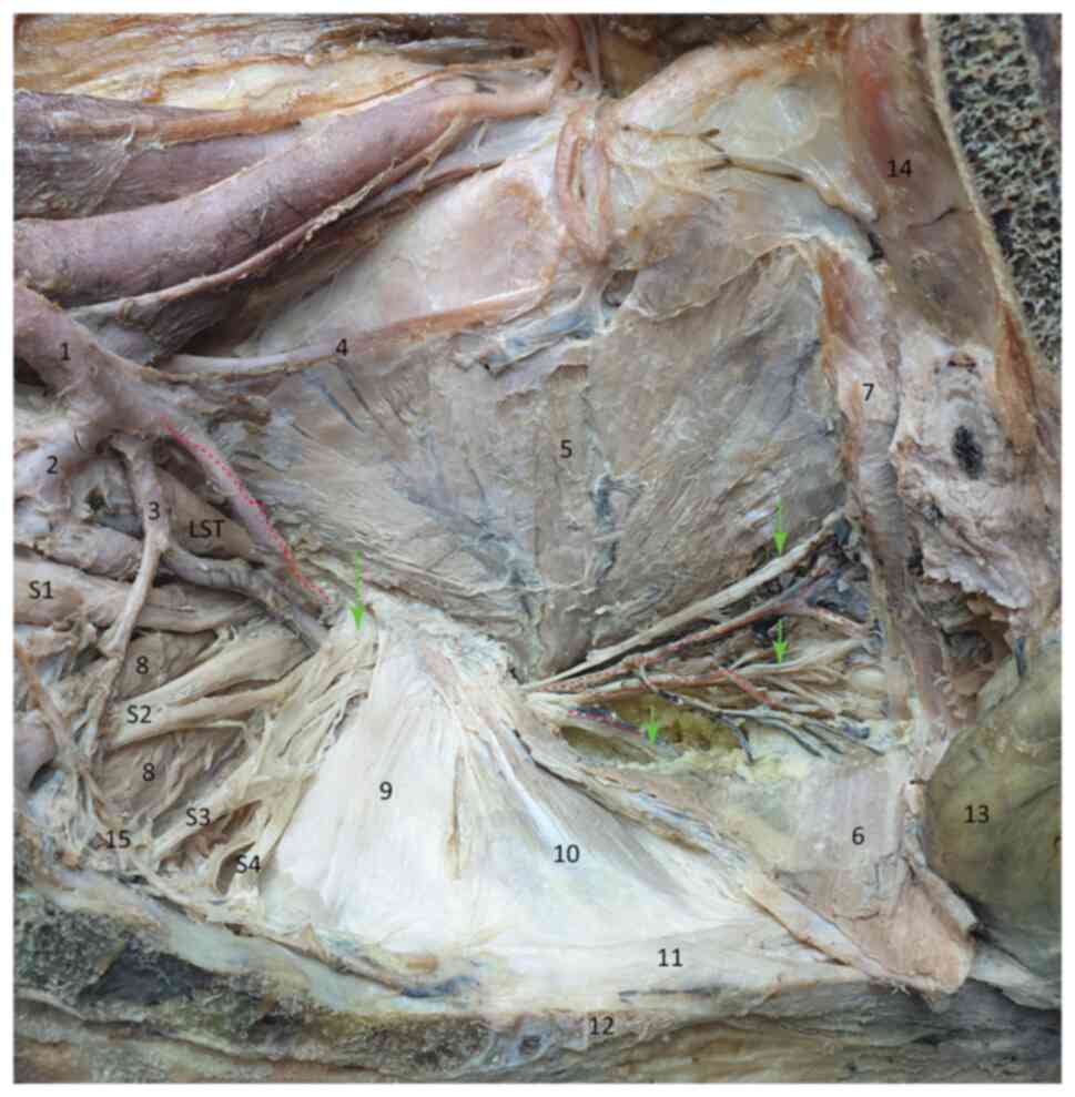
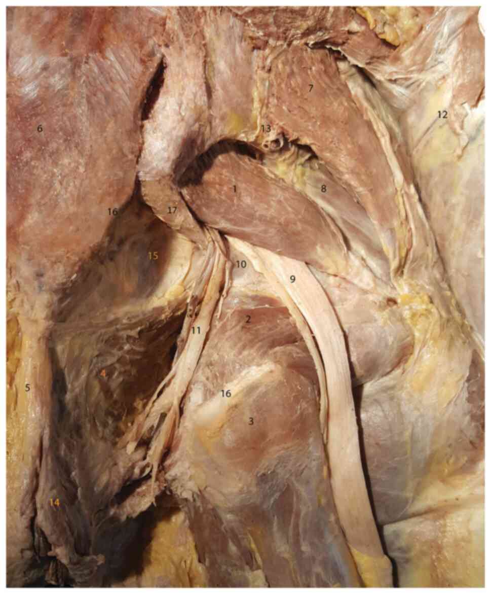
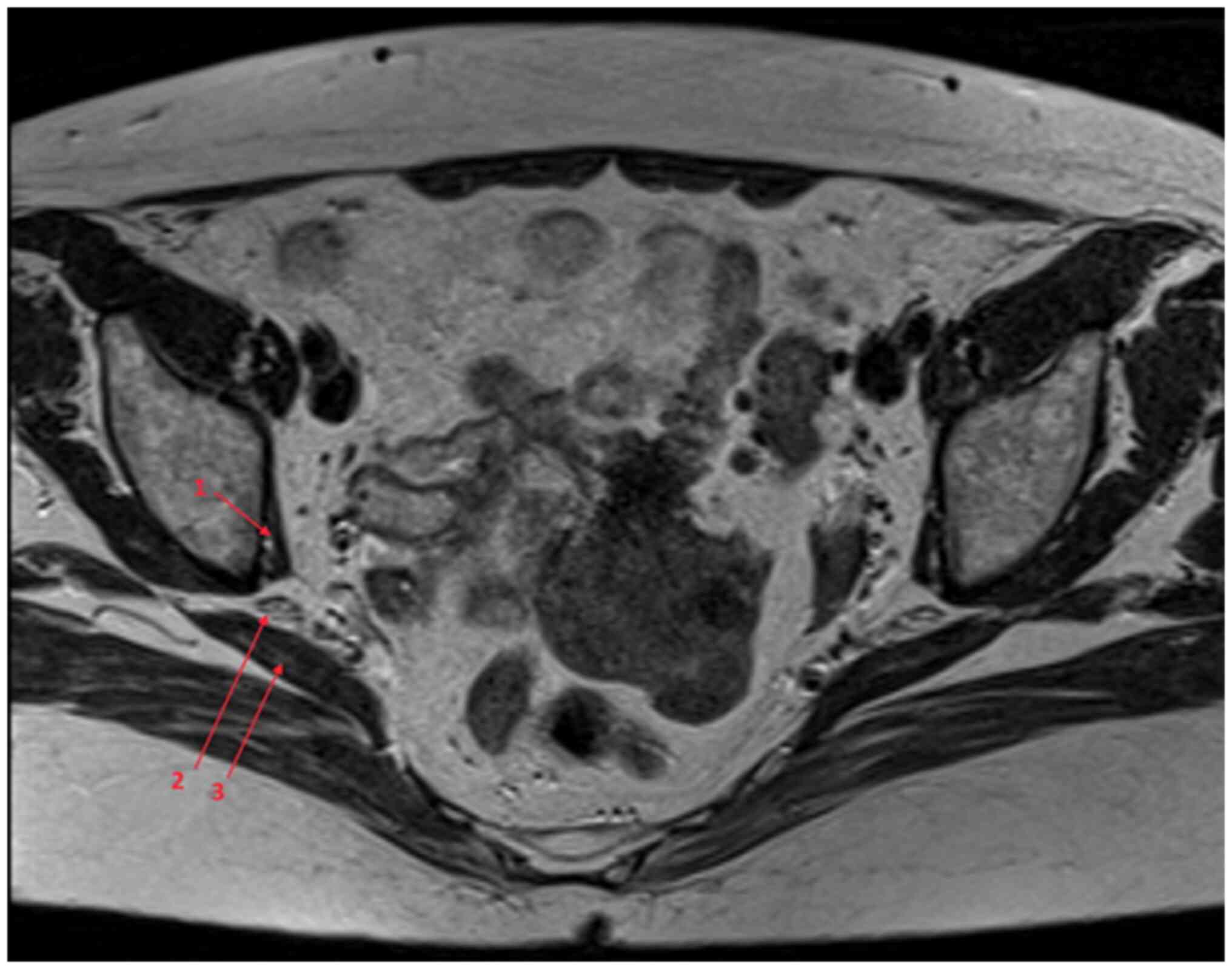
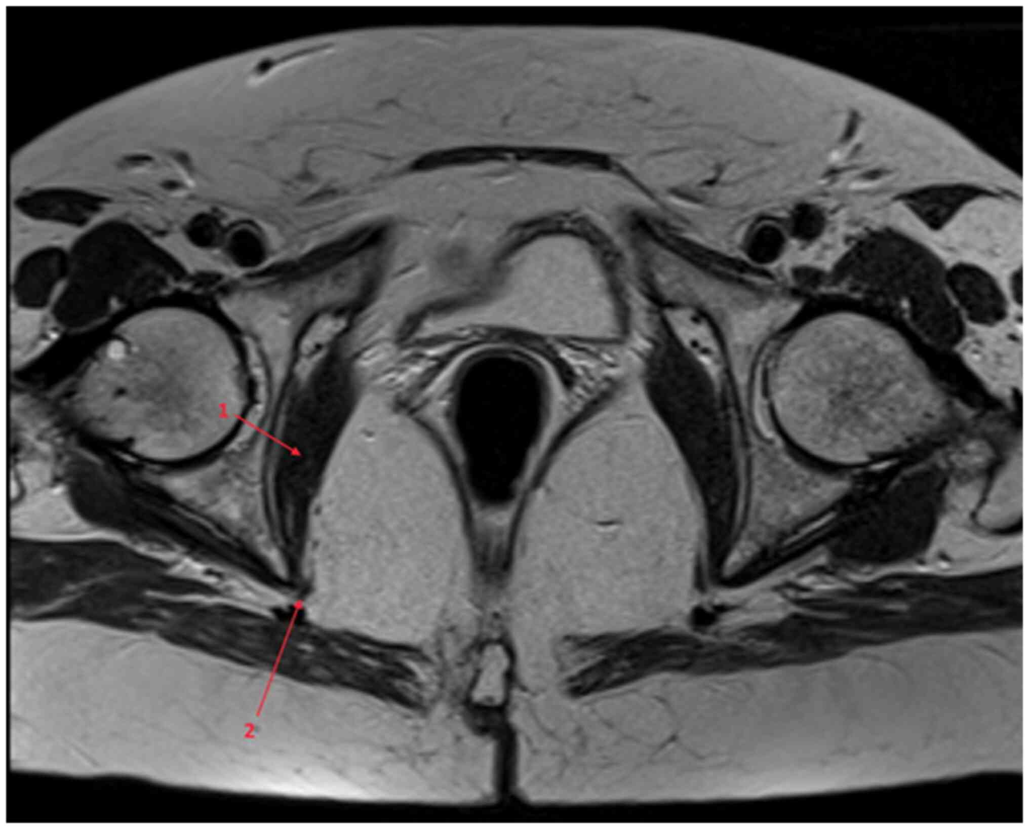
The piriformis syndrome is one of the most commonly misdiagnosed causes of lower back and gluteal pain caused by the compression of the sciatic nerve and the internal pudendal neurovascular bundle by the piriformis muscle. Although this syndrome was first suggested over 90 years ago, its diagnosis still represents a challenge for clinicians. In the present study, dissection was used to determine the intra‑ and extrapelvic anatomical course of the internal pudendal nerve and the data were compared with the information obtainable through MRI examination, in order to identify the piriformis syndrome and to differentiate it from other causes of internal pudendal neuralgia. Thorough dissections of the pelvis and deep gluteal region were conducted on female cadavers, which were correlated with MRI scans, in order to describe the course of the internal pudendal nerve in contact with the piriformis muscle. The dissection findings and MRI scans obtained allowed us to describe and demonstrate the compression points along the course of the sciatic nerve and the internal pudendal bundle, the anatomical correlations between the piriformis muscle and the nervous structures around it, emphasizing the areas most susceptible to possible nerve impingement syndromes. In the anatomic trajectory of the sciatic nerve and the internal pudendal bundle there are multiple contact points with anatomical structures that may lead to compression of the nerve structures, generating symptoms that comprise the piriformis syndrome. The present study sought to establish clear osseous landmarks that may help evaluate these associations and possible nerve compressions on pelvic MRI examination.
View Figures
View References

Related Articles

Journal Cover

February-2022
Volume 23 Issue 2

Print ISSN: 1792-0981
Online ISSN:1792-1015

Sign up for eToc alerts

Recommend to Library

Copy and paste a formatted citation
x
Spandidos Publications style
Goidescu O, Enyedi M, Tulin A, Tulin R, Vacaroiu IA, Nica AE, Dragos D, Ionescu D, Georgescu D, Miron A, Miron A, et al: Overview of the anatomical basis of the piriformis syndrome‑dissection with magnetic resonance correlation. Exp Ther Med 23: 113, 2022
APA
Goidescu, O., Enyedi, M., Tulin, A., Tulin, R., Vacaroiu, I.A., Nica, A.E. ... Filipoiu, F. (2022). Overview of the anatomical basis of the piriformis syndrome‑dissection with magnetic resonance correlation. Experimental and Therapeutic Medicine, 23, 113. https://doi.org/10.3892/etm.2021.11036
MLA
Goidescu, O., Enyedi, M., Tulin, A., Tulin, R., Vacaroiu, I. A., Nica, A. E., Dragos, D., Ionescu, D., Georgescu, D., Miron, A., Filipoiu, F."Overview of the anatomical basis of the piriformis syndrome‑dissection with magnetic resonance correlation". Experimental and Therapeutic Medicine 23.2 (2022): 113.
Chicago
Goidescu, O., Enyedi, M., Tulin, A., Tulin, R., Vacaroiu, I. A., Nica, A. E., Dragos, D., Ionescu, D., Georgescu, D., Miron, A., Filipoiu, F."Overview of the anatomical basis of the piriformis syndrome‑dissection with magnetic resonance correlation". Experimental and Therapeutic Medicine 23, no. 2 (2022): 113. https://doi.org/10.3892/etm.2021.11036