|
1
|
Goldstein JL and Brown MS: Regulation of
the mevalonate pathway. Nature. 343:425–430. 1990.PubMed/NCBI View
Article : Google Scholar
|
|
2
|
Bathaie SZ, Ashrafi M, Azizian M and
Tamanoi F: Mevalonate Pathway and Human Cancers. Curr Mol
Pharmacol. 10:77–85. 2017.PubMed/NCBI View Article : Google Scholar
|
|
3
|
Yeganeh B, Wiechec E, Ande SR, Sharma P,
Moghadam AR, Post M, Freed DH, Hashemi M, Shojaei S, Zeki AA, et
al: Targeting the mevalonate cascade as a new therapeutic approach
in heart disease, cancer and pulmonary disease. Pharmacol Ther.
143:87–110. 2014.PubMed/NCBI View Article : Google Scholar
|
|
4
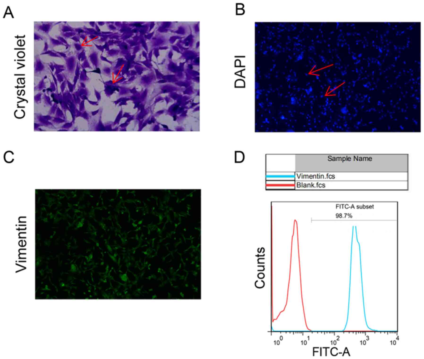
|
Ference BA, Ray KK, Catapano AL, Ference
TB, Burgess S, Neff DR, Oliver-Williams C, Wood AM, Butterworth AS,
Di Angelantonio E, et al: Mendelian Randomization Study of ACLY and
Cardiovascular Disease. N Engl J Med. 380:1033–1042.
2019.PubMed/NCBI View Article : Google Scholar
|
|
5
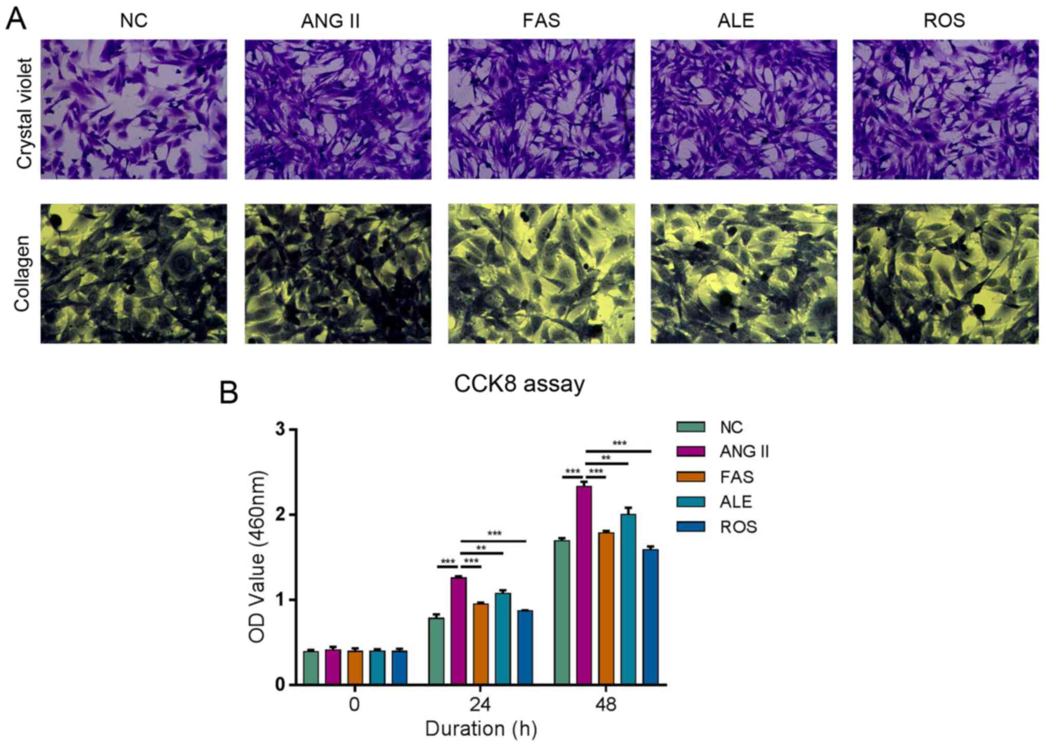
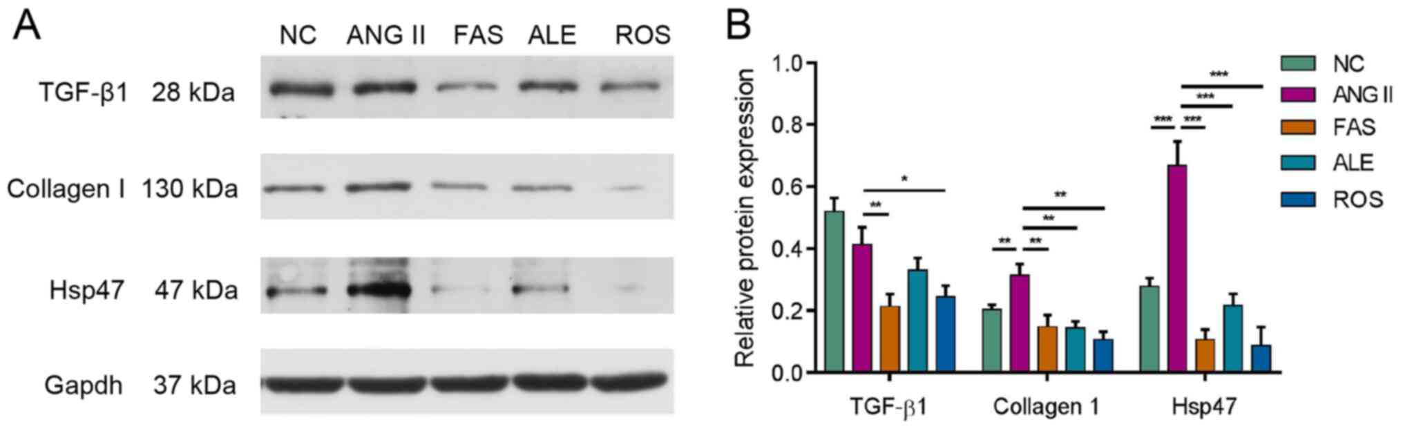
|
Raper A, Kolansky DM and Cuchel M:
Treatment of familial hypercholesterolemia: Is there a need beyond
statin therapy? Curr Atheroscler Rep. 14:11–16. 2012.PubMed/NCBI View Article : Google Scholar
|
|
6
|
Oesterle A, Laufs U and Liao JK:
Pleiotropic effects of statins on the cardiovascular system. Circ
Res. 120:229–243. 2017.PubMed/NCBI View Article : Google Scholar
|
|
7
|
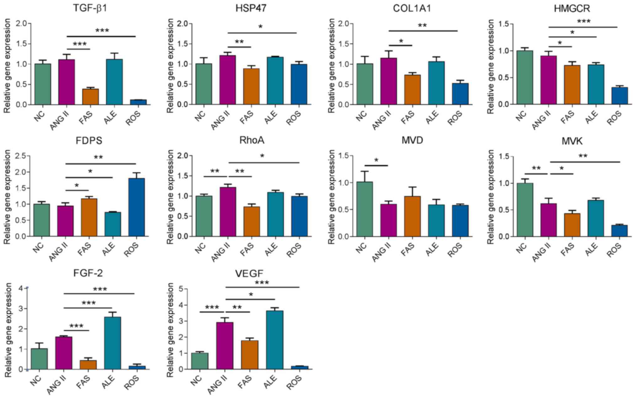
Steffens S and Mach F: Anti-inflammatory
properties of statins. Semin Vasc Med. 4:417–422. 2004.PubMed/NCBI View Article : Google Scholar
|
|
8
|
Greenwood J, Steinman L and Zamvil SS:
Statin therapy and autoimmune disease: From protein prenylation to
immunomodulation. Nat Rev Immunol. 6:358–370. 2006.PubMed/NCBI View
Article : Google Scholar
|
|
9
|
Ward NC, Watts GF and Eckel RH: Statin
toxicity. Circ Res. 124:328–350. 2019.PubMed/NCBI View Article : Google Scholar
|
|
10
|
Schleyer T, Hui S, Wang J, Zhang Z, Knapp
K, Baker J, Chase M, Boggs R and Simpson RJ Jr: Quantifying unmet
need in statin-treated hyperlipidemia patients and the potential
benefit of further LDL-C reduction through an EHR-based
retrospective cohort study. J Manag Care Spec Pharm. 25:544–554.
2019.PubMed/NCBI View Article : Google Scholar
|
|
11
|
Toth PP and Banach M: Statins: Then and
now. Methodist Debakey Cardiovasc J. 15:23–31. 2019.PubMed/NCBI View Article : Google Scholar
|
|
12
|
Nochioka K, Sakata Y, Miyata S, Miura M,
Takada T, Tadaki S, Ushigome R, Yamauchi T, Takahashi J and
Shimokawa H: CHART-2 Investigators. Prognostic impact of statin use
in patients with heart failure and preserved ejection fraction.
Circ J. 79:574–582. 2015.PubMed/NCBI View Article : Google Scholar
|
|
13
|
Kjekshus J, Apetrei E, Barrios V, Böhm M,
Cleland JG, Cornel JH, Dunselman P, Fonseca C, Goudev A, Grande P,
et al: CORONA Group: Rosuvastatin in older patients with systolic
heart failure. N Engl J Med. 357:2248–2261. 2007.PubMed/NCBI View Article : Google Scholar
|
|
14
|
Liu G, Zheng XX, Xu YL, Ru J, Hui RT and
Huang XH: Meta-analysis of the effect of statins on mortality in
patients with preserved ejection fraction. Am J Cardiol.
113:1198–1204. 2014.PubMed/NCBI View Article : Google Scholar
|
|
15
|
Xu H, Qing T, Shen Y, Huang J, Liu Y, Li
J, Zhen T, Xing K, Zhu S and Luo M: RNA-seq analyses the effect of
high-salt diet in hypertension. Gene. 677:245–250. 2018.PubMed/NCBI View Article : Google Scholar
|
|
16
|
Li X, Han J, Li L, Wang KJ and Hu SJ:
Effect of farnesyltransferase inhibition on cardiac remodeling in
spontaneously hypertensive rats. Int J Cardiol. 168:3340–3347.
2013.PubMed/NCBI View Article : Google Scholar
|
|
17
|
Zhao CZ, Zhao XM, Yang J, Mou Y, Chen B,
Wu HD, Dai DP, Ding J and Hu SJ: Inhibition of farnesyl
pyrophosphate synthase improves pressure overload induced chronic
cardiac remodeling. Sci Rep. 6(39186)2016.PubMed/NCBI View Article : Google Scholar
|
|
18
|
Yang J, Mou Y, Wu T, Ye Y, Jiang JC, Zhao
CZ, Zhu HH, Du CQ, Zhou L and Hu SJ: Cardiac-specific
overexpression of farnesyl pyrophosphate synthase induces cardiac
hypertrophy and dysfunction in mice. Cardiovasc Res. 97:490–499.
2013.PubMed/NCBI View Article : Google Scholar
|
|
19
|
Perez-Calahorra S, Laclaustra M,
Marco-Benedi V, Pinto X, Sanchez-Hernandez RM, Plana N, Ortega E,
Fuentes F and Civeira F: Comparative efficacy between atorvastatin
and rosuvastatin in the prevention of cardiovascular disease
recurrence. Lipids Health Dis. 18(216)2019.PubMed/NCBI View Article : Google Scholar
|
|
20
|
Wander GS, Hukkeri MYK, Yalagudri S,
Mahajan B and Panda AT: Rosuvastatin: Role in Secondary Prevention
of Cardiovascular Disease. J Assoc Physicians India. 66:70–74.
2018.PubMed/NCBI
|
|
21
|
Han J, Jiang DM, Ye Y, Du CQ, Yang J and
Hu SJ: Farnesyl pyrophosphate synthase inhibitor, ibandronate,
improves endothelial function in spontaneously hypertensive rats.
Mol Med Rep. 13:3787–3796. 2016.PubMed/NCBI View Article : Google Scholar
|
|
22
|
Du CQ, Yang L, Yang J, Han J, Hu XS, Wu T
and Hu SJ: Inhibition of farnesyl pyrophosphate synthase prevents
norepinephrine-induced fibrotic responses in vascular smooth muscle
cells from spontaneously hypertensive rats. Hypertens Res.
37:26–34. 2014.PubMed/NCBI View Article : Google Scholar
|
|
23
|
Oh KS, Oh BK, Park CH, Seo HW, Kang NS,
Lee JH, Lee JS and Ho Lee B: Cardiovascular effects of a novel
selective Rho kinase inhibitor,
2-(1H-indazole-5-yl)amino-4-methoxy-6-piperazino triazine (DW1865).
Eur J Pharmacol. 702:218–226. 2013.PubMed/NCBI View Article : Google Scholar
|
|
24
|
Huang YY, Wu JM, Su T, Zhang SY and Lin
XJ: Fasudil, a Rho-kinase inhibitor, exerts cardioprotective
function in animal models of myocardial ischemia/reperfusion
injury: a meta-analysis and review of preclinical evidence and
possible mechanisms. Front Pharmacol. 9(1083)2018.PubMed/NCBI View Article : Google Scholar
|
|
25
|
Landry NM, Rattan SG and Dixon IMC: An
improved method of maintaining primary murine cardiac fibroblasts
in two-dimensional cell culture. Sci Rep. 9(12889)2019.PubMed/NCBI View Article : Google Scholar
|
|
26
|
Ackers-Johnson M, Li PY, Holmes AP,
O'Brien SM, Pavlovic D and Foo RS: A simplified, Langendorff-free
method for concomitant isolation of viable cardiac myocytes and
nonmyocytes from the adult mouse heart. Circ Res. 119:909–920.
2016.PubMed/NCBI View Article : Google Scholar
|
|
27
|
Livak KJ and Schmittgen TD: Analysis of
relative gene expression data using real-time quantitative PCR and
the 2(-Delta Delta C(T)) μethod. Methods. 25:402–408.
2001.PubMed/NCBI View Article : Google Scholar
|
|
28
|
Alonso R, Cuevas A and Cafferata A:
Diagnosis and management of statin intolerance. J Atheroscler
Thromb. 26:207–215. 2019.PubMed/NCBI View Article : Google Scholar
|
|
29
|
Krähenbühl S, Pavik-Mezzour I and von
Eckardstein A: Unmet νeeds in LDL-C lowering: when statins won't
do! Drugs. 76:1175–1190. 2016.PubMed/NCBI View Article : Google Scholar
|
|
30
|
Sethunath V, Hu H, De Angelis C,
Veeraraghavan J, Qin L, Wang N, Simon LM, Wang T, Fu X, Nardone A,
et al: Targeting the mevalonate pathway to overcome acquired
anti-HER2 treatment resistance in breast cancer. Mol Cancer Res.
17:2318–2330. 2019.PubMed/NCBI View Article : Google Scholar
|
|
31
|
Longo J, Mullen PJ, Yu R, van Leeuwen JE,
Masoomian M, Woon DTS, Wang Y, Chen EX, Hamilton RJ, Sweet JM, et
al: An actionable sterol-regulated feedback loop modulates statin
sensitivity in prostate cancer. Mol Metab. 25:119–130.
2019.PubMed/NCBI View Article : Google Scholar
|
|
32
|
Wu CK, Yeh CF, Chiang JY, Lin TT, Wu YF,
Chiang CK, Kao TW, Hung KY and Huang JW: Effects of atorvastatin
treatment on left ventricular diastolic function in peritoneal
dialysis patients-The ALEVENT clinical trial. J Clin Lipidol.
11:657–666. 2017.PubMed/NCBI View Article : Google Scholar
|
|
33
|
Beck AL, Otto ME, D'Avila LB, Netto FM,
Armendaris MK and Sposito AC: Diastolic function parameters are
improved by the addition of simvastatin to enalapril-based
treatment in hypertensive individuals. Atherosclerosis.
222:444–448. 2012.PubMed/NCBI View Article : Google Scholar
|
|
34
|
Akahori H, Tsujino T, Naito Y, Matsumoto
M, Sasaki N, Iwasaku T, Eguchi A, Sawada H, Hirotani S and Masuyama
T: Atorvastatin ameliorates cardiac fibrosis and improves left
ventricular diastolic function in hypertensive diastolic heart
failure model rats. J Hypertens. 32:1534–1541; discussion 1541.
2014.PubMed/NCBI View Article : Google Scholar
|
|
35
|
Choi SY, Park JS, Roh MS, Kim CR, Kim MH
and Serebruany V: Inhibition of angiotensin II-induced cardiac
fibrosis by atorvastatin in adiponectin knockout mice. Lipids.
52:415–422. 2017.PubMed/NCBI View Article : Google Scholar : Erratum in: Lipids
52: 1061, 2017.
|
|
36
|
Vasan RS, Benjamin EJ and Levy D:
Congestive heart failure with normal left ventricular systolic
function. Clinical approaches to the diagnosis and treatment of
diastolic heart failure. Arch Intern Med. 156:146–157.
1996.PubMed/NCBI
|
|
37
|
Fukuta H, Goto T, Wakami K and Ohte N: The
effect of statins on mortality in heart failure with preserved
ejection fraction: A meta-analysis of propensity score analyses.
Int J Cardiol. 214:301–306. 2016.PubMed/NCBI View Article : Google Scholar
|
|
38
|
Florkowski CM, Molyneux SL and George PM:
Rosuvastatin in older patients with systolic heart failure. N Engl
J Med. 358(1301): author reply 1301. 2008.PubMed/NCBI View Article : Google Scholar
|
|
39
|
Ito S and Nagata K: Roles of the
endoplasmic reticulum-resident, collagen-specific molecular
chaperone Hsp47 in vertebrate cells and human disease. J Biol Chem.
294:2133–2141. 2019.PubMed/NCBI View Article : Google Scholar
|
|
40
|
Brown KE, Broadhurst KA, Mathahs MM, Brunt
EM and Schmidt WN: Expression of HSP47, a collagen-specific
chaperone, in normal and diseased human liver. Lab Invest.
85:789–797. 2005.PubMed/NCBI View Article : Google Scholar
|
|
41
|
Nishino T, Miyazaki M, Abe K, Furusu A,
Mishima Y, Harada T, Ozono Y, Koji T and Kohno S: Antisense
oligonucleotides against collagen-binding stress protein HSP47
suppress peritoneal fibrosis in rats. Kidney Int. 64:887–896.
2003.PubMed/NCBI View Article : Google Scholar
|
|
42
|
Hagiwara S, Iwasaka H, Matsumoto S and
Noguchi T: Introduction of antisense oligonucleotides to heat shock
protein 47 prevents pulmonary fibrosis in
lipopolysaccharide-induced pneumopathy of the rat. Eur J Pharmacol.
564:174–180. 2007.PubMed/NCBI View Article : Google Scholar : Retraction in: Eur
J Pharmacol 792: 80, 2016.
|
|
43
|
Sauk JJ, Nikitakis N and Siavash H: Hsp47
a novel collagen binding serpin chaperone, autoantigen and
therapeutic target. Front Biosci. 10:107–118. 2005.PubMed/NCBI View Article : Google Scholar
|
|
44
|
Sun S and McKenna CE: Farnesyl
pyrophosphate synthase modulators: A patent review (2006-2010).
Expert Opin Ther Pat. 21:1433–1451. 2011.PubMed/NCBI View Article : Google Scholar
|
|
45
|
Dhar MK, Koul A and Kaul S: Farnesyl
pyrophosphate synthase: A key enzyme in isoprenoid biosynthetic
pathway and potential molecular target for drug development. N
Biotechnol. 30:114–123. 2013.PubMed/NCBI View Article : Google Scholar
|
|
46
|
Vukelic S, Stojadinovic O, Pastar I,
Vouthounis C, Krzyzanowska A, Das S, Samuels HH and Tomic-Canic M:
Farnesyl pyrophosphate inhibits epithelialization and wound healing
through the glucocorticoid receptor. J Biol Chem. 285:1980–1988.
2010.PubMed/NCBI View Article : Google Scholar
|
|
47
|
Hooff GP, Wood WG, Müller WE and Eckert
GP: Isoprenoids, small GTPases and Alzheimer's disease. Biochim
Biophys Acta. 1801:896–905. 2010.PubMed/NCBI View Article : Google Scholar
|
|
48
|
Chen GP, Yao L, Lu X, Li L and Hu SJ:
Tissue-specific effects of atorvastatin on
3-hydroxy-3-methylglutarylcoenzyme A reductase expression and
activity in spontaneously hypertensive rats. Acta Pharmacol Sin.
29:1181–1186. 2008.PubMed/NCBI View Article : Google Scholar
|
|
49
|
Chen B, Zhong LY, Yang JX, Pan YY, Chen F,
Yang J, Wu T and Hu SJ: Alteration of mevalonate pathway related
enzyme expressions in pressure overload-induced cardiac hypertrophy
and associated heart failure with preserved ejection fraction. Cell
Physiol Biochem. 32:1761–1775. 2013.PubMed/NCBI View Article : Google Scholar
|
|
50
|
Holstein SA: A patent review of
bisphosphonates in treating bone disease. Expert Opin Ther Pat.
29:315–325. 2019.PubMed/NCBI View Article : Google Scholar
|
|
51
|
Sing CW, Wong AY, Kiel DP, Cheung EY, Lam
JK, Cheung TT, Chan EW, Kung AW, Wong IC and Cheung CL: Association
of alendronate and risk of cardiovascular events in patients with
hip fracture. J Bone Miner Res. 33:1422–1434. 2018.PubMed/NCBI View Article : Google Scholar
|
|
52
|
Yang J, Chen YN, Xu ZX, Mou Y and Zheng
LR: Alteration of RhoA prenylation ameliorates cardiac and vascular
remodeling in spontaneously hypertensive rats. Cell Physiol
Biochem. 39:229–241. 2016.PubMed/NCBI View Article : Google Scholar
|
|
53
|
Susic D, Varagic J, Ahn J, Slama M and
Frohlich ED: Beneficial pleiotropic vascular effects of
rosuvastatin in two hypertensive models. J Am Coll Cardiol.
42:1091–1097. 2003.PubMed/NCBI View Article : Google Scholar
|
|
54
|
Schaefer A, Reinhard NR and Hordijk PL:
Toward understanding RhoGTPase specificity: Structure, function and
local activation. Small GTPases. 5(6)2014.PubMed/NCBI View Article : Google Scholar
|
|
55
|
Vesterinen HM, Currie GL, Carter S, Mee S,
Watzlawick R, Egan KJ, Macleod MR and Sena ES: Systematic review
and stratified meta-analysis of the efficacy of RhoA and Rho kinase
inhibitors in animal models of ischaemic stroke. Syst Rev.
2(33)2013.PubMed/NCBI View Article : Google Scholar
|
|
56
|
Sun Z, Wu X, Li W, Peng H, Shen X, Ma L,
Liu H and Li H: RhoA/rock signaling mediates peroxynitrite-induced
functional impairment of Rat coronary vessels. BMC Cardiovasc
Disord. 16(193)2016.PubMed/NCBI View Article : Google Scholar
|
|
57
|
Antoniu SA: Targeting RhoA/ROCK pathway in
pulmonary arterial hypertension. Expert Opin Ther Targets.
16:355–363. 2012.PubMed/NCBI View Article : Google Scholar
|
|
58
|
Loirand G, Sauzeau V and Pacaud P: Small G
proteins in the cardiovascular system: Physiological and
pathological aspects. Physiol Rev. 93:1659–1720. 2013.PubMed/NCBI View Article : Google Scholar
|
|
59
|
Sah VP, Minamisawa S, Tam SP, Wu TH, Dorn
GW II, Ross J Jr, Chien KR and Brown JH: Cardiac-specific
overexpression of RhoA results in sinus and atrioventricular nodal
dysfunction and contractile failure. J Clin Invest. 103:1627–1634.
1999.PubMed/NCBI View Article : Google Scholar
|
|
60
|
Shimizu T and Liao JK: Rho kinases and
cardiac remodeling. Circ J. 80:1491–1498. 2016.PubMed/NCBI View Article : Google Scholar
|
|
61
|
Hasegawa S, Hasegawa Y and Miura M:
Current therapeutic drugs against cerebral vasospasm after
subarachnoid hemorrhage: a comprehensive review of basic and
clinical studies. Curr Drug Deliv. 14:843–852. 2017.PubMed/NCBI View Article : Google Scholar
|