Introduction
The generic term tympanoplasty describes procedures
that address the status of the middle ear from the tympanic
membrane to the vestibule. A successful tympanoplasty requires a
mobile tympanic membrane (TM) and a secure sound-conducting
mechanism (1) within the middle
ear. Ossiculoplasty, also known as ossicular chain reconstruction
(OCR) is the process of recreating an interrupted ossicular chain
and re-establishing the sound-transforming mechanism to provide a
mobile connection from the tympanic membrane through an aerated
middle ear space to the perilymph (2). The purpose of modern OCR is to obtain
improved hearing, especially for conversational speech by restoring
the stable sound transfer mechanism, which is achieved by coupling
the tympanic membrane with a mobile stapes footplate via a
reconstructed ossicular chain (3).
The treatment of conductive hearing loss of various aetiology, due
to neuroendocrine dysfunctions during pregnancy and post-partum
(4), is currently based on
replacing the affected ossicles with ossicular prosthesis in the
attempt to give the patient better functional results and a higher
level of social integration. The integrity of the auditory system
is one of the prerequisites for the acquisition and the proper
development of oral language and a person suffering from hypoacusis
is more likely to have poorer professional results than their
colleagues, will be less competitive on the labour market and will
have smaller chances to complete higher education (5-7).
The term histological results refers to integration
of the implant or rejection by the receiving organism. Long term
defines a period of more than 5 years after implantation, but for
the present study the time span was prolonged to 10 years or even
longer. Synthetic or artificial bio-materials are those materials
that are purposely introduced inside the human body in order to
replace an organ, a tissue or a specific function.
Ossicular reconstruction materials are categorized
as autografts, homografts, and alloplastic prosthetics. Each of
these materials has advantages and disadvantages for prosthesis use
(1).
Hydroxyapatite (HA), which is the mineral matrix of
living bone, was introduced for OCR in 1984 by Grote (calcium
phosphate ceramic) (8). It is a
bioactive material that can achieve integration with surrounding
bone and tissue. For instance, a collagen-hydroxyapatite composite
material, characterized by a strong interaction between the
collagen fibers and the hydroxyapatite crystals, can be
successfully used as a bone substitute (9). In order to overcome the disadvantage
of brittleness which makes HA technically difficult to sculpt,
various composite materials that include HA were identified
(silastic or polyethylene).
Ceramic implants are biocompatible and react with
surrounding tissues and bone and were used, with various results,
worldwide. In the former communist bloc, including Romania,
bio-ceramic implants were extensively used, throughout the 1990s
and even well into the 2000s due to economic reasons. The advantage
of ceramic implants is that they are not as harsh and can be placed
in direct contact with the TM, without interposing cartilage
(10,11). However, its brittle nature made it
difficult to handle and shape (10,11).
Bio-vitro-ceramic PAW-1 is a solid,
bioreactive, synthetic biomaterial, comprising fluro-hydroxyapatite
and wollastonite microcrystals encompassed into a vitreous mass
(glass); the material is obtained by controlled crystalization of a
glass from the Silicium-Calcium-Magnesium-Phosphorus system with
minute additions of Borum trioxyde and molecular fluoride. It
represents a locally developed product (12). Bio-materials, both bio-reactive or
bio-active are those that react physically or chemically to water
solutions, cells and tissues of the recipient organism, creating
physical and chemical bounds with these. A direct integration into
the recipient structures results.
There are four main classes of synthetic
bio-materials: Bio-tolerated (noble metals, alloys, plastics);
bio-inert (carbon fibres, frialit, corindon, ruby, inert ceramic);
bio-active (bio-glass, bio-ceramic, bio-vitro-ceramic,
hydrogels, microspheres); composite bio-materials (association of
two or three bio-materials from different classes).
Materials and methods
General
A retrospective non-controlled study was conducted
by making a random selection of 200 long-term patients. The basic
statistical criterion for the selection was post-operative time
span. Of the 200 patients, 108 patients with both radical and
partial mastoidectomy were included. OCR was performed in a 3-year
period (1993-1996). Synthetic prosthetics PAW1 of autochthonous
origin were used. Data analysis began in 2004 giving a mean
follow-up period of 9.12 years, thereby allowing it to be
considered for long-term evaluation. All patients were clinically
evaluated (microscope otoscopy), both before and after the surgery,
as follows: During the first 6 months, every month; for the next 6
months, every 2 months; in the 2nd year, every 6 months; for the
next 2 years, every 8 months; starting from the 5th year, every
12-16 months; at any other time when an otorrhea episode
occurred.
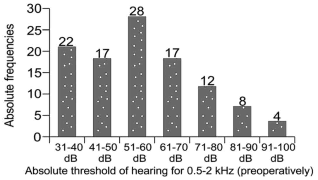
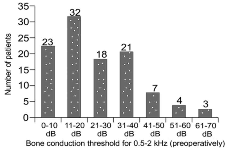
Absolute threshold
Absolute measurement of the absolute threshold of
hearing and absolute threshold of bone conduction (between 0.5 and
2 kHz) was performed and used as a statistical indicator for
rejection of prosthesis (Figs. 1
and 2). All ossicular
reconstructions were performed at the end of a partial or radical
mastoidectomy. The entire cohort had a long-term follow-up, as
follows: 7 years (n=1), 8 years (n=25), 9 years (n=38), 10 years
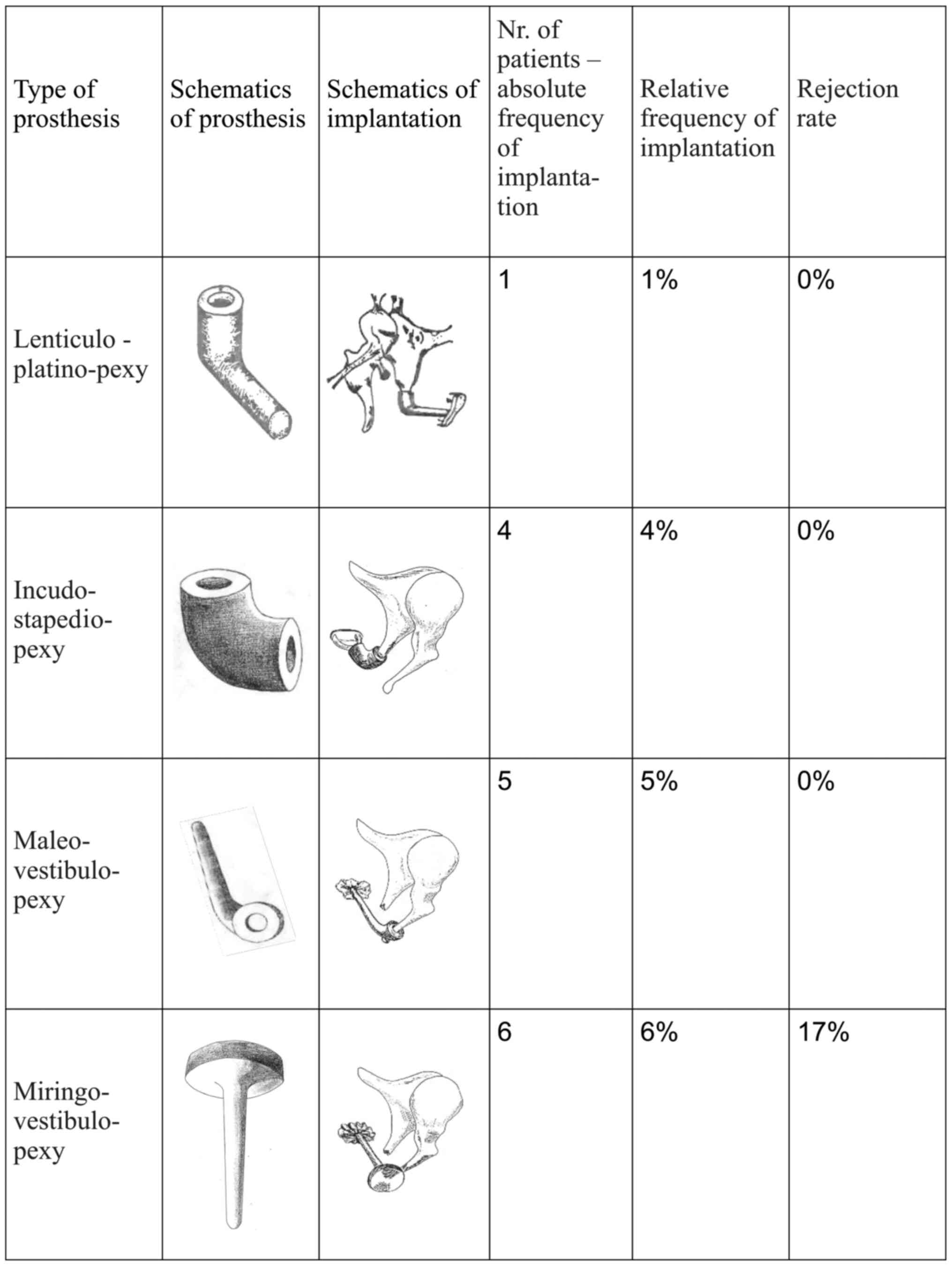
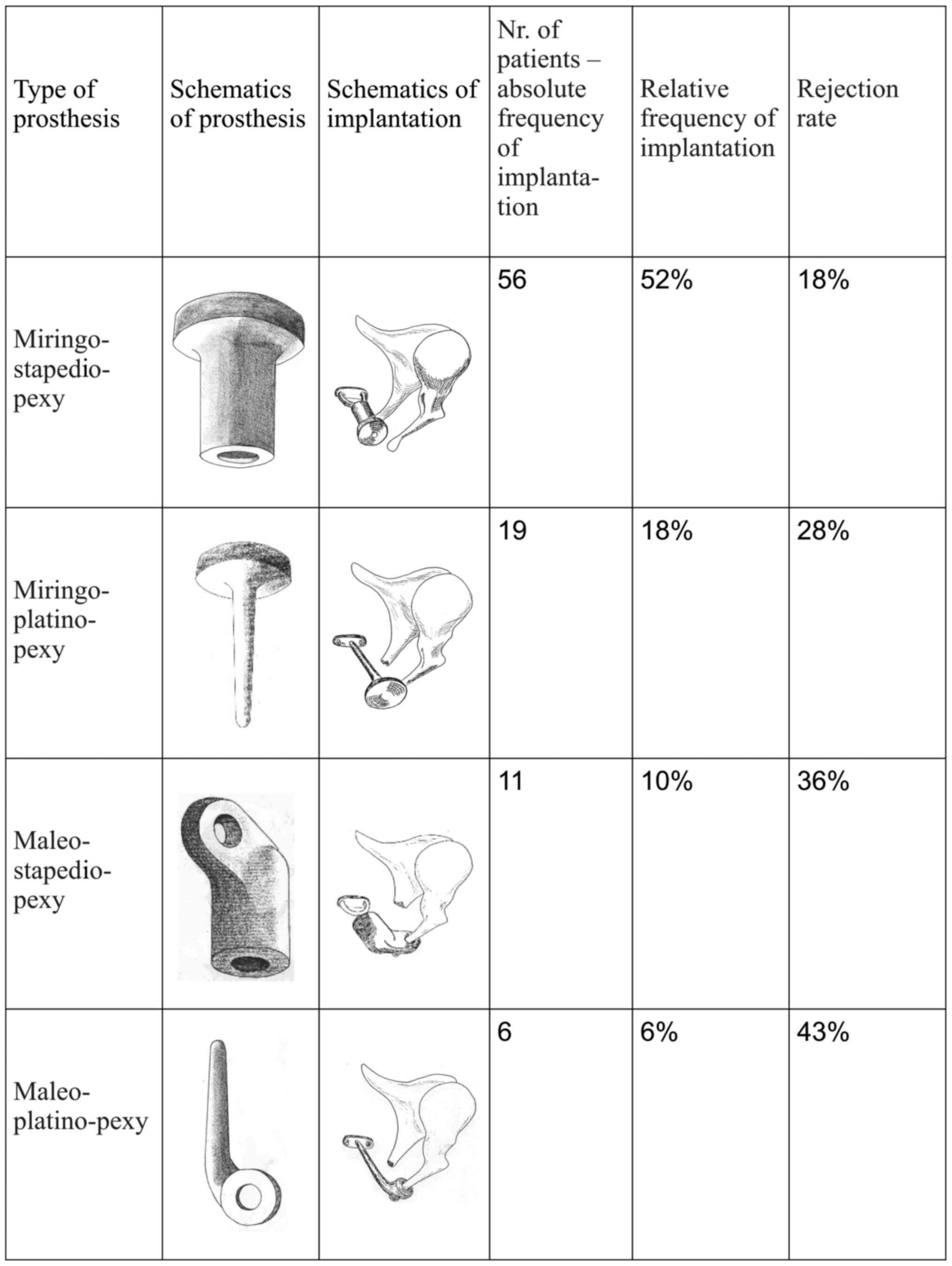
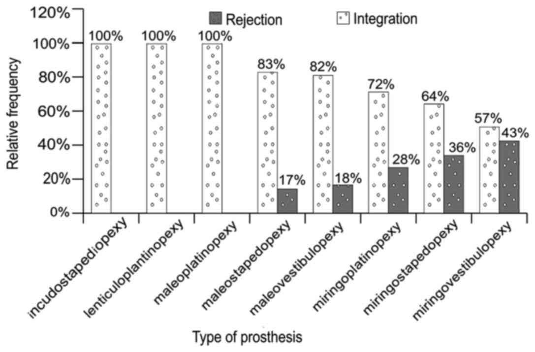
(n=38), and 11 years (n=6). The types of prosthesis used, the
absolute and relative frequencies of use and the rejection rate for
each type of prosthesis are presented in Figs. 3 and 4. The type of prosthesis was also
correlated to the intraoperative status of the ossicular chain
(malleus, incus, stapes) as shown in Figs.
5-7.
Statistical analysis
Data were analyzed using SPSS ver.15. Measurement
data were assessed as percentage, mean, or standard deviation (SD).
Parametric tests (Student's t-test) or non-parametric tests
(Mann-Whitney) were also applied. P
Results and Discussion
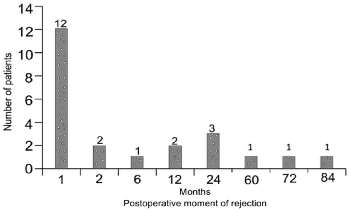
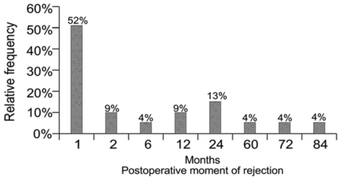
The distribution of absolute and relative
frequencies of rejection moments after the surgery are presented in
Figs. 8 and 9. The rejection rate after 9.12 years was
21% (n=23). Histological integration rate was 79% (n=85).
Over 50% of all rejections occur in short time span
of one month post-operatively and the bulk of the rejections (74%)
occur during the first year. Although ejections no longer occur
after 2 years after the surgical intervention, we found them even
after a 7-year period. Insufficient follow-up may explain this
result and allows us to define the rejection rate as an
exponentially decreasing function (Fig. 10).
The success rate of the mastoidectomy (performed
simultaneously with the implantation) is congruent to the rate of
the successful implants (83 successful vs. 25 unsuccessful). A
similar rate of success regarding mastoidectomy was recorded for
the entire initial cohort of 200 patients: 77.5% success (155
patients) vs. 22.5% failure (45 patients).
Our main aim was to define the situations and
factors that influence the ossiculoplasty results and could yield a
type of histological prognosis.
Based on the selection criteria of the studied group
and by performing statistical analysis of the correlation of
histological results and statistically significant variables we can
formulate pertinent conclusions regarding the implantation success
of bioceramic materials.
Those variables, in regard to the time variable are
structured as follows:
Before surgery variables
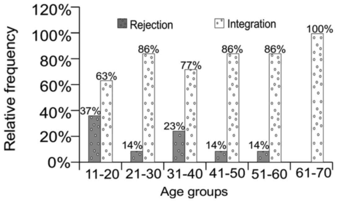
Age group: 11-20 years (n=27), 21-30 years (n=29),
31-40 years (n=22), 41-50 years (n=22), 51-60 years (n=7), 61-70
years (n=1) (Fig. 16); clinical
stage of disease: complicated (n=10), not complicated (n=98) (data
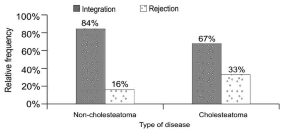
not shown); type of disease: cholesteatoma (n=75),
non-cholesteatoma (n=33) (Fig.
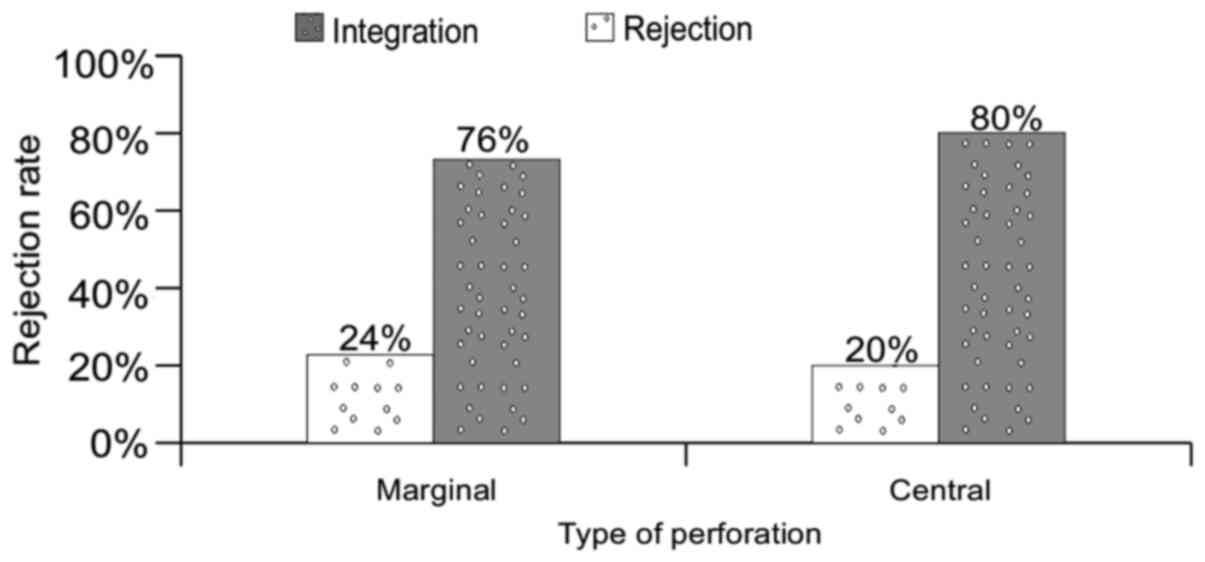
11); type of tympanic membrane perforation: marginal (n=38),
central (n=70) (Fig. 12); surgery
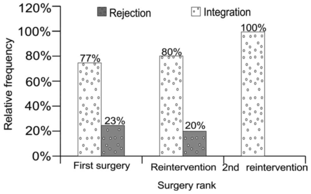
rank: first intervention (n=83), re-intervention (n=20), 2nd
re-intervention (n=5) (Fig.
17).
Intraoperative data
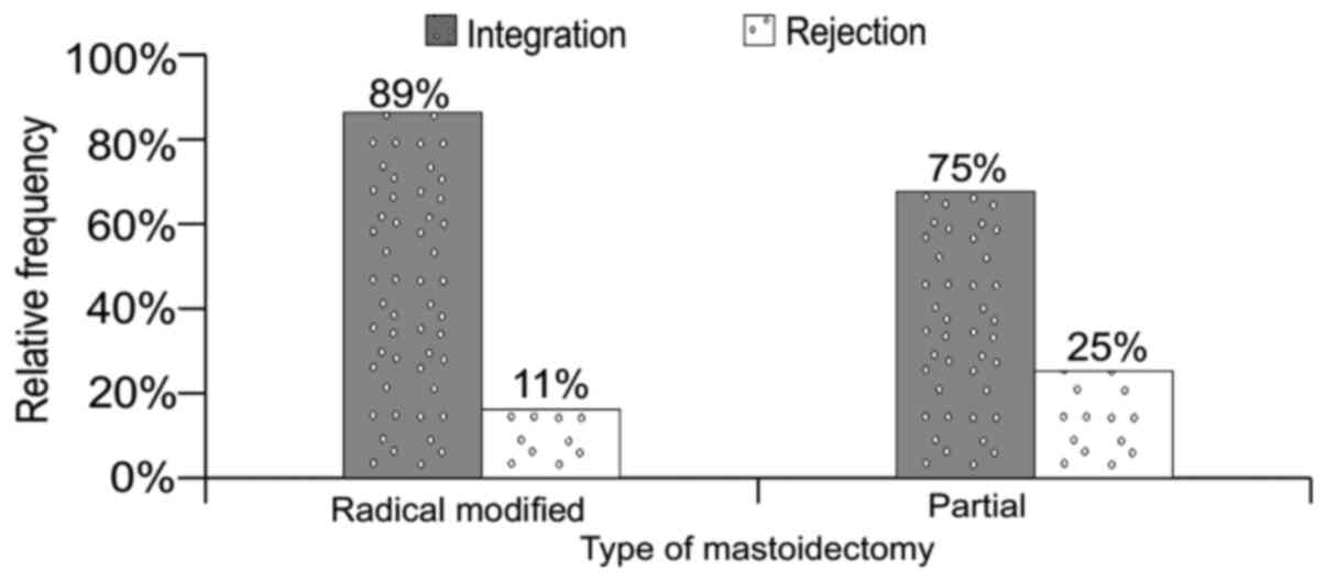
Type of mastoidectomy: modified radical (n=27),
partial (n=81) (Fig. 18); Stapes
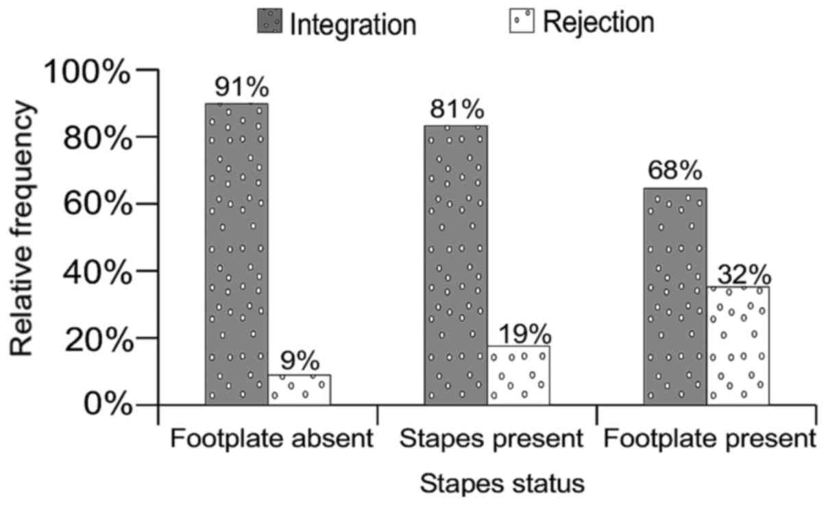
status: footplate lysis (n=11), footplate present and mobile
(n=12), footplate present and fixed (n=13), stapes present and
mobile (n=36) (Fig. 13), stapes
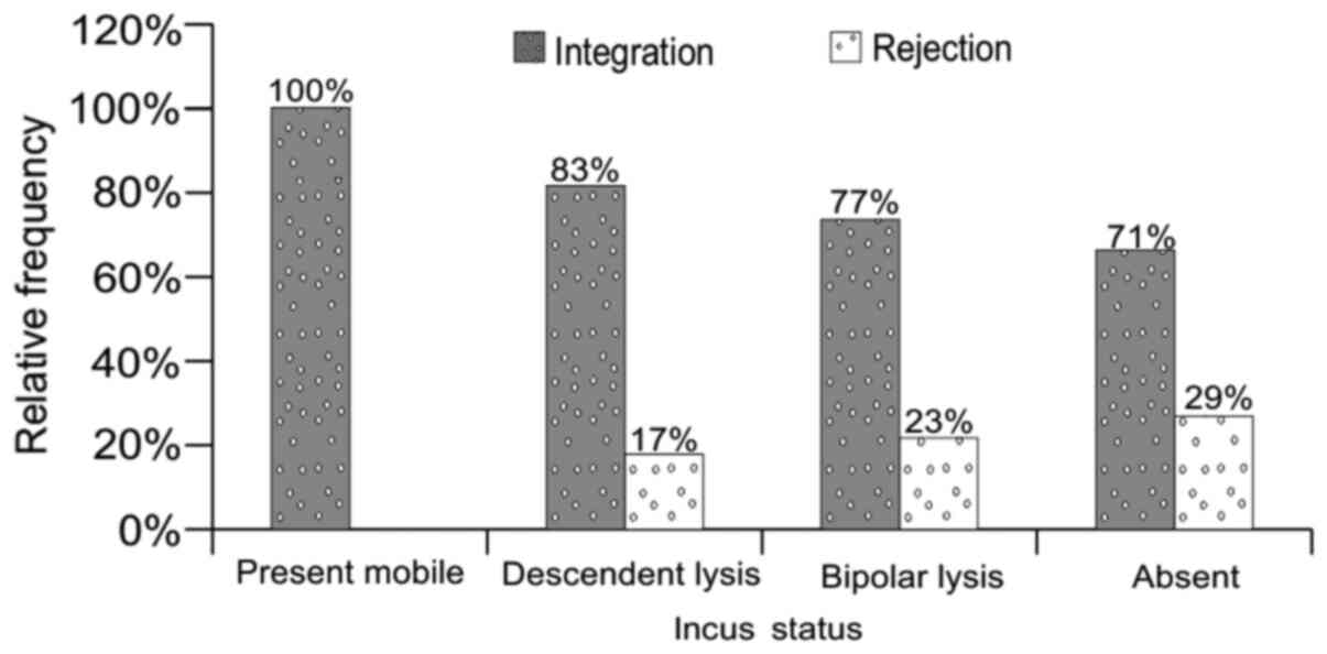
present and fixed (n=36); Incus status: present and mobile (n=2),
bipolar lysis (n=13), absent (n=34), descendent lysis (n=59)
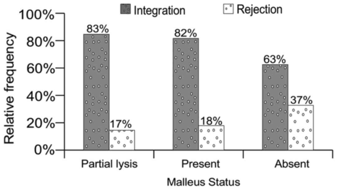
(Fig. 14); Malleus status: present
and mobile (n=76), present and fixed (n=1), malleus mallei lysis
(n=3), malleus head lysis (n=9), absent (n=19) (Fig. 15); Total lesional score: 0 (n=79),
1-10 (n=20), 11-20 (n=4), 21-30 (n=4), 41-50 (n=1) (data not
shown); Type of prosthesis (3,4,25).
Follow-up data
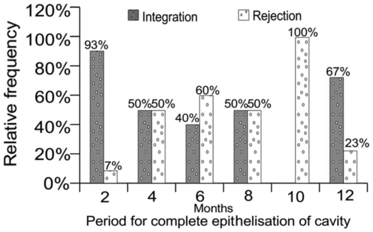
Period for complete epithelisation of cavity
(Fig. 23): 2, 4, 6, 8, 10 and 12
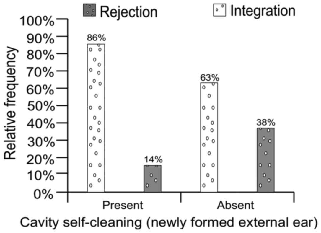
months; Cavity self-cleansing: present or absent (Fig. 24); Mastoidectomy result (dainage
effect): failure or success (Fig.
22).
Bio ceramics were developed as a solution to improve
alloplastic biocompatibility. HA has many of the ideal
characteristics required to be a good prosthesis. It is extremely
biocompatible, exhibits a very low extrusion rate, has no
transmittal of disease, and provides good sound transmission
(13). HA forms a chemical bond
with living bone and shows little biodegradation (14). It can also be placed in direct
contact to the tympanic membrane or cartilage struts.
The general factors involved in OCR failure are:
Problems with the design and function of the prosthesis, middle ear
disease (mastoiditis) or Eustachian tube dysfunction. Each of these
conditions eventually results in poor contact between the footplate
and the graft (15). Although an
optimally positioned prosthesis can migrate postoperatively and
dislocate from the malleus handle, TM or stapes, the surgeon should
provide the prerequisite for an optimal hearing outcome by placing
the prosthesis properly (16).
Perforation of the tympanic membrane, with or without extrusion of
the prosthesis, may also occur (17). We found the influence of the
clinical stage of the disease (complicated or non-complicated) to
be paradoxal. A pertinent explanation for this is not readilly
present and we tend to dismiss it as prognostic criteria.
The type of disease correlates to the expectations
that the presence of cholesteatoma is a negative prognostic factor
in general (Fig. 11). The type of
tympanic perforation influences the rejection rate by 4% (Fig. 12). Eustachian tube dysfunction can
favorize graft retraction and increases tension against the
prosthesis therefore causing tympanic membrane perforation and
prosthesis extrusion. Sustained tension may break the prosthesis or
result in partial or complete extrusion.
It is difficult to interpret the status of the
stapes as a factor on the rejection rate (Fig. 13). On the other hand, the status of
the incus, as found intraoperatively, has predictive significance
as seen in Fig. 14, which means
that there is a direct causal relationship defined by an analitical
function. The status of the malleus has a comparable predictive
significance but does not have the same mathematical accuracy as
the one mentioned above (Fig.
15).
Austin defines four groups in which the incus had
been partially or completely eroded to emphasize the importance of
malleus handle and stapes superstructure presence for OCR (18): A, malleus handle and stapes
superstructure present (60% occurrence); B, malleus handle present,
stapes superstructure absent (23%); C, malleus handle absent,
stapes superstructure present (8%); D, malleus handle and stapes
superstructure absent (8%).
As early as 1973, Bellucci noted a relationship
between the OCR results and middle-ear status (19).
Kartush devised a scoring system called the middle
ear risk index (MERI) to determine the probability of success in
hearing restoration surgery (20-22).
It takes into consideration the various stages of middle ear
disease and ossicular status, including Austin's: otorrhea,
perforation, cholesteatoma, middle ear granulation or effusion,
previous surgery.
Black proposed a new evaluation system called SPITE
for preoperative predictive factors of poor outcome (23): S, surgical-complexity of the
surgery; necessity of scutum and tympanic membrane repair; P,
prosthetic-absence of malleus or stapes; presentation of a 50 dB
air bone gap; I, infection-chronic otorrhea; myringitis; T,
tissue-poor general condition of tissue, referring to extremes of
youth (under 5 years of age) or advanced age (over 70 years);
meatoplasty required; poor mucosa of the middle ear; E, eustachian
tube dysfunction/middle ear effusion present; severely collapsed
tympanic membrane.
Loss of the stapes superstructure was found by
several researchers to be associated with poorer outcome in
ossiculoplasty (24,25).
Dornhoffer and Gardner suggested the ossicular
outcomes parameters staging (OOPS): middle ear factors (drainage,
mucosa ossicles); surgical factors (type of surgery, rank of
surgery) (26).
Grote used for the first time referred to HA as OCR
material with excellent long-term hearing results (8).
Goldenberg's study from 1992 reported low extrusion
rates for hydroxyapatite (2.6%) and plastipore (6.5%) (27).
Numerous other studies have reported results of
ossiculoplasty with HA or HAPEX prosthesis, with similar rejection
results as our findings. Pasha et al studied 33
hydroxyapatite PORPs and TORPs. Hearing results were evaluated by
postoperative mean ABG. The use of incus struts provided the best
functional results. These patients had lower MERI scores in general
and malleus handle present. In addition, PORPs extruded vs. no
incus struts or TORPs (28).
House and Teufert, after an extensive study of 1,210
OCRs with HA and plastipore total ossicular replacement prostheses
(TORPs) or partial ossicular replacement prostheses (PORPs)
reported 63% implant integration. Hearing results were improved for
first surgery patients and no chronic otitis media, when a
cartilage graft was used, and for Plasti-Pore rather than
hydroxyapatite (29).
Iurato et al (30), in an extensive review of the
literature, investigates results of OCR in Austin-Kartush type A
patients. After a minimum follow up of 12 months, incus
interposition showed an 84% success rate vs. 82% for allograft
(ceramics or HA) PORP.
Rondini-Gilli et al reported 100 patients
with HA PORP (n=65) or TORP (n=35). Ten percent (10%) of cases
presented extrusion or displacement of the implants, more commonly
when no cartilage cap was placed (31).
Dornhoffer and Gardner reconstructed 200 ears with
HAPEX PORP or TORP and concluded that the significant prognostic
factors were: The state of the ossicular chain, the state of the
middle ear mucosa, presence of otorrhea, previous mastoidectomy,
and revision surgery (25). Other
authors compared the results of HA and titanium implants.
Truy et al (32) published a success rate of 55% for HA
TORP, 51% for Titanium TORP, 67% for HA PORP, 72% for Titanium PORP
in a retrospective comparison of hydroxyapatite vs. titanium TORP
and PORP.
Coffey et al conducted a study on 105 cases
(80 with titanium and 25 with non-titanium implants) and reported a
success rate of 50% for non-titanium cases and 77.1% for titanium
cases; extrusion was observed with two non-titanium prostheses
(8.0%) and three titanium prostheses (3.8%) (20,33).
Gardner et al, in a retrospective study
comparing titanium reconstructions to non-titanium reconstructions,
reported the successful rehabilitation of conductive hearing loss
in 70% of PORPs vs. 44% of TORPs when titanium prostheses were used
as compared to 48 and 21% of non-titanium-based partial and total
reconstructions (34).
Emir et al (35) reviewed 304 cases of ossiculoplasty
with intact canal wall. The success rate for autologous incus
interposition was 58 vs. 56% for plastipore PORPs, while 9.3% of
implants extruded.
We should also note that numerous studies reported
similar rejection rates for the modern and largely used titanium
implants.
Martin and Harner (36) reviewed 68 cases of OCR using a
titanium TORP or PORP. Closure of the ABG to within 20 dB was
obtained in 57%.
Dalchow et al published results from 1,304
implanted titanium TORPs and PORPs. His overall success rate was
76% (37).
Neff et al (38) studied results for 18 OCR with
titanium TORP and reported an 89% surgical success rate for an
average follow-up time of 8 months.
De Vos et al (39) reported a success rate of 60% for
both PORP and TORP on 149 ears, all implanted with titanium
prosthesis. The extrusion rate was 3.5% and displacement of the
prosthesis occurred in 4.3%.
Vassbotn et al (40) published a report on 73 OCR with
titanium prostheses (38 PORPs vs. 35 TORPs). After a mean follow up
of 14 months, the success rate was 77% for the entire cohort.
The influence of the age group on the rejection rate
was direct and analytic (Fig. 16).
In other words, the rejection rate was determined by an analytical
function of the gravity of the disease which is inversly
proportional to the patient's age and directly proportional to the
degree of the hearing loss.
The rank of the surgery is also paradoxal in
influencing the rejection rate, since a superior rank means better
mastoid drainage and elimination of leassions and therefore better
premises for histologic integration of implants (Fig. 17). The same discussion could be
applied when discussing the type of surgery: Partial surgery
increased the chance for remanent lessions (Fig. 18).
The total lesional score refers to all lesions found
pre-, intra- and post-surgery, except those of the middlea ear
mucosa. This score is therefore insignificant for the rejection
rate and we should also consider that medical research can
sometimes require a high degree of abstraction (41).
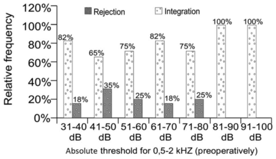
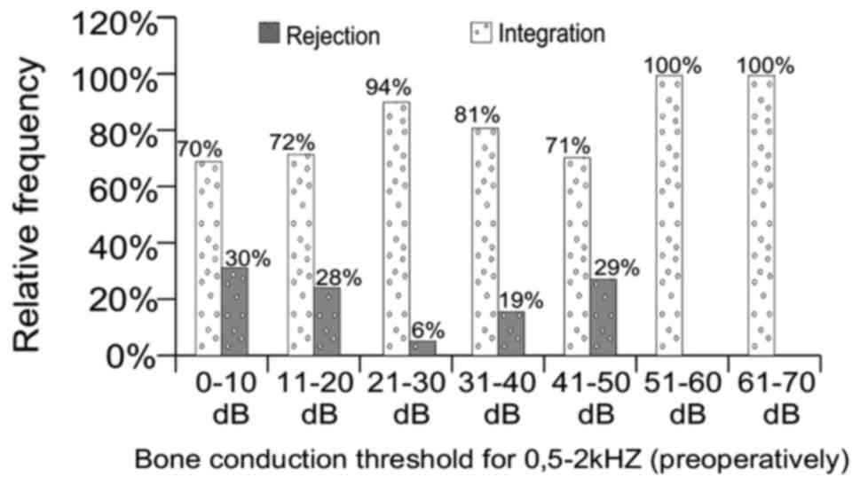
The pre-surgery absolute threshold of hearing and
threshold of BC do not seem to have a statistical effect on the
rejection rate (Figs. 19 and
20). Notably, the value of 6%
rejection rate for BC threshold between 21-30 dB is not consistent
with the other results that are closer to 30% suggesting that,
under certain conditions, some connection exists between these
factors. A valid explanation for this discrepancy remains to be
determined in future studies.
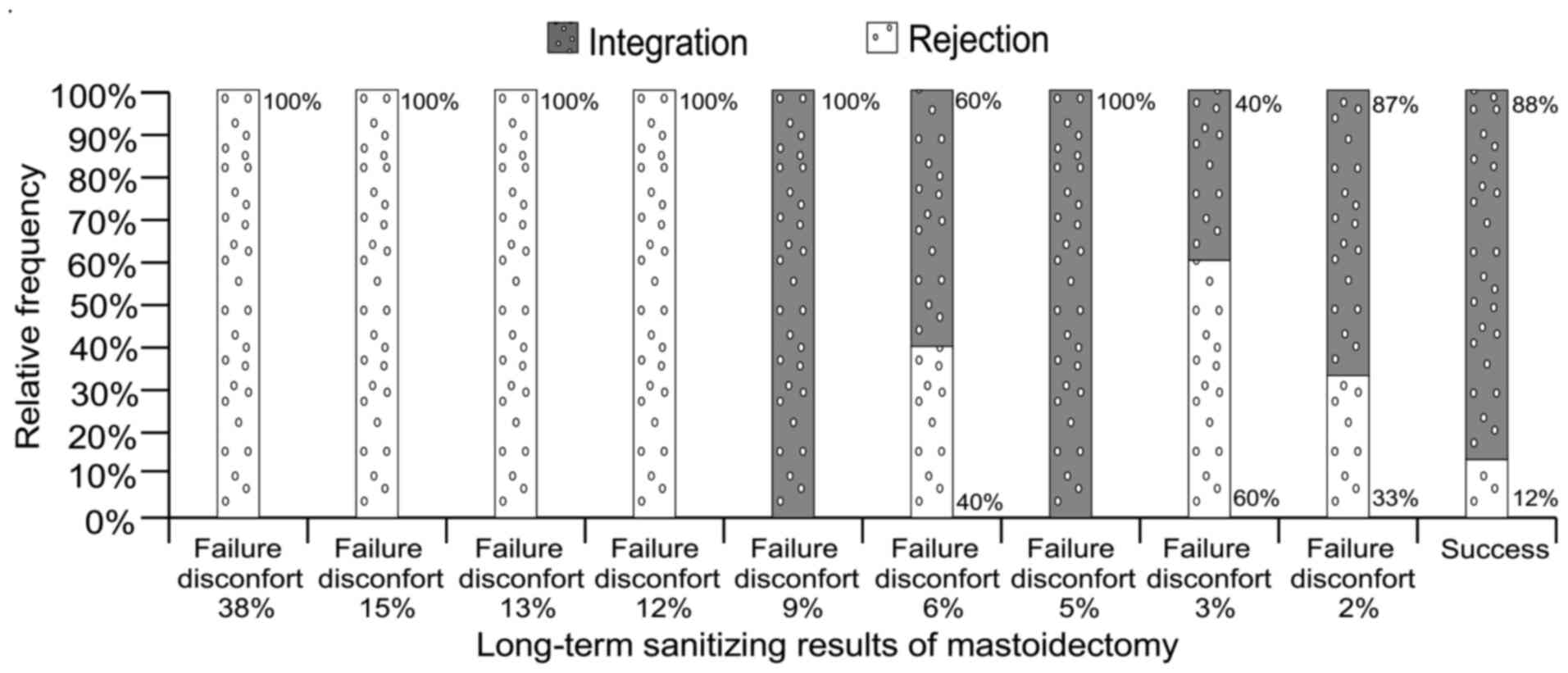
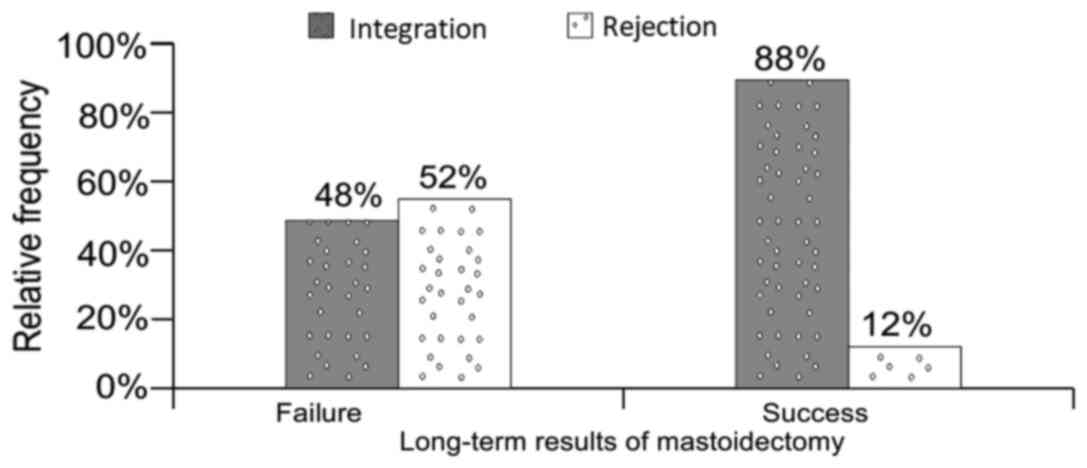
The long-term results of mastoidectomy have direct
bearing on the results of implantation as shown in Figs. 21 and 22, as does the period of epithelisation
after surgery and the rate of self-cleansing of the cavity
(Figs. 23 and 24).
It is difficult to describe the influence of the
type of prosthesis but we can nevertheless state that the smaller
the part of ossicular chain replaced is, the smaller the rejection
rate is (Figs. 3, 4 and 25).
These findings are in accordance with the results from medical
literature, which report improved functional results for PORP
implantations as compared to TORP. Notably, the physical tension
involved in all these interventions that have a high impact on the
quality of life of the patient should be considered (42).
Ho et al reported achieving an air-bone gap
lower than 20 dB in 64% of PORP and 45% of TORP implants (43).
Schmerber et al published a retrospective
chart review of 111 OCRs with titanium prostheses. For PORP 77% of
the cases were reported as successful vs. 52% for TORP (44).
Vassbotn et al (40) reported 89% of PORP implants as
successful vs. 63% of TORPs (29)
while Siddiq and Raut, in a prospective study of 33 OCRs assessed
the early results of titanium PORP and TORP in chronic ear disease
and concluded that PORPs had a higher success rate (85%) than TORPs
(46%) (45).
A large number of materials is available for
reconstructing the ossicular chain. Therefore, the surgeon should
assess the method and the type of prosthesis to be used very
carefully in order to obtain the best hearing result with the
smallest risk of complications. It is clear that optimal results
depend, not only on the choice of material and design of the
prosthesis, but also on the status of the middle ear in which it is
placed, and also on the expertise of the surgeon and the techniques
to be used.
Hydroxyapatite has many of the ideal characteristics
required to be a good prosthesis with a high degree of
biocompatibility, very low extrusion rate, low risk of disease
transmission and good functional results. Of note, bioceramic
implants are cheaper and can be produced locally, which is a great
advantage for struggling economies.
The general factors that showed statistical
influence in OCR failure are: Prior to surgery factors (age group,
type of lesion-cholesteatoma, type of TM perforation); during
surgery factors (type of surgery, state of incus, type of
prosthesis); after surgery factors (results of mastoidectomy,
period of complete epidermal growth, self-cleansing of cavity).
Additional causes of functional failure include
improperly sized prosthesis (too short), sliding of the prosthesis,
stapes anterior crural fracture, and contraction and movement of
the healing tympanic membrane.
Acknowledgements
Not applicable.
Funding
No funding was received.
Availability of data and materials
Not applicable.
Authors' contributions
HM and MR are responsible for the original idea,
conception, patient selection and care, operations, data collection
and editing structure. MR is also responsible for first use of the
implant material. HM and MR also contributed to design of
prosthesis and final study. AIM and AMD gathered the medical
information and reviewed the final version of the article. All
authors read and approved the final manuscript.
Ethics approval and consent to
participate
For this study, the agreement was obtained from the
Research Ethics Committee of the Faculty of Medicine, Titu
Maiorescu University.
Patient consent for publication
All patients provided informed consent and approved
the publication of data.
Competing interests
The authors declare that they have no competing
interests.
References
|
1
|
Bojrab DI and Babu SC: Ossiculoplasty I.
In: Middle Ear and Mastoid Surgery. Habermann RSII (ed.) Thieme
Verlag, New York-Stuttgart, pp151-158, 2004.
|
|
2
|
Hirsch BE: Ossicular chain reconstruction.
In: Head and neck surgery. Myers E (ed.) Saunders Elsevier,
pp1147-1162, 2008.
|
|
3
|
Colletti V, Carner M and Colletti L: TORP
vs. round-window implant for hearing restoration of patients with
extensive ossicular chain defect. Acta Otolaryngol. 129:449–452.
2009.PubMed/NCBI View Article : Google Scholar
|
|
4
|
Trifu S, Vladuti A and Popescu A:
Neuroendocrine aspects of pregnancy and postpartum depression. Acta
Endocrinol (Bucharest). 15:410–415. 2019.PubMed/NCBI View Article : Google Scholar
|
|
5
|
Mocanu H and Oncioiu I: The influence of
clinical and environmental risk factors in the etiology of
congenital sensorineural hearing loss in the Romanian population.
Iran J Publ Health. 48:2301–2303. 2019.PubMed/NCBI
|
|
6
|
Mocanu H: The role of perinatal hearing
screening in the normal development of the infant's language. In:
Debating Globalization. Identity, Nation and Dialogue fourth
Edition. Boldea I and Sigmirean C (eds). Arhipeleag XXI Press,
Tirgu Mures, pp562-569, 2017.
|
|
7
|
Mocanu H: The economic impact of early
diagnosis of congenital hearing loss. 4th International Conference
Globalization, Intercultural Dialogue and National. Targu Mures,
Romania.
|
|
8
|
Grote JJ: Tympanoplasty with calcium
phosphate. Arch Otolaryngol. 110:197–199. 1984.PubMed/NCBI View Article : Google Scholar
|
|
9
|
Ficai A, Andronescu E, Ghitulicã C, Voicu
G, Trandafir V, Mânzu D, Ficai M and Pall S: Colagen/hydroxyapatite
interactions in composite biomaterials. Mat Plast. 46:11–15.
2009.
|
|
10
|
Niparko JK, Kemink JL, Graham MD and
Kartush JM: Bioactive glass ceramic in ossicular reconstruction: A
preliminary report. Laryngoscope. 98:822–825. 1988.PubMed/NCBI View Article : Google Scholar
|
|
11
|
McElveen JT, Cunningham CD and Sheehy JL:
Ossicular reconstruction. In: Otologic Surgury. Saunders Elsevier,
pp161-171, 2001.
|
|
12
|
Rădulescu M and Popescu-Negreanu T:
Timpanoplastie cu biovitroceramica PAW-1. Oto-Rino-Laringologia XL:
97-102, 1995.
|
|
13
|
Bojrab DI, Causse JB, Battista RA, Vincent
R, Gratacap B and Vandeventer G: Ossiculoplasty with composite
prostheses. Overview and analysis. Otolaryngol Clin North Am.
27:759–776. 1994.PubMed/NCBI
|
|
14
|
Hörman K and Donath K: Is hydroxylapatite
ceramic an adequate biomaterial in ossicular reconstruction? Am J
Otol. 8:402–409. 1987.PubMed/NCBI
|
|
15
|
Sellari-Franceschini S, Piragine F,
Bruschini P and Berrettini S: TORPs and PORPs: Causes of failure.
Am J Otol. 8:551–552. 1987.PubMed/NCBI
|
|
16
|
Neudert M, Bornitz M, Mocanu H,
Lasurashvili N, Beleites T, Offergeld C and Zahnert T: Feasibility
study of a mechanical real-time feedback system for optimizing the
sound transfer in the reconstructed middle ear. Otol Neurotol.
39:e907–e920. 2018.PubMed/NCBI View Article : Google Scholar
|
|
17
|
Batti JS and Bluestone CD: Ossiculoplasty.
In: Surgical Atlas of Pediatric Otolaryngology. Bluestone CD and
Rosenfeld R (eds). 2nd edition. BC Decker Inc, Hamilton, ON,
pp75-89, 2002.
|
|
18
|
Austin DF: Ossicular reconstruction.
Otolaryngol Clin North Am. 5:145–160. 1972.PubMed/NCBI
|
|
19
|
Bellucci RJ: Dual classification of
tympanoplasty. Laryngoscope. 83:1754–1758. 1973.PubMed/NCBI
|
|
20
|
Kartush JM: Ossicular chain
reconstruction: Capitulum to malleus. Otolaryngol Clin North Am.
27:689–715. 1994.PubMed/NCBI
|
|
21
|
Simon C and Makishima T: Ossicular
reconstruction. In: Grand rounds presentation. The University of
Texas Medical Branch, Department of Otolaryngology, 2009.
|
|
22
|
Trifu S: Neuroendocrine insights into
burnout syndrome. Acta Endocrinol (Buchar). 15:404–405.
2019.PubMed/NCBI View Article : Google Scholar
|
|
23
|
Black B: Ossiculoplasty prognosis: The
spite method of assessment. Am J Otol. 13:544–551. 1992.PubMed/NCBI
|
|
24
|
Mills RP: The influence of pathological
and technical variables on hearing results in ossiculoplasty. Clin
Otolaryngol Allied Sci. 18:202–205. 1993.PubMed/NCBI View Article : Google Scholar
|
|
25
|
Smyth GD and Patterson CG: Results of
middle ear reconstruction: Do patients and surgeons agree? Am J
Otol. 6:276–279. 1985.PubMed/NCBI
|
|
26
|
Dornhoffer JL and Gardner E: Prognostic
factors in ossiculoplasty: A statistical staging system. Otol
Neurotol. 22:299–304. 2001.PubMed/NCBI View Article : Google Scholar
|
|
27
|
Goldenberg RA: Hydroxylapatite ossicular
replacement prostheses: Results in 157 consecutive cases.
Laryngoscope. 102:1091–1096. 1992.PubMed/NCBI View Article : Google Scholar
|
|
28
|
Pasha R, Hill SL III and Burgio DL:
Evaluation of hydroxyapatite ossicular chain prostheses.
Otolaryngol Head Neck Surg. 123:425–429. 2000.PubMed/NCBI View Article : Google Scholar
|
|
29
|
House JW and Teufert KB: Extrusion rates
and hearing results in ossicular reconstruction. Otolaryngol Head
Neck Surg. 125:135–141. 2001.PubMed/NCBI View Article : Google Scholar
|
|
30
|
Iurato S, Marioni G and Onofri M: Hearing
results of ossiculoplasty in Austin-Kartush group A patients. Otol
Neurotol. 22:140–144. 2001.PubMed/NCBI View Article : Google Scholar
|
|
31
|
Rondini-Gilli E, Grayeli AB, Borges
Crosara PF, El Garem H, Mosnier I, Bouccara D and Sterkers O:
Ossiculoplasty with total hydroxylapatite prostheses anatomical and
functional outcomes. Otol Neurotol. 24:543–547. 2003.PubMed/NCBI View Article : Google Scholar
|
|
32
|
Truy E, Naiman AN, Pavillon C, Abedipour
D, Lina-Granade G and Rabilloud M: Hydroxyapatite versus titanium
ossiculoplasty. Otol Neurotol. 28:492–498. 2007.PubMed/NCBI View Article : Google Scholar
|
|
33
|
Coffey CS, Lee FS and Lambert PR: Titanium
versus nontitanium prostheses in ossiculoplasty. Laryngoscope.
118:1650–1658. 2008.PubMed/NCBI View Article : Google Scholar
|
|
34
|
Gardner EK, Jackson CG and Kaylie DM:
Results with titanium ossicular reconstruction prostheses.
Laryngoscope. 114:65–70. 2004.PubMed/NCBI View Article : Google Scholar
|
|
35
|
Emir H, Kizilkaya Kaptan Z, Göcmen H,
Uzunkulaoglu H, Tuzuner A, Bayiz U and Samim E: Ossiculoplasty with
intact stapes: Analysis of hearing results according to the middle
ear risk index. Acta Otolaryngol. 129:1088–1094. 2009.PubMed/NCBI View Article : Google Scholar
|
|
36
|
Martin AD and Harner SG: Ossicular
reconstruction with titanium prosthesis. Laryngoscope. 114:61–64.
2004.PubMed/NCBI View Article : Google Scholar
|
|
37
|
Dalchow CV, Grun D and Stupp HF:
Reconstruction of the ossicular chain with titanium implants.
Otolaryngol Head Neck Surg. 125:628–630. 2001.PubMed/NCBI View Article : Google Scholar
|
|
38
|
Neff BA, Rizer FM, Schuring AG and Lippy
WH: Tympano-ossiculoplasty utilizing the Spiggle and Theis titanium
total ossicular replacement prosthesis. Laryngoscope.
113:1525–1529. 2003.PubMed/NCBI View Article : Google Scholar
|
|
39
|
De Vos C, Gersdorff M and Gérard JM:
Prognostic factors in ossiculoplasty. Otol Neurotol. 28:61–67.
2007.PubMed/NCBI View Article : Google Scholar
|
|
40
|
Vassbotn FS, Møller P and Silvola J:
Short-term results using Kurz titanium ossicular implants. Eur Arch
Otorhinolaryngol. 264:21–25. 2007.PubMed/NCBI View Article : Google Scholar
|
|
41
|
Alecu I, Mocanu H and Călin IE:
Intellectual mobility in higher education system. Rom J Mil Med.
120:16–21. 2017.
|
|
42
|
Trifu S, Delcuescu C and Boer CM:
Psychosomatics and psychical tension (clinical research). Procedia
Soc Behav Sci. 33:128–132. 2012.
|
|
43
|
Ho SY, Battista RA and Wiet RJ: Early
results with titanium ossicular implants. Otol Neurotol.
24:149–152. 2003.PubMed/NCBI View Article : Google Scholar
|
|
44
|
Schmerber S, Troussier J, Dumas G,
Lavieille JP and Nguyen DQ: Hearing results with the titanium
ossicular replacement prostheses. Eur Arch Otorhinolaryngol.
263:347–354. 2006.PubMed/NCBI View Article : Google Scholar
|
|
45
|
Siddiq MA and Raut VV: Early results of
titanium ossiculoplasty using the Kurz titanium prosthesis-a UK
perspective. J Laryngol Otol. 121:539–544. 2007.PubMed/NCBI View Article : Google Scholar
|