Introduction
Sepsis is a systemic inflammatory response syndrome
in which pathogenic microorganisms invade the circulatory system,
grow and multiply to produce endotoxins and exotoxins, and
potentially induce injury to multiple organs (1). In newborns, the source of the pathogen
may be an in utero infection, maternal flora infection or
postpartum infection from the hospital or community (2). Neonatal sepsis (NS) is a leading cause
of morbidity and mortality in newborns (3), and it may be categorized as
early-onset NS (EOS) or late-onset NS (LOS) (4). The morbidity and mortality of NS have
been reduced due to progress made in neonatal care (5). However, there have been minimal
advances in the specific clinical management and accuracy of
diagnostic testing options over the last several decades (6). Numerous sepsis biomarkers have been
evaluated for their potential in early detection of NS, but to
date, no ideal biomarker has met all of the basic criteria for
being an ideal indicator (7). The
most commonly used biomarkers are C-reactive protein (CRP) and
procalcitonin (PCT), but both of them have demonstrated varying
sensitivity, specificity and positive and negative predictive
values in different studies (7).
Blood culture has been considered as a gold standard method, but
the identification of EOS is complicated by a high rate of
false-negative results (8).
MicroRNAs (miRNAs/miRs) are a class of small (~22
nucleotides), non-coding, single-stranded RNAs that regulate gene
expression by binding the 3'-untranslated region (3'-UTR) of target
mRNAs for mRNA degradation or translational suppression (9). They have been indicated to be
frequently dysregulated in cancers and serve as attractive targets
for prognostication and therapeutic applications (10). miRNAs have been used in fingerprint
diagnosis of sepsis and have been identified as potential
biomarkers of sepsis (11). In the
pathogenesis of sepsis, certain miRNAs, such as miR-125b and
miR-142-3p, have been demonstrated to regulate inflammation
(12,13). In addition, Chen et al
(14) performed miRNA expression
profiling using a microarray and determined that altered miRNAs
(including miR-1184) may modulate the immune response during NS in
a way that represses the inflammatory response. However, the
association between miR-1184 and NS remains to be fully elucidated.
Interleukin-16 (IL-16) is a cytokine associated with various
inflammatory processes (15) and
has been documented to facilitate the production of
pro-inflammatory cytokines in monocytes (16). Studies have reported that IL-16 may
be used as a target for certain miRNAs in cancer (17,18).
Furthermore, the study by Chen et al (14) used a protein chip and preliminarily
determined that downregulation of miR-1184 in patients with NS was
associated with increased protein levels of IL-16.
Lipopolysaccharide (LPS) has been widely used to induce
inflammatory responses that mimic the pathological processes of
inflammation in human inflammatory diseases, including sepsis
(19). However, the clinical value
and biological function of the aberrant miR-1184 in NS remain to be
fully elucidated.
The present study sought to explore the diagnostic
value of serum miR-1184 by comparing the serum levels of miR-1184
between subjects with NS and healthy newborns. The effect of
miR-1184 on the LPS-induced inflammatory response was analyzed in
monocytes. The current study simultaneously upregulated miR-1184
and IL-16 levels in the cell model, then determined whether IL-16
was involved in the regulation of miR-1184 during the inflammatory
response by detecting changes in inflammatory factor levels.
Therefore, the present study may provide novel insight into the
regulation of inflammation in the pathogenesis of NS.
Materials and methods
Patients and blood sample
collection
The experimental protocols were approved by the
Ethics Committee of Weifang People's Hospital (Weifang, China) and
the parents of each neonate had provided written informed consent.
Blood samples were collected from 72 neonates with NS (including 6
cases of EOS and 66 cases of LOS) and 56 neonates with respiratory
infection or pneumonia who visited Weifang People's Hospital
between April 2015 and December 2018. The latter were selected as
controls as it is critical to differentiate between sepsis and
respiratory infection to provide more effective treatment. Serum
was isolated from the blood of all participants by centrifugation
and stored at -80˚C for further use. None of the neonates had
received any therapy prior to blood collection. The patients with
NS were diagnosed according to the criteria established at the 2003
Kunming Neonatal Sepsis Definitions Conference (20) and the neonates with respiratory
infection or pneumonia served as controls without any symptoms or
signs of sepsis. The inclusion criteria for patients with NS were
as follows: i) Term birth; ii) no antibiotic treatment prior to
sample collection; iii) no inflammation or antibiotic therapy prior
to delivery; iv) no congenital malformations; v) Apgar score of
>6 at 5 min. The diagnosis of NS mainly relied on the clinical
manifestations and the detection of blood pathogens. The
clinicopathological characteristics of the neonates included are
listed in Table I.
 | Table IBaseline characteristics of the study
population. |
Table I
Baseline characteristics of the study
population.
| Feature | Controls
(n=56) | NS (n=72) | P-value |
|---|
| Age (days) | 12.8±4.6 | 12.8±4.2 | 0.988 |
| Sex
(female/male) | 24/32 | 33/39 | 0.737 |
| Body weight
(kg) | 3.7±0.3 | 3.6±0.3 | 0.225 |
| WBC
(x109/l) | 10.7
(8.1-13.5) | 11.7
(8.2-14.7) | 0.130 |
| CRP (mg/l) | 11.8
(9.7-14.1) | 13.9
(9.7-17.4) | 0.016 |
| PCT (ng/ml) | 2.0 (1.3-2.8) | 4.5 (2.8-5.8) | <0.001 |
Monocyte culture and LPS
induction
Monocytes were collected from blood samples of
subjects with NS according to a previously described method
(21). In brief, the blood samples
were settled with 4.5% dextran 500 (1:5; Amersham Biosciences) to
separate leukocytes from red blood cells. The monocytes were then
obtained using density gradient centrifugation and the cell purity
was confirmed by FACSCalibur flow cytometry (BD Biosciences) based
on the detection of the specific cell markers CD14 (cat. no.
562691; 1:20; BD Biosciences) and CD15 (cat. no. 560827; 1:20; BD
Biosciences). The extracted monocytes were cultured in RPMI-1640
medium supplemented with 10% FBS (both from Gibco; Thermo Fisher
Scientific, Inc.) in a humidified atmosphere with 5% CO2
at 37˚C. The monocytes were cultured for 7 days for further use. To
explore the effects of miR-1184 on LPS-induced inflammatory
response, the monocytes were stimulated using LPS (Sigma-Aldrich;
Merck KGaA) for 4 h. All of the experiments were performed at least
3 times.
Cell transfection
miR-1184 mimics (5'-CCUGCAGCGACU UGAUGGCUUCC-3') and
mimics negative control (NC) (5'-UUCUCCGAACGUGUCACGU-3'), which
were obtained from GenePharma, were used for cell transfection to
regulate the expression of miR-1184 in monocytes. IL-16
overexpression vectors pcDNA3.1-IL-16 were constructed to
upregulate IL-16 expression in monocytes and pcDNA3.1 empty vector
(Invitrogen; Thermo Fisher Scientific, Inc.) was used as a control
for pcDNA3.1-IL-16 (Shanghai GenePharma Co., Ltd.). The above
vectors were separately transfected into monocytes using
Lipofectamine 2000 (Thermo Fisher Scientific, Inc.) according to
the manufacturer's protocols. Cells were collected after
transfection for 48 h at 37˚C and used for the subsequent
analyses.
RNA extraction and reverse
transcription-quantitative PCR (RT-qPCR)
The collected blood was centrifuged to isolate the
serum samples. TRIzol® reagent (Invitrogen; Thermo
Fisher Scientific, Inc.) was used to extract total RNA from serum
or monocyte samples, including miRNA. A NanoDrop 2000 (Thermo
Fisher Scientific, Inc.) was used to evaluate the purity and
concentration of the RNA. The complementary DNA was then
synthesized from the obtained RNA by RT using a PrimeScript RT
reagent kit (Takara Bio, Inc.) according to the manufacturer's
protocol.
The serum levels of miR-1184 and mRNA serum/monocyte
levels of IL-16 were measured using RT-qPCR, which was performed
using a SYBR Green I Master Mix kit (Invitrogen; Thermo Fisher
Scientific, Inc.) and a 7300 Real-Time PCR System (Applied
Biosystems; Thermo Fisher Scientific, Inc.). All of the procedures
were performed according to the manufacturer's instructions. U6 was
used as an endogenous control for miR-1184 and GAPDH was used as an
internal control for IL-16. The primers used for analysis were as
follows: miR-1184 forward, 5'-GCCGAGCCTGCA GCGACTTG-3' and reverse,
5'-CTCAACTGGTGTCGT GGA-3'; IL-16 forward, 5'-GGAATCGTGCTTCAGACC
CA-3' and reverse, 5'-CTCTGGGCTCCTTTGTCAGG-3'; U6 forward,
5'-CTCGCTTCGGCAGCACA-3' and reverse, 5'-AA CGCTTCACGAATTTGCGT-3';
GAPDH forward, 5'-GCT CCCTCTTTCTTTGCAGC-3' and reverse, 5'-GTTGTC
ATGGATGACCTTGGC-3'. The relative expression values were calculated
using the 2-ΔΔCq method (22).
ELISA
To evaluate the status regarding the inflammatory
response, the levels of pro-inflammatory cytokines, including
IL-1β, IL-6 and TNF-α, in both serum samples and monocytes were
examined by ELISA. For cell lysis, 100 µl RIPA lysis buffer (0.5 M
Tris-HCl, pH 7.4, 1.5 M NaCl, 2.5% deoxycholic acid, 10% NP-40 and
10 mM EDTA) was added to per 1x10⁶ cells. The serum and
cell lysate were examined using ELISA kits for IL-1β (cat. no.
557953), IL-6 (cat. no. 555220) and TNF-α (cat. no. 555212; all
from BD Biosciences) according to the manufacturer's protocols. The
absorbance value at 450 nm was detected with a microplate reader
(Bio-Rad Laboratories, Inc.). The concentration of the cytokines in
cell lysate was quantified using standard curves.
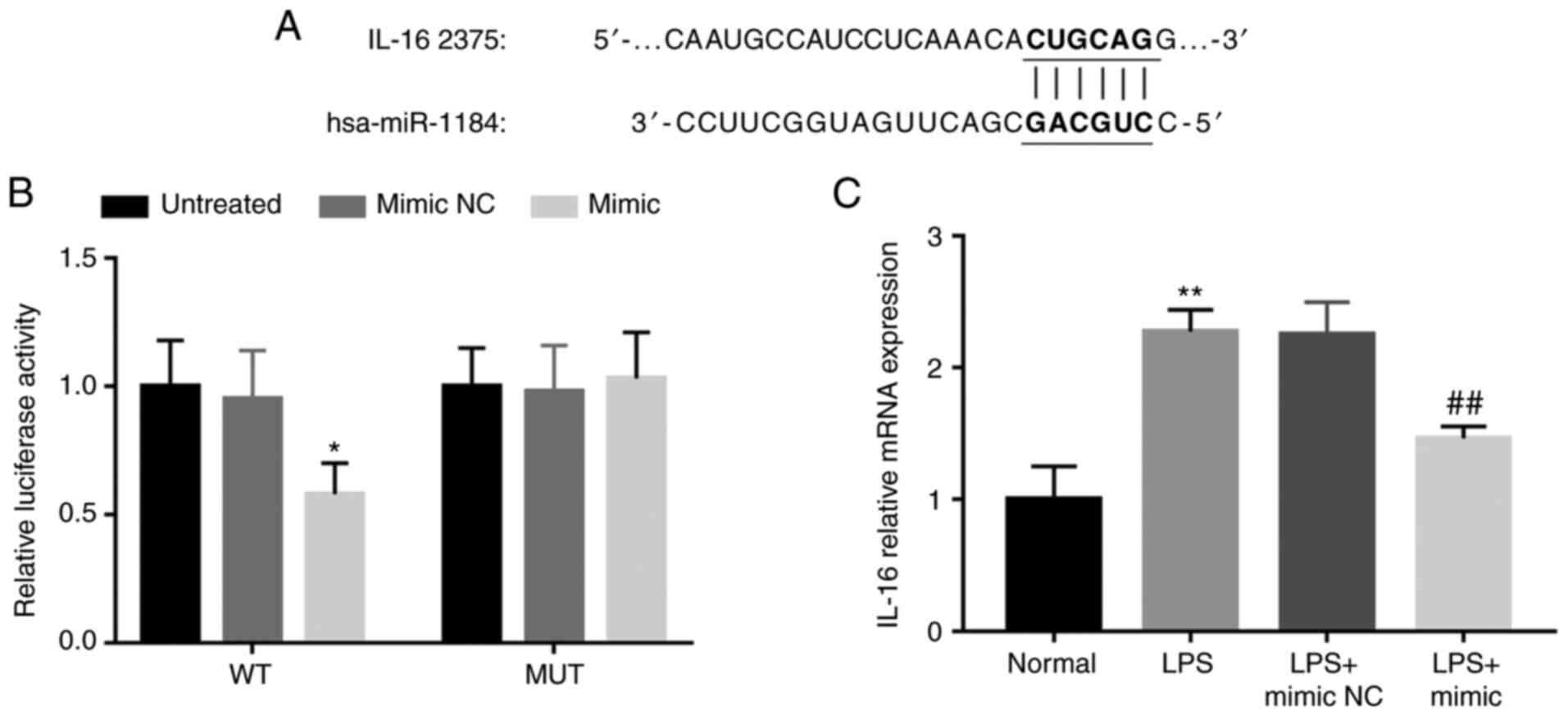
Luciferase reporter assay
The putative binding site of miR-1184 at the 3'-UTR
of IL-16 was predicted using miRanda (http://www.microrna.org/microrna/home.do). To confirm
whether there was a direct interaction between miR-1184 and IL-16,
a luciferase reporter assay was performed. The wild-type (WT)
3'-UTR containing the binding site for miR-1184 was amplified by
PCR, and the mutant-type (MUT) 3'-UTR was generated using a
QuickMutation kit (Beyotime Institute of Biotechnology). The 3'-UTR
sequences were inserted in the pGL-control vector (Promega, Corp.).
The vectors constructed were separately transfected into monocytes
together with either miR-1184 mimics or mimics NC using
Lipofectamine 3000 (Thermo Fisher Scientific, Inc.) according to
the manufacturer's protocols. After 48 h of transfection, the
activity of luciferase was evaluated using a Dual-Luciferase
Reporter Assay System (Promega, Corp.) and normalized to
Renilla luciferase activity.
Statistical analysis
All statistical analyses were performed by using
SPSS 21.0 software (IBM Corp.) and GraphPad Prism 7.0 software
(GraphPad Software, Inc.). Values are expressed as the mean ±
standard deviation, number or median (interquartile range).
Differences between two groups were analyzed using Student's
t-test, the χ2 or the Mann-Whitney U-test. One-way ANOVA
followed by Tukey's test were used to compare differences among
multiple groups. A receiver operating characteristic (ROC) curve
was plotted to evaluate the ability of miR-1184 to differentiate
between patients with NS and neonates with respiratory
infection/pneumonia. Pearson's correlation analysis was performed
to determine the correlation coefficient. P<0.05 was considered
to indicate statistical significance.
Results
Baseline characteristics of patients
with NS and controls
In the present study, a total of 72 neonates with NS
and 56 newborns with respiratory infection/pneumonia as controls
were enrolled. Their baseline characteristics are summarized in
Table I. The neonates with NS
included 33 females and 39 males with an average age of 12.8±4.2
days. The controls included 24 females and 32 males with an average
age of 12.8±4.6 days. There were no significant differences in age,
gender, body weight and white blood cell (WBC) count (all
P>0.05) between the cases of NS and controls, whereas the
patients with NS had markedly higher levels of CRP and PCT than the
controls (both P<0.05).
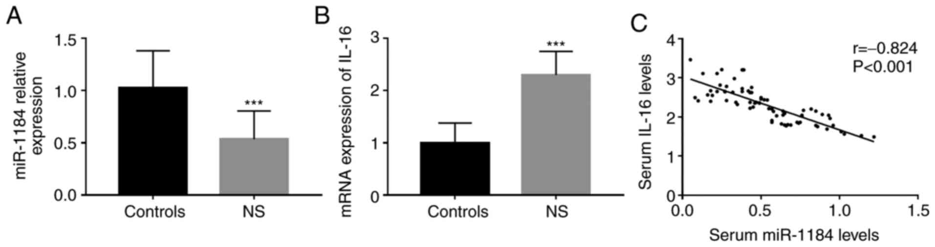
Serum levels of miR-1184 and IL-16 and
their correlation in patients with NS
The serum levels of miR-1184 and IL-16 mRNA in
neonates with sepsis and controls were measured using RT-qPCR. As
presented in Fig. 1A and B, the neonates with NS had significantly
lower miR-1184 levels and higher IL-16 mRNA levels than those in
the control group (P<0.001 for each). Furthermore, a negative
correlation between the serum levels of miR-1184 and IL-16 was
observed in neonates with NS (r=-0.824, P<0.001; Fig. 1C).
Correlation between miR-1184 and
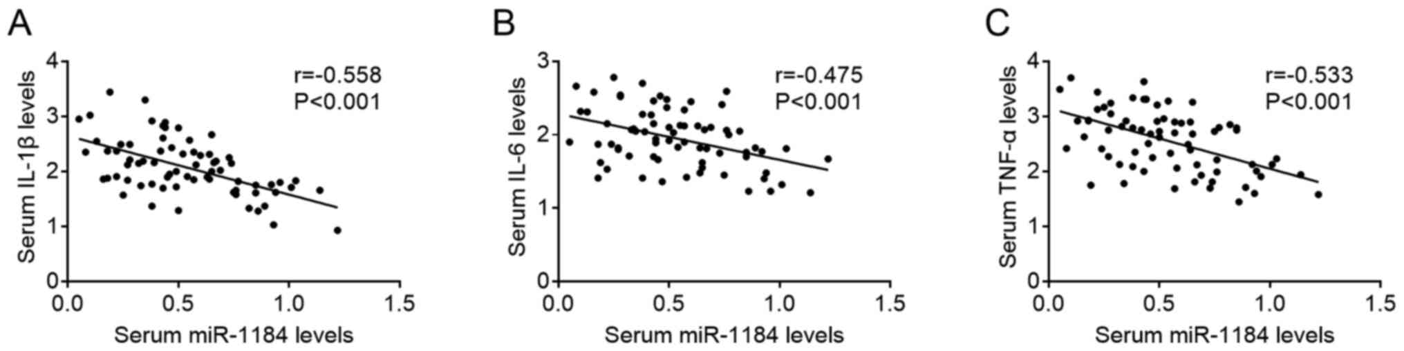
inflammatory response in patients with NS
The inflammatory response in patients with NS was
evaluated by measuring the levels of serum pro-inflammatory
cytokines, including IL-1β, IL-6 and TNF-α. The results presented
in Table II and Fig. 2 indicated that the serum levels of
miR-1184 were negatively correlated with the serum levels of IL-1β,
IL-6 and TNF-α, which suggested a potential relationship between
miR-1184 and the inflammation in the progression of NS.
| Table IICorrelation between serum miR-1184
levels and pro-inflammatory cytokines in neonates with sepsis. |
Table II
Correlation between serum miR-1184
levels and pro-inflammatory cytokines in neonates with sepsis.
| | miR-1184 |
|---|
| Cytokine | r-value | P-value |
|---|
| IL-1β | -0.558 | <0.001 |
| IL-6 | -0.475 | <0.001 |
| TNF-α | -0.533 | <0.001 |
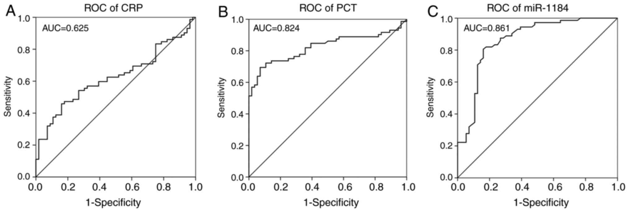
Diagnostic value of miR-1184 for
NS
Due to the significant dysregulation of miR-1184 in
the serum of patients with NS, the diagnostic significance of serum
miR-1184 was assessed in the present study. The ROC curves for CRP
(Fig. 3A) and PCT (Fig. 3B), which are two widely used markers
to diagnose sepsis, were first constructed. The area under the
curve (AUC) for CRP was 0.625 with a sensitivity of 45.83% and a
specificity of 83.93% at a cutoff value of 15.370. The AUC for PCT
was 0.824 with a sensitivity of 69.44% and a specificity of 92.86%
at a cutoff value of 3.375. The ROC curve for the levels of
miR-1184 is presented in Fig. 3C;
the AUC was 0.861 with a sensitivity and specificity of 80.56 and
83.93%, respectively, at a cutoff value of 1.307.
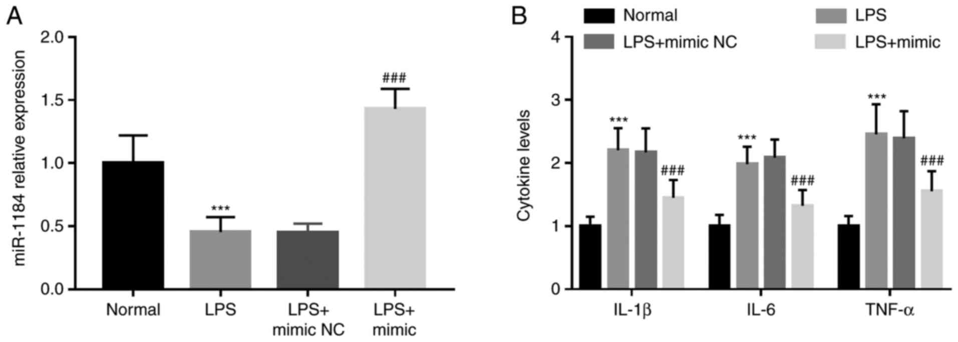
miR-1184 ameliorates LPS-induced
inflammation in monocytes
In the present study, monocytes were induced with
LPS to establish an in vitro model of sepsis with active
inflammation. Similar to the results in neonates with sepsis, the
expression levels of miR-1184 were also downregulated in cells
after LPS induction compared with those in untreated cells
(P<0.001; Fig. 4A). Following
transfection of the miR-1184 mimics, the expression levels of
miR-1184 were confirmed to be significantly upregulated in the
monocytes (P<0.001; Fig. 4A).
Furthermore, overexpression of miR-1184 in monocytes reversed the
effect of LPS to increase the levels of IL-1β, IL-6 and TNF-α (all
P<0.001; Fig. 4B), which meant
that miR-1184 was able to weaken the LPS-induced inflammatory
response.
miR-1184 directly regulates IL-16
expression in monocytes
In order to further confirm the direct interaction
between miR-1184 and IL-16, a luciferase reporter assay was
performed. A binding site for miR-1184 was identified in the 3'-UTR
of IL-16 (Fig. 5A). A subsequent
luciferase reporter assay indicated that miR-1184 mimics markedly
inhibited the relative luciferase activity in the WT group
(P<0.05), while there was no change in the MUT group (Fig. 5B). In addition, the LPS-induced
elevation of IL-16 mRNA expression in monocytes was inhibited by
overexpression of miR-1184 (P<0.01; Fig. 5C). These data suggested that
miR-1184 directly regulates IL-16 in monocytes.
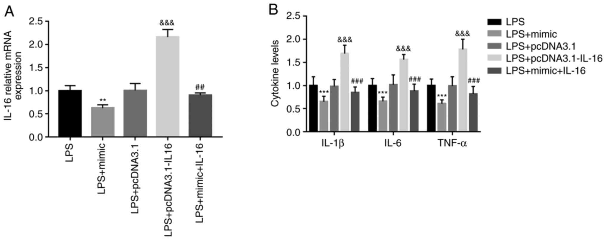
Effect of miR-1184/IL-16 on
LPS-induced inflammatory response in monocytes
To verify whether IL-16 mediated the regulation of
miR-1184 on LPS-induced inflammation, IL-16, which was inhibited by
miR-1184 mimics, was overexpressed using pcDNA3.1-IL-16 (P<0.01;
Fig. 6A). Regarding the
inflammatory responses in LPS-induced monocytes, the results
suggested that the inhibition of IL-1β, IL-6 and TNF-α following
transfection with miR-1184 mimics was abolished by simultaneous
overexpression of IL-16 (P<0.001; Fig. 6B).
Discussion
Numerous studies have indicated that miRNAs have
pivotal roles in the development of various human diseases,
including cancer (23),
neurodegenerative diseases (24)
and metabolic diseases (25). There
are various functional miRNAs that are associated with inflammatory
disease progression by regulating the inflammatory response,
including NS. For instance, miR-223 is an anti-inflammatory miRNA
that is mainly expressed in myeloid cells (26). miR-144 was observed to induce
autophagy and inflammatory responses in microglia after cerebral
hemorrhage by regulating mTOR (27). Overexpression of miR-300 enhanced
autophagy by targeting nicotinamide phosphoribosyltransferase to
reduce the inflammatory response (28). miR-26a was reported to participate
in the pathogenesis of NS by targeting its downstream
IL-16(29). These studies highlight
the key roles of miRNAs in the pathogenesis of inflammatory
diseases, including NS, by regulating inflammatory responses. In
the present study, subjects with NS and neonates with respiratory
tract infection or pneumonia were used as controls, but healthy
neonates were not recruited. Consistent with the results of a
previous study by Chen et al (14), who reported that miR-1184 may
directly regulate IL-16 and was downregulated in neonates with
sepsis compared with uninfected neonates. The results of the
present study revealed that miR-1184 expression was decreased in
neonates with sepsis compared with neonates exhibiting respiratory
tract infection or pneumonia, and decreased in LPS-induced
monocytes compared with normal cells. Therefore, the abnormal
expression of miR-1184 in NS suggested that miR-1184 may be
involved in the development of NS.
Accurate diagnosis is the first and most important
prerequisite for the efficient treatment of diseases. To improve
the treatment of infectious diseases in neonates, it is of value to
distinguish cases of NS from neonates with pneumonia/respiratory
tract infection (30). CRP and PCT
were established as biomarkers for NS and are widely used for this
purpose, but elevated CRP and PCT may also be detected in certain
infectious and inflammatory diseases other than NS, such as
pneumonia (31) and respiratory
tract infection (32); thus, their
application specificity is limited. At present, the existing
diagnostic methods for NS are limited due to their poor sensitivity
and specificity. Thus, novel biomarkers with high sensitivity and
specificity are needed to improve the diagnosis of NS. miRNAs are
stable and can be easily detected from body fluids, such as serum
and tears, meaning they may be detected as biomarkers in biological
fluids and archival tissues (33,34).
Plasma miR-1290 was reported as a novel and specific biomarker for
early diagnosis of necrotizing enterocolitis (35). miR-15a and miR-16 have been
evaluated in blood samples from patients with NS and had were
indicated to be diagnostic and prognostic biomarkers for NS
(11). In the present study, no
significant differences were observed in age, sex, body weight and
WBC between the controls and subjects with NS. However, the cases
with NS had significantly higher levels of CRP and PCT than the
controls, suggesting that the levels of CRP and PCT may be able to
screen NS cases from the neonates with pneumonia/respiratory tract
infection. Thus, ROC curve analysis for CRP and PCT to distinguish
NS from respiratory conditions in neonates was performed. Given the
significant downregulation of miR-1184 in serum samples of patients
with NS, a ROC curve for miR-1184 was also constructed to compare
its diagnostic value with that of CRP and PCT. The analysis
indicated that abnormal expression of miR-1184, which may be used
as a biomarker for the diagnosis of NS, had a relatively high
diagnostic accuracy in newborns with considerable sensitivity and
specificity, and had a better diagnostic performance compared with
CRP and PCT. The present study may provide novel and effective
diagnostic biomarkers for NS. Previous studies have indicated that
biomarkers used for EOS and LOS may not be the same. For instance,
a previous study reported differentially expressed plasma soluble
CD14 subtypes between EOS and LOS, and emphasized the importance to
notice differences when treating EOS and LOS (36). Furthermore, miR-23b expression,
which may serve as a good marker, was upregulated in EOS but
downregulated in LOS, and its expression was altered during
different neonatal periods (37).
In the present study, only 6 cases of EOS were included in the NS
group, which was not sufficient to distinguish EOS from LOS for
analysis. Considering the potential differences in the progression
of EOS and LOS, future studies by our group will expand the sample
size and perform analyses in EOS and LOS separately.
NS is characterized by an inflammatory response. To
mimic the activated inflammation in sepsis, LPS-induced monocytes
have been widely used to investigate the molecules involved in the
inflammation that is part of the pathogenesis of sepsis (38). Zhang et al (39) suggested that the circulating levels
of miR-23b were decreased in patients with sepsis and were able to
inhibit the LPS-induced inflammatory response in monocytes.
Overexpression of miR-181a inhibited the LPS-induced inflammatory
response at least partially by targeting Toll-like receptor
4(30). The present study
determined that the expression of miR-1184 was also downregulated
in LPS-induced monocytes and overexpression of miR-1184 reversed
the promoting effect of LPS on the inflammatory response in
monocytes. These results suggested that miR-1184 may be involved in
NS progression by regulating the inflammatory response and may be
used as a therapeutic target.
The C-terminal fragment IL-16C is a bioactive
protein that is a secreted pro-inflammatory cytokine (40). IL-16 has been reported to be
upregulated in patients with NS (41). A previous study suggested that IL-16
was a target of miR-1184 and was inhibited by miR-1184(14). In the present study, the results of
a luciferase reporter assay and the relative mRNA expression data
of IL-16 in monocytes indicated that miR-1184 directly regulates
the expression of IL-16 in monocytes. Furthermore, the present
study simultaneously upregulated miR-1184 and IL-16 levels in
LPS-induced monocytes to determine whether IL-16 was involved in
the regulation of miR-1184 during the inflammatory response by
detecting the changes in inflammatory factor levels. The results
indicated that in monocytes co-transfected with miR-1184 and IL-16
overexpression vectors, the inhibitory effect of miR-1184 mimics on
inflammation was abolished by overexpression of IL-16. In addition,
serum miR-1184 in patients with NS was negatively correlated with
IL-16. These results suggested that the inhibitory effect of
miR-1184 on inflammation in NS may be achieved by inhibiting IL-16.
IL-16 has been indicated to facilitate the production of
pro-inflammatory cytokines in monocytes, such as IL-1β, IL-6, IL-15
and TNF-α (16). In addition, Huang
et al (42) reported that
IL-16 regulated IL-10, IL-1a and IL-6 expression and served as a
mediator of miR-145-3p to regulate macrophage polarization. Thus,
IL-16 may also act as an important molecule in the pathogenesis of
NS and the miR-1184/IL-16 axis may be a novel therapeutic target
for the treatment of NS.
In conclusion, the present study indicated that
downregulated miR-1184 in the serum of neonates with sepsis may be
closely linked to NS-related inflammation and may be used as a
candidate biomarker for the diagnosis of NS. Overexpression of
miR-1184 in monocytes may improve the LPS-induced inflammatory
response by targeting IL-16, suggesting that the miR-1184/IL-16
axis may provide a novel therapeutic target for the treatment of
NS. A limitation of the present study is the use of only in
vitro experiments to evaluate the regulatory effects of
miR-1184 on NS-related inflammation, with no in vivo animal
experiments (43), which may be one
of the limitations of the present study. In addition, the sample
size was relatively small, which may limit the accuracy of the
clinical study data and the understanding of the potential role of
the miR-1184/IL-16 axis in cases with EOS or LOS individually.
Thus, further investigations in a large study cohort are
necessary.
Acknowledgements
Not applicable.
Funding
Funding: No funding received.
Availability of data and materials
The datasets used and/or analyzed during the current
study are available from the corresponding author on reasonable
request.
Authors' contributions
DW and LH designed and conceived the study,
conducted clinical and cellular experiments, analyzed the data,
wrote the manuscript, and confirmed the authenticity of the raw
data. All authors read and approved the final manuscript.
Ethics approval and consent to
participate
Written informed consent was obtained from the
parents of each patient and the experimental procedures were
approved by the Ethics Committee of Weifang People's Hospital
(Weifang, China).
Patient consent for publication
Not applicable.
Competing interests
The authors declare that they have no competing
interests.
References
|
1
|
Huang H and Tu L: Expression of S100
family proteins in neonatal rats with sepsis and its significance.
Int J Clin Exp Pathol. 8:1631–1639. 2015.PubMed/NCBI
|
|
2
|
Shane AL, Sánchez PJ and Stoll BJ:
Neonatal sepsis. Lancet. 390:1770–1780. 2017.PubMed/NCBI View Article : Google Scholar
|
|
3
|
Aydemir C, Aydemir H, Kokturk F, Kulah C
and Mungan AG: The cut-off levels of procalcitonin and C-reactive
protein and the kinetics of mean platelet volume in preterm
neonates with sepsis. BMC Pediatr. 18(253)2018.PubMed/NCBI View Article : Google Scholar
|
|
4
|
Giannoni E, Agyeman PKA, Stocker M,
Posfay-Barbe KM, Heininger U, Spycher BD, Bernhard-Stirnemann S,
Niederer-Loher A, Kahlert CR, Donas A, et al: Swiss Pediatric
Sepsis Study: Neonatal sepsis of early onset, and hospital-acquired
and community-acquired late onset: a prospective population-based
cohort study. J Pediatr. 201:106–114.e4. 2018.PubMed/NCBI View Article : Google Scholar
|
|
5
|
Tzialla C, Manzoni P, Achille C, Bollani
L, Stronati M and Borghesi A: New diagnostic possibilities for
neonatal sepsis. Am J Perinatol. 35:575–577. 2018.PubMed/NCBI View Article : Google Scholar
|
|
6
|
Wynn JL: Defining neonatal sepsis. Curr
Opin Pediatr. 28:135–140. 2016.PubMed/NCBI View Article : Google Scholar
|
|
7
|
Sharma D, Farahbakhsh N, Shastri S and
Sharma P: Biomarkers for diagnosis of neonatal sepsis: a literature
review. J Matern Fetal Neonatal Med. 31:1646–1659. 2018.PubMed/NCBI View Article : Google Scholar
|
|
8
|
Memar MY, Alizadeh N, Varshochi M and
Kafil HS: Immunologic biomarkers for diagnostic of early-onset
neonatal sepsis. J Matern Fetal Neonatal Med. 32:143–153.
2019.PubMed/NCBI View Article : Google Scholar
|
|
9
|
Vishnoi A and Rani S: miRNA biogenesis and
regulation of diseases: an overview. Methods Mol Biol. 1509:1–10.
2017.PubMed/NCBI View Article : Google Scholar
|
|
10
|
Mishra S, Yadav T and Rani V: Exploring
miRNA based approaches in cancer diagnostics and therapeutics. Crit
Rev Oncol Hematol. 98:12–23. 2016.PubMed/NCBI View Article : Google Scholar
|
|
11
|
Wang X, Wang X, Liu X, Wang X, Xu J, Hou
S, Zhang X and Ding Y: miR-15a/16 are upreuglated in the serum of
neonatal sepsis patients and inhibit the LPS-induced inflammatory
pathway. Int J Clin Exp Med. 8:5683–5690. 2015.PubMed/NCBI
|
|
12
|
Huang HC, Yu HR, Huang LT, Huang HC, Chen
RF, Lin IC, Ou CY, Hsu TY and Yang KD: miRNA-125b regulates TNF-α
production in CD14+ neonatal monocytes via post-transcriptional
regulation. J Leukoc Biol. 92:171–182. 2012.PubMed/NCBI View Article : Google Scholar
|
|
13
|
Huang HC, Yu HR, Hsu TY, Chen IL, Huang
HC, Chang JC and Yang KD: MicroRNA-142-3p and let-7g negatively
regulates augmented IL-6 production in neonatal polymorphonuclear
leukocytes. Int J Biol Sci. 13:690–700. 2017.PubMed/NCBI View Article : Google Scholar
|
|
14
|
Chen J, Jiang S, Cao Y and Yang Y: Altered
miRNAs expression profiles and modulation of immune response genes
and proteins during neonatal sepsis. J Clin Immunol. 34:340–348.
2014.PubMed/NCBI View Article : Google Scholar
|
|
15
|
Ahmad SF, Ansari MA, Nadeem A, Bakheet SA,
Al-Ayadhi LY and Attia SM: Elevated IL-16 expression is associated
with development of immune dysfunction in children with autism.
Psychopharmacology (Berl). 236:831–838. 2019.PubMed/NCBI View Article : Google Scholar
|
|
16
|
Mathy NL, Scheuer W, Lanzendörfer M,
Honold K, Ambrosius D, Norley S and Kurth R: Interleukin-16
stimulates the expression and production of pro-inflammatory
cytokines by human monocytes. Immunology. 100:63–69.
2000.PubMed/NCBI View Article : Google Scholar
|
|
17
|
Zhang F, Yang C, Xing Z, Liu P, Zhang B,
Ma X, Huang L and Zhuang L: LncRNA GAS5-mediated miR-1323 promotes
tumor progression by targeting TP53INP1 in hepatocellular
carcinoma. OncoTargets Ther. 12:4013–4023. 2019.PubMed/NCBI View Article : Google Scholar
|
|
18
|
Smith S, Wu PW, Seo JJ, Fernando T, Jin M,
Contreras J, Montano EN, Gabhann JN, Cunningham K, Widaa A, et al:
IL-16/miR-125a axis controls neutrophil recruitment in
pristane-induced lung inflammation. JCI Insight.
3(120798)2018.PubMed/NCBI View Article : Google Scholar
|
|
19
|
Plotnikov EY, Brezgunova AA, Pevzner IB,
Zorova LD, Manskikh VN, Popkov VA, Silachev DN and Zorov DB:
Mechanisms of LPS-induced acute kidney injury in neonatal and adult
rats. Antioxidants. 7(E105)2018.PubMed/NCBI View Article : Google Scholar
|
|
20
|
Subspecialty Group of Neonatology
Pediatric Society Chinese Medical Association; Editorial Board
Chinese Journal of Pediatrics. Protocol for diagnosis and treatment
of neonatal septicemia. Zhonghua Er Ke Za Zhi. 41:897–899.
2003.PubMed/NCBI(In Chinese).
|
|
21
|
Yu HR, Chen RF, Hong KC, Bong CN, Lee WI,
Kuo HC and Yang KD: IL-12-independent Th1 polarization in human
mononuclear cells infected with varicella-zoster virus. Eur J
Immunol. 35:3664–3672. 2005.PubMed/NCBI View Article : Google Scholar
|
|
22
|
Livak KJ and Schmittgen TD: Analysis of
relative gene expression data using real-time quantitative PCR and
the 2(-Delta Delta C(T)) Method. Methods. 25:402–408.
2001.PubMed/NCBI View Article : Google Scholar
|
|
23
|
Rupaimoole R and Slack FJ: MicroRNA
therapeutics: Towards a new era for the management of cancer and
other diseases. Nat Rev Drug Discov. 16:203–222. 2017.PubMed/NCBI View Article : Google Scholar
|
|
24
|
Ferrante M and Conti GO: Environment and
neurodegenerative diseases: an update on miRNA role. MicroRNA.
6:157–165. 2017.PubMed/NCBI View Article : Google Scholar
|
|
25
|
Correia de Sousa M, Gjorgjieva M, Dolicka
D, Sobolewski C and Foti M: Deciphering miRNAs' action through
miRNA editing. Int J Mol Sci. 20(E6249)2019.PubMed/NCBI View Article : Google Scholar
|
|
26
|
Jeffries J, Zhou W, Hsu AY and Deng Q:
miRNA-223 at the crossroads of inflammation and cancer. Cancer
Lett. 451:136–141. 2019.PubMed/NCBI View Article : Google Scholar
|
|
27
|
Yu A, Zhang T, Zhong W, Duan H, Wang S, Ye
P, Wang J, Zhong S and Yang Z: miRNA-144 induces microglial
autophagy and inflammation following intracerebral hemorrhage.
Immunol Lett. 182:18–23. 2017.PubMed/NCBI View Article : Google Scholar
|
|
28
|
Li Y, Ke J, Peng C, Wu F and Song Y:
MicroRNA-300/NAMPT regulates inflammatory responses through
activation of AMPK/mTOR signaling pathway in neonatal sepsis.
Biomed Pharmacother. 108:271–279. 2018.PubMed/NCBI View Article : Google Scholar
|
|
29
|
Cheng Q, Tang L and Wang Y: Regulatory
role of miRNA-26a in neonatal sepsis. Exp Ther Med. 16:4836–4842.
2018.PubMed/NCBI View Article : Google Scholar
|
|
30
|
Liu G, Liu W and Guo J: Clinical
significance of miR-181a in patients with neonatal sepsis and its
regulatory role in the lipopolysaccharide-induced inflammatory
response. Exp Ther Med. 19:1977–1983. 2020.PubMed/NCBI View Article : Google Scholar
|
|
31
|
Guo S, Mao X and Liang M: The moderate
predictive value of serial serum CRP and PCT levels for the
prognosis of hospitalized community-acquired pneumonia. Respir Res.
19(193)2018.PubMed/NCBI View Article : Google Scholar
|
|
32
|
Kim HS, Won S, Lee EK, Chun YH, Yoon JS,
Kim HH and Kim JT: Pentraxin 3 as a clinical marker in children
with lower respiratory tract infection. Pediatr Pulmonol. 51:42–48.
2016.PubMed/NCBI View Article : Google Scholar
|
|
33
|
To KK, Tong CW, Wu M and Cho WC: MicroRNAs
in the prognosis and therapy of colorectal cancer: From bench to
bedside. World J Gastroenterol. 24:2949–2973. 2018.PubMed/NCBI View Article : Google Scholar
|
|
34
|
Benz F, Roy S, Trautwein C, Roderburg C
and Luedde T: Circulating microRNAs as biomarkers for sepsis. Int J
Mol Sci. 17(E78)2016.PubMed/NCBI View Article : Google Scholar
|
|
35
|
Ng PC, Chan KYY, Yuen TP, Sit T, Lam HS,
Leung KT, Wong RPO, Chan LCN, Pang YLI, Cheung HM, et al: Plasma
miR-1290 is a novel and specific biomarker for early diagnosis of
necrotizing enterocolitis-biomarker discovery with prospective
cohort evaluation. J Pediatr. 205:83–90.e10. 2019.PubMed/NCBI View Article : Google Scholar
|
|
36
|
van Maldeghem I, Nusman CM and Visser DH:
Soluble CD14 subtype (sCD14-ST) as biomarker in neonatal
early-onset sepsis and late-onset sepsis: A systematic review and
meta-analysis. BMC Immunol. 20(17)2019.PubMed/NCBI View Article : Google Scholar
|
|
37
|
Fatmi A, Rebiahi SA, Chabni N, Zerrouki H,
Azzaoui H, Elhabiri Y, Benmansour S, Ibáñez-Cabellos JS, Smahi MC,
Aribi M, et al: miRNA-23b as a biomarker of culture-positive
neonatal sepsis. Mol Med. 26(94)2020.PubMed/NCBI View Article : Google Scholar : Erratum in: Mol Med
26: 129 2020.
|
|
38
|
Schüller SS, Wisgrill L, Herndl E,
Spittler A, Förster-Waldl E, Sadeghi K, Kramer BW and Berger A:
Pentoxifylline modulates LPS-induced hyperinflammation in monocytes
of preterm infants in vitro. Pediatr Res. 82:215–225.
2017.PubMed/NCBI View Article : Google Scholar
|
|
39
|
Zhang W, Lu F, Xie Y, Lin Y, Zhao T, Tao
S, Lai Z, Wei N, Yang R, Shao Y, et al: miR-23b negatively
regulates sepsis-induced inflammatory responses by targeting ADAM10
in human THP-1 monocytes. Mediators Inflamm.
2019(5306541)2019.PubMed/NCBI View Article : Google Scholar
|
|
40
|
Roth S, Solbach W and Laskay T: IL-16 and
MIF: Messengers beyond neutrophil cell death. Cell Death Dis.
7(e2049)2016.PubMed/NCBI View Article : Google Scholar
|
|
41
|
Sugitharini V, Prema A and Berla Thangam
E: Inflammatory mediators of systemic inflammation in neonatal
sepsis. Inflamm Res. 62:1025–1034. 2013.PubMed/NCBI View Article : Google Scholar
|
|
42
|
Huang Y, Du KL, Guo PY, Zhao RM, Wang B,
Zhao XL and Zhang CQ: IL-16 regulates macrophage polarization as a
target gene of mir-145-3p. Mol Immunol. 107:1–9. 2019.PubMed/NCBI View Article : Google Scholar
|
|
43
|
Xia D, Yao R, Zhou P, Wang C, Xia Y and Xu
S: LncRNA NEAT1 reversed the hindering effects of miR-495-3p/STAT3
axis and miR-211/PI3K/AKT axis on sepsis-relevant inflammation. Mol
Immunol. 117:168–179. 2020.PubMed/NCBI View Article : Google Scholar
|